 Open access peer-reviewed chapter


Abstract
miRNA-mRNA interaction depends on multiple factors such as 3’UTR isoforms, the cell and tissue-specific expression levels of RNA-binding proteins, the sequence context around the mRNA target site, and other mechanisms. Genetic polymorphisms within miRNAs and their target sites appear to be among the most important ones because they influence the mode and outcome of miRNA-mRNA interaction universally and irreversibly. SNP disruption of miRNAs and their binding sites, as well as conformational changes preventing the access of the miRNA to its target site, are adopted as the most credible mechanistic explanations of SNP-mediated effects. The occurrence of multiple SNPs within the same miRNA-binding site implies their combinatorial mode of action. The presence of the repetitive (homologous) binding sites for the same miRNA on its mRNA target may both enhance the miRNA targeting and provide for the backup target site instead of the one disrupted by SNP, thus rescuing the miRNA functionality. While being underexplored, the multiple genetic polymorphisms within the miRNA-binding sites, as well as homologous miRNA-binding sites, may be considered as additional factors influencing miRNA-mediated regulation of gene expression.
Keywords
- microRNA
- mRNA
- target
- single-nucleotide polymorphism
- homologous microRNA-binding sites
1. Introduction
Since the beginning of the 2000s, miRNAs stay in the focus of every aspect of medical and biomedical research [1, 2]. miRNAs are involved in a wide range of biological processes such as cell differentiation [3, 4, 5, 6, 7] and reprogramming [8], cellular senescence [9] and cell death [10, 11, 12, 13, 14, 15, 16], tissue and organ development [5, 17, 18, 19, 20] and regeneration [21, 22, 23, 24, 25, 26], cell signaling [27, 28], oxidative stress and metabolism [3, 29, 30, 31, 32, 33, 34, 35, 36, 37], mitochondrial dysfunctions [22, 38, 39], hormonal regulation [16, 40, 41] and adaptive responses [17], brain function [42, 43], inflammation [31, 44, 45, 46], immune response and the effects of the microbiome [46, 47, 48, 49, 50, 51, 52, 53], viral infections and latency [43, 54, 55, 56, 57, 58, 59, 60, 61], DNA damage and repair [22, 62, 63, 64, 65, 66, 67], genomic balance [68] and genomic instability [65, 69], etc.
miRNAs deregulation promotes the biological processes resulting in various human diseases [41, 70, 71, 72, 73, 74, 75, 76, 77, 78, 79, 80, 81, 82, 83, 84] including oncogenesis [6, 7, 35, 37, 54, 59, 84, 85, 86, 87, 88, 89, 90, 91, 92, 93, 94, 95, 96, 97, 98, 99, 100, 101, 102, 103, 104, 105, 106, 107, 108, 109, 110, 111, 112, 113, 114, 115, 116, 117, 118, 119, 120]. miRNAs are recognized as important factors that are involved in tumor cell invasiveness [121], metastases [90, 121, 122, 123, 124, 125, 126, 127, 128, 129], cancer cachexia [130], drug resistance [131, 132, 133, 134, 135, 136], sensitivity to chemotherapy [65, 137, 138], and radiosensitivity [138, 139, 140]. miRNAs are actively explored as both therapeutic agents [32, 112, 141, 142, 143, 144, 145, 146, 147, 148, 149, 150, 151, 152, 153, 154, 155, 156, 157, 158, 159, 160, 161, 162, 163, 164] and targets [28, 42, 161, 165, 166, 167, 168, 169, 170, 171, 172, 173, 174]. A growing number of reports point to the usefulness of miRNAs, and specifically circulating miRNAs [175, 176] as diagnostic and prognostic biomarkers [51, 70, 71, 73, 110, 112, 155, 160, 161, 174, 177, 178, 179, 180, 181, 182, 183, 184, 185, 186, 187, 188, 189, 190, 191, 192, 193, 194, 195, 196, 197, 198, 199, 200, 201, 202, 203, 204, 205, 206, 207, 208, 209, 210, 211, 212, 213, 214, 215, 216, 217, 218, 219, 220, 221, 222, 223] related to clinical and forensic [177, 224] studies, as well as normal physiological conditions, such as, diet [225] and increased physical activity [226, 227].
Deep sequencing and computational approaches indicate that miRNA genes comprise about 0.5–1% of the predicted genes in animals and humans [228]. One mRNA can be targeted by hundreds of miRNAs [84, 229], and a single miRNA can potentially recognize hundreds of different target transcripts [229, 230], which often share pathways to ensure their impact [52]. Therefore, miRNAs may be considered as global regulators of gene expression [231] with the vast
Identification and characterization of the factors influencing miRNA functionality are essential to elucidate the mechanisms of miRNA activity and to explain and predict the effects of miRNAs for clinical applications. Genetic variations of both miRNAs and their target sites may have a significant impact on the efficacy of miRNA targeting. It is growingly recognized that naturally occurring variations in miRNAs and their target genes contribute to phenotypic complexity and may be associated with human pathologies [240, 241], including cancer [231, 242, 243, 244]. We present information about two underexplored SNP-related mechanisms influencing miRNA-mRNA interaction: the multiple SNPs within the single miRNA target site and the multiple (homologous) target sites for the same miRNA.
2. miRNA properties and biogenesis
miRNAs belong to the category of small noncoding RNA molecules [2, 245] ranging between 19 and 24 nucleotides in length [229]. miRNAs have been detected in all biological species, including viruses [55, 56, 57, 58, 59, 60, 246]. Of note, bacteria do have short RNA sequences with the miRNA-similar functions, yet they are not considered as true miRNAs [247]. Bioinformatics analysis indicates the existence of miRNAs derived from transposable elements [248, 249]. Although miRNAs are usually located in the cytoplasm, several studies have detected a regulatory role of miRNAs in other cell compartments such as mitochondria
Circulating miRNA may be released by different types of cells and delivered into recipient cells for functional purposes, acting as cell-to-cell signaling mediators [251]. miRNA may be freed from dying cells or selectively sorted into the secreted small vesicles called exosomes [158, 229, 252, 253, 254, 255]. Such exosomal miRNAs are regarded as promising clinical biomarkers (see above). miRNA can undergo both vertical and horizontal transmissions among distinct species, remarkably through feeding between plants and animals [229, 245].
The biogenesis of canonical animal miRNAs starts with the transcription of the pri-miRNA by RNA Pol II [256, 257]. The pri-miRNA has the length of about several thousands of nucleotides [258], the 7-methylguanosine, and 3′- poly(A) tail [259]. The pri-miRNA forms a stem-loop structure, which is recognized and cleaved by
The core miRNA function is posttranscriptional regulation of the expression of targeted genes. It is achieved through miRNA-mediated RNA silencing [269]. miRNAs, as a part of the RISC, trigger the various forms of the translational repression of the target mRNA
While target repression is universally recognized as the dominant mode of miRNA action, some reports indicate that miRNAs can stabilize the targeted transcript [234, 272, 273, 274, 275, 276]. MiRNAs can also regulate gene expression both indirectly by targeting the mRNAs of transcription factors and directly after being transported back to the nucleus and binding to the complementary sequence in the promoter [232, 234, 250, 262, 276]. miRNAs can also perform other noncanonical functions acting as ligands for toll-like receptors [277].
3. MiRNA target recognition
miRNAs bind to the complementary sequences of the host mRNA as a part of the RISC with its core component
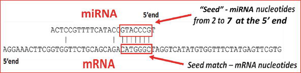
Figure 1.
miRNA-mRNA interaction in animal cells (developed by F. Ahrend).
Canonical miRNA-binding sites are positioned in the 3’UTR of the targeted mRNA sequence and classified upon the extent and location of matching miRNA nucleotides: 6mers perfectly pair to nucleotides 2–7 on the 5′-end of miRNA, 7-merA1 and 7mer8—additional pairing with miRNA nucleotide 1 or 8 respectively, and 8mer sites match miRNA nucleotides 1–8 [261]. The efficacy of these sites is usually augmented by additional pairing at the 3′ end of miRNAs. In addition to the binding site at the miRNA 5′ end, pairing around the binding sites contributes to the targeting efficacy [233, 261, 280]. The so-called 3′-supplementary sites with atypical elaboration of the 6mer, 7mer, and 8mer sites (≥3–4 pairs), and 3′-compensatory sites stretching for more than 4–5 pairs, while the seed region is mismatched are called atypical sites [84, 233, 261]. The supplementary pairing is believed to enhance the miRNA-mRNA interaction with the greater specificity. However, in animal cells, the high complementarity beyond the seed may cause TDMD [281], the common mechanism of destabilization of the short RNA molecules including miRNAs, which is observed in many diseases [84]. In general, the miRNA-mRNA complementarity with nucleotides at 3′ end or with nucleotides in the center (centered pairing) in the absence of the perfectly matched seed nucleotides is believed to be much less effective in regulating gene expression [84, 233, 261, 282, 283, 284]. The noncanonical seed-matching sites may also be in the protein-coding regions of the target mRNAs and even in the 5’UTR mRNA sequence [84, 232, 233, 285, 286].
4. Factors influencing miRNA targeting
miRNA-mRNA interaction depends on a range of factors. The posttranscriptional modification resulting in miRNA methylation causes structural changes that affect AGO binding [232]. miRNA sponges [287, 288, 289] bind miRNAs, thus preventing them from interacting with mRNA targets. miRNAs competition and cross talk with RNA-binding proteins [84, 232, 233, 236, 270], whose expression levels may depend on the cellular context and vary between the tissues [261, 290], also add up to the spectrum of various regulatory outcomes beside the dominating mechanism of miRNA targeting with moderate target repression [84]. miRNA targeting is known to depend on 3’UTR isoforms: 3’UTR size, alternative cleavage, and the location of target sequences within AU-rich regions [233, 261, 282, 284, 290, 291, 292].
The extent of complementary identity between miRNA and its mRNA targets appears to be one of the crucial factors, which determine the mode of target repression. The target cleavage by RISC is typical for plants, where the pairwise alignment is characterized by high complementarity. In opposite, the limited seed matching in animals often leads to translational inhibition [270]. It has been suggested that the above-mentioned noncanonical stabilization of the targeted transcript by miRNAs may depend on the complementarity between the miRNA and its target mRNA [275]. On the other hand, the extensive base-pairing may also cause TDMD [84, 293]. In this regard, genetic polymorphisms within miRNAs and their target sites appear to be among the most important mechanisms that ubiquitously and irreversibly define the mode and outcome of miRNA-mRNA interaction.
5. miRNAs and genetic polymorphisms
SNPs are genetic variations (nucleotide substitutions and indels) in DNA sequence [294, 295]. SNPs within the mRNA noncoding regions do not change the protein sequence, but they may significantly alter gene expression by affecting transcription, RNA processing, translation, and interaction of mRNA with noncoding regulatory RNAs [296]. While most SNPs are likely to be functionally neutral, some of them may represent causative links with human diseases [297]. SNPs in miRNA-precursor flanking regions, promoters of miRNA-encoding genes, and in the genes involved in miRNA biogenesis (transcription and RNA processing) may result in higher or lower miRNA expression profiles [298, 299, 300, 301, 302], which, in turn, represent the unique disease-specific signatures that can identify cancer types [231, 240, 243, 303]. SNPs within mature miRNA sequences, specifically in their seed regions, dissimilarities in mature miRNAs due to variable cleavage sites for
Within the mRNA targets, SNPs can either create or destroy miRNA-binding sites. It may impair miRNA ability to target oncogenes and, in opposite, render tumor-suppressor genes susceptible to miRNA-mediated inhibition (Figure 2). SNPs located both within and beyond the seed-target regions [322] can affect miRNA-mRNA interaction by destabilizing the mRNA target molecule, slowing down its ability for ribosomal loading, and altering the secondary RNA structure, which in turn, may have an impact on the availability of miRNA-binding sites [284, 290, 323]. Disruption or creation of miRNA-binding sites is recognized as one of the most credible mechanistic explanations of SNP-mediated effects [240, 241].
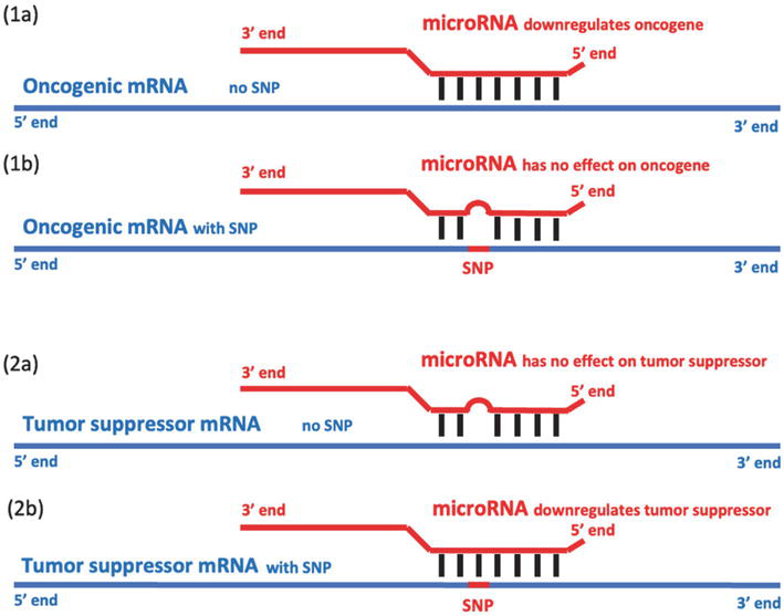
Figure 2.
The hypothetical effects of SNP on miRNA-mRNA interaction and cancer development.
6. Multiple SNPs within miRNA-binding sites
A considerable number of genome-wide association studies demonstrated links between the SNPs within the miRNA target sites and predisposition to various human diseases. The attention was mostly devoted to SNPs, whose population frequency was well above 1%. Still, miRNA target sites may harbor multiple SNPs, although most of them are of low population frequency [324]. Even though the probability of their coincidental occurrence within the site is low, the frequency of some SNPs may vary significantly due to their ethnic disparities [325]. It has been suggested that, at least for some genes, it is not a separately taken SNP, but the combinatorial effect of several SNPs that may determine the outcome and efficacy of miRNA-mediated regulation of gene expression [326]. The location of SNPs (seed-corresponding region, centered, or 3′ end position) should also be taken into consideration.
Thus, the presence of additional, even rare (low frequency) SNPs within miRNA target sites, may modify (weaken or enhance) the effects of the SNPs, which occur at higher frequencies. Consequently, multiple SNPs may increase the probability of the site disruption, but may be also neutral, and even enhancing (Figure 3). This situation is less typical for the protein-coding regions due to the selection pressure resulting in the fewer SNPs. In the case of the overlapping miRNA-binding sites, the outcome is less clear due to the co-targeting effect—additive repression by more than one miRNA [84, 232]. Beside this, the increased complementarity between miRNA and its target in animals may result in miRNA decay (see above about TDMD).
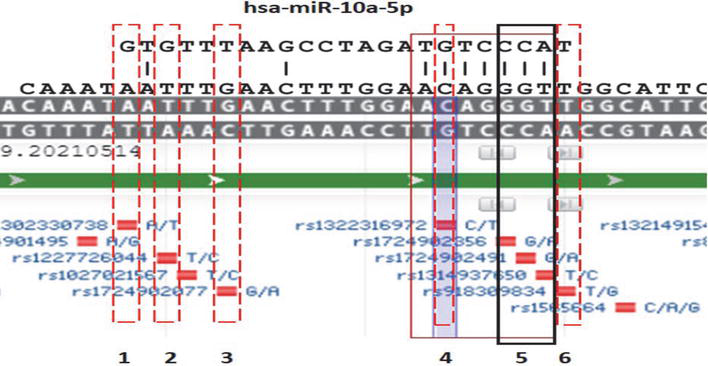
Figure 3.
The examples of disrupting, enhancing, and neutral SNPs within a single target site. The image shows the fragment of the human PDGFRA DNA sequence corresponding to the mRNA 3’UTR region, the miRNA-10a-5P binding site.
SNPs within the single target site may be independent (Figure 4) and mutually exclusive if they are overlapping (Figure 5). Of note, SNP frequencies in the databases reflect the experimental NextGen sequencing data, and the possible SNP overlap is not taken into consideration. If mutually exclusive SNPs are represented by the overlapping indel and nucleotide substitution, the latter one is “shadowed” by the indel (Figure 5), and its real population frequency may be higher than that presented in the database.
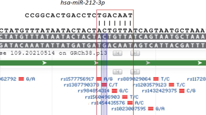
Figure 4.
The fragment of the human PDGFRA DNA sequence corresponding to the mRNA 3’UTR region, the miRNA-212-3P binding site. SNPs rs1577756917, rs1387790378, rs984854314, rs1560496903, and rs1454435576 represent independent events (developed by M. Giurgiu).
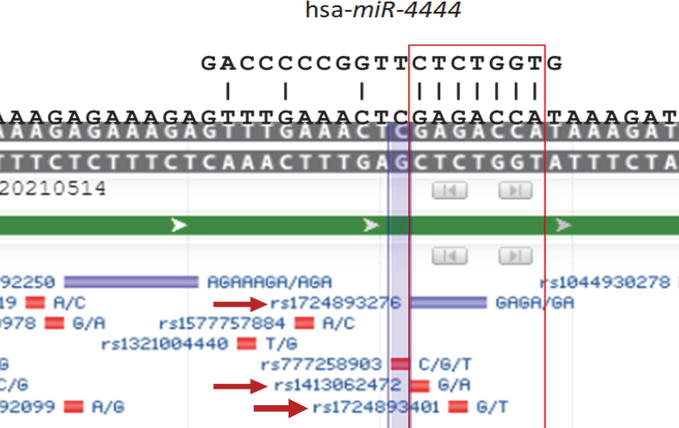
Figure 5.
The fragment of the human PDGFRA DNA sequence corresponding to the mRNA 3’UTR region, the miRNA-4444 binding site. SNPs rs1724893276 and rs1413062472, as well as rs1724893401, represent mutually exclusive events. rs1724893276 is an indel, and in the case of the deletion, neither rs1413062472 nor s172489 3401 (nucleotide substitutions) will be present (developed by M. Giurgiu).
7. Homologous miRNA-binding sites
It has been reported that some mRNAs may have more than one binding (seed-matching) site for the miRNAs [329, 330, 331, 332, 333, 334, 335, 336]. These sites have the analogous seed-binding motif, but the different nucleotide content outside (Figure 6). Such multiple sites are proposed to act synergistically [330, 333, 334, 335, 337, 338], which, in turn, may depend on the distance between the sites [330, 338], as well as the activity and structural variations of miRNA-Ago complexes [336, 339]. It would be rational to suggest that if SNPs disrupt one of the miRNA-binding sites, the other sites for the same miRNA could preserve the miRNA-mediated control of gene expression. Such homologous miRNA-binding sites are present in almost all human genes, and their numbers are significant and directly proportional to the length of the 3’UTR [340].
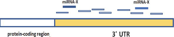
Figure 6.
The repeated (homologous) target sites for miRNA-X (developed by A. Kofman).
8. Bioinformatic tools
Whereas miRNAs and SNPs are both subjects of intense studies, there are few resources that allow to investigate the potential effects of SNPs on miRNA functionality.
9. Conclusions
The ability of miRNAs to regulate the variety of genes in all tissues, as well as their presence in all biological species and their promise for clinical applications as novel diagnostics and therapeutics prompt further exploration of the factors influencing miRNA activity and functions. It is possible to assume that the presence of SNPs within miRNAs and their binding sites represents a powerful mechanism that can influence all biological processes in which miRNAs are involved. However, the growing number of reports indicates that the presence of SNPs cannot be interpreted as unalterable situation defining the biological outcome of miRNA targeting. The combinatorial effect of multiple SNPs located within the same target site, as well as beyond the target site, yet within the region where additional complementary matches between miRNA and its target can be formed, the circumstances where some SNPs may be mutually exclusive, and the homologous sites, which hypothetically can be utilized by miRNAs as an alternative target if the other sites are disrupted or become inaccessible, all these additional factors related to miRNA functionalities are anticipated of being studied with the development of the appropriate biocomputing tools and mathematical models.
Abbreviations
| miRNA | microRNA |
| mRNA | messenger RNA |
| SNP | single-nucleotide polymorphism |
| Pol | polymerase |
| pri-miRNA | primary miRNA transcript |
| pre-miRNA | precursor miRNA |
| AGO | argonaute proteins |
| RISC | RNA-induced silencing complex |
| UTR | untranslated region |
| 3′ | 3-prime |
| 5′ | 5-prime |
| Indel | insertion/deletion |
| PDGFRA | platelet-derived growth factor receptor alpha |
| TDMD | target-directed miRNA degradation |
References
- 1.
Arteaga-Vazquez M, Caballero-Perez J, Vielle-Calzada JP. A family of microRNAs present in plants and animals. Plant Cell. 2006; 18 (12):3355-3369 - 2.
Ambros V. The functions of animal microRNAs. Nature. 2004; 431 (7006):350-355 - 3.
Singh V. Intracellular metabolic reprogramming mediated by micro-RNAs in differentiating and proliferating cells under non-diseased conditions. Molecular Biology Reports. 2021; 48 (12):8123-8140 - 4.
Galagali H, Kim JK. The multifaceted roles of microRNAs in differentiation. Current Opinion in Cell Biology. 2020; 67 :118-140 - 5.
Zare A et al. Epigenetic modification factors and microRNAs network associated with differentiation of embryonic stem cells and induced pluripotent stem cells toward cardiomyocytes: A Review. Life (Basel). 2023; 13 (2):569. DOI: 10.3390/life13020569 - 6.
Souza OF, Popi AF. Role of microRNAs in B-cell compartment: Development, proliferation and Hematological diseases. Biomedicines. 2022; 10 (8):2004. DOI: 10.3390/biomedicines10082004 - 7.
Katsaraki K et al. MicroRNAs: Tiny regulators of gene expression with pivotal roles in Normal B-cell development and B-cell chronic lymphocytic Leukemia. Cancers (Basel). 2021; 13 (4):593. DOI: 10.3390/cancers13040593 - 8.
Pascale E et al. MicroRNA roles in cell reprogramming mechanisms. Cells. 2022; 11 (6):940. DOI: 10.3390/cells11060940 - 9.
Lettieri-Barbato D et al. MicroRNAs, long non-coding RNAs, and circular RNAs in the redox control of cell senescence. Antioxidants (Basel). 2022; 11 (3):480. DOI: 10.3390/antiox11030480 - 10.
Ferris WF. The role and interactions of programmed cell death 4 and its regulation by microRNA in transformed cells of the gastrointestinal tract. Frontiers in Oncology. 2022; 12 :903374 - 11.
Lamberti MJ et al. Damage-associated molecular patterns modulation by microRNA: Relevance on immunogenic cell death and cancer treatment outcome. Cancers (Basel). 2021; 13 (11):2566. DOI: 10.3390/cancers13112566 - 12.
Palumbo S et al. Emerging roles of microRNA in modulating cell-death processes in malignant glioma. Journal of Cellular Physiology. 2014; 229 (3):277-286 - 13.
Chen CH, Guo M, Hay BA. Identifying microRNA regulators of cell death in drosophila. Methods in Molecular Biology. 2006; 342 :229-240 - 14.
Bai X et al. Role of microRNA-34b-5p in cancer and injury: How does it work? Cancer Cell International. 2022; 22 (1):381 - 15.
Azam INA et al. Roles of microRNAs in regulating apoptosis in the pathogenesis of endometriosis. Life (Basel). 2022; 12 (9):1321. DOI: 10.3390/life12091321 - 16.
Matarrese P et al. The sex-related interplay between TME and cancer: On the critical role of Estrogen, MicroRNAs and autophagy. Cancers (Basel). 2021; 13 (13):3287. DOI: 10.3390/cancers13133287 - 17.
DeVeale B, Swindlehurst-Chan J, Blelloch R. The roles of microRNAs in mouse development. Nature Reviews Genetics. 2021; 22 (5):307-323 - 18.
Raza SHA et al. The role of MicroRNAs in muscle tissue development in beef cattle. Genes (Basel). 2020; 11 (3):295. DOI: 10.3390/genes11030295 - 19.
Boshtam M et al. Crosstalk of transcriptional regulators of adaptive immune system and microRNAs: An insight into differentiation and development. Cells. 2023; 12 (4):635. DOI: 10.3390/cells12040635 - 20.
Zolboot N et al. MicroRNAs instruct and maintain cell type diversity in the nervous system. Frontiers in Molecular Neuroscience. 2021; 14 :646072 - 21.
Mahtal N et al. MicroRNAs in kidney injury and disease. Nature Reviews Nephrology. 2022; 18 (10):643-662 - 22.
Rodrigues SC, Cardoso RMS, Duarte FV. Mitochondrial microRNAs: A putative role in tissue regeneration. Biology (Basel). 2021; 9 (12):486. DOI: 10.3390/biology9120486 - 23.
Cione E et al. Liver damage and microRNAs: An update. Current Issues in Molecular Biology. 2022; 45 (1):78-91 - 24.
Maries L et al. MicroRNAs-the heart of post-myocardial infarction Remodeling. Diagnostics (Basel). 2021; 11 (9):1675. DOI: 10.3390/diagnostics11091675 - 25.
Theofilis P et al. The impact of proangiogenic microRNA modulation on blood flow recovery following hind limb ischemia. A systematic review and meta-analysis of animal studies. Vascular Pharmacology. 2021; 141 :106906 - 26.
Tanase DM et al. Current knowledge of MicroRNAs (miRNAs) in acute coronary syndrome (ACS): ST-elevation myocardial infarction (STEMI). Life (Basel). 2021; 11 (10):1057. DOI: 10.3390/life11101057 - 27.
Vaghf A et al. The role of microRNAs in diseases and related signaling pathways. Molecular Biology Reports. 2022; 49 (7):6789-6801 - 28.
Humphries BA, Wang Z, Yang C. MicroRNA regulation of the small rho GTPase regulators-complexities and opportunities in targeting cancer metastasis. Cancers (Basel). 2020; 12 (5):1092. DOI: 10.3390/cancers12051092 - 29.
Tarlton JMR, Patterson S, Graham A. MicroRNA sequences modulated by Beta cell lipid metabolism: Implications for type 2 diabetes mellitus. Biology (Basel). 2021; 10 (6):534. DOI: 10.3390/biology10060534 - 30.
Wallace SR, Pagano PJ, Kracun D. MicroRNAs in the regulation of NADPH oxidases in vascular diabetic and ischemic pathologies: A case for alternate inhibitory strategies? Antioxidants (Basel). 2023; 12 (1):70. DOI: 10.3390/antiox12010070 - 31.
Kolodziej F et al. MicroRNAs as the sentinels of redox and hypertrophic signalling. International Journal of Molecular Sciences. 2022; 23 (23):14716. DOI: 10.3390/ijms232314716 - 32.
Gibson MS, Noronha-Estima C, Gama-Carvalho M. Therapeutic metabolic reprograming using microRNAs: From cancer to HIV infection. Genes (Basel). 2022; 13 (2):273. DOI: 10.3390/genes13020273 - 33.
Virga F et al. MicroRNA-mediated metabolic shaping of the tumor microenvironment. Cancers (Basel). 2021; 13 (1):127. DOI: 10.3390/cancers13010127 - 34.
Vezza T et al. MicroRNAs and oxidative stress: An intriguing crosstalk to Be exploited in the Management of Type 2 diabetes. Antioxidants (Basel). 2021; 10 (5):802. DOI: 10.3390/antiox10050802 - 35.
Shiah SG, Chou ST, Chang JY. MicroRNAs: Their role in metabolism, tumor microenvironment, and therapeutic implications in head and neck squamous cell carcinoma. Cancers (Basel). 2021; 13 (22):5604. DOI: 10.3390/cancers13225604 - 36.
Azizi M, Othman I, Naidu R. The role of MicroRNAs in lung cancer metabolism. Cancers (Basel). 2021; 13 (7):1716. DOI: 10.3390/cancers13071716 - 37.
Wai Hon K et al. Insights into the role of microRNAs in colorectal cancer (CRC). Metabolism. Cancers (Basel). 2020; 12 (9):2462. DOI: 10.3390/cancers12092462 - 38.
Ortega MA et al. The regulatory role of mitochondrial MicroRNAs (MitomiRs) in breast cancer: Translational implications present and future. Cancers (Basel). 2020; 12 (9):2443. DOI: 10.3390/cancers12092443 - 39.
Tan WL et al. An insight into the associations between microRNA expression and mitochondrial functions in cancer cell and cancer stem cell. Molecular Biology Reports. 2023; 50 (6):5395-5405 - 40.
Macias S, Michlewski G, Cáceres JF. Hormonal regulation of MicroRNA biogenesis. Molecular Cell. 2009; 36 (2):172-173 - 41.
Kim IK et al. The role of Epicardial adipose tissue-derived MicroRNAs in the regulation of cardiovascular disease: A narrative review. Biology (Basel). 2023; 12 (4):498. DOI: 10.3390/biology12040498 - 42.
Brennan GP, Henshall DC. MicroRNAs as regulators of brain function and targets for treatment of epilepsy. Nature Reviews Neurology. 2020; 16 (9):506-519 - 43.
Cao DD, Li L, Chan WY. MicroRNAs: Key regulators in the central nervous system and their implication in neurological diseases. International Journal of Molecular Sciences. 2016; 17 (6):842. DOI: 10.3390/ijms17060842 - 44.
Brites D. Regulatory function of microRNAs in microglia. Glia. 2020; 68 (8):1631-1642 - 45.
Lee A, Kim SN. Serum MicroRNA on inflammation: A literature review of mouse model studies. Biomarkers. 2020; 25 (7):513-524 - 46.
Maranini B et al. microRNAs and inflammatory immune response in SARS-CoV-2 infection: A narrative review. Life (Basel). 2022; 12 (2):288. DOI: 10.3390/life12020288 - 47.
Bi K et al. MicroRNAs regulate intestinal immunity and gut microbiota for gastrointestinal health: A comprehensive review. Genes (Basel). 2020; 11 (9):1075. DOI: 10.3390/genes11091075 - 48.
Allegra A et al. Interactions between the MicroRNAs and microbiota in cancer development: Roles and therapeutic opportunities. Cancers (Basel). 2020; 12 (4):805. DOI: 10.3390/cancers12040805 - 49.
Zhou H et al. MicroRNAs with multiple targets of immune checkpoints, as a potential sensitizer for immune checkpoint inhibitors in breast cancer treatment. Cancers (Basel). 2023; 15 (3):824. DOI: 10.3390/cancers15030824 - 50.
Mourenza A et al. Understanding microRNAs in the context of infection to find new treatments against human bacterial pathogens. Antibiotics (Basel). 2022; 11 (3):356. DOI: 10.3390/antibiotics11030356 - 51.
Sarshar M et al. Fecal microRNAs as innovative biomarkers of intestinal diseases and effective players in host-microbiome interactions. Cancers (Basel). 2020; 12 (8):2174. DOI: 10.3390/cancers12082174 - 52.
Cho S, Tai JW, Lu LF. MicroRNAs and their Targetomes in tumor-immune communication. Cancers (Basel). 2020; 12 (8):2025. DOI: 10.3390/cancers12082025 - 53.
De Silva S, Tennekoon KH, Karunanayake EH. Interaction of gut microbiome and host microRNAs with the occurrence of colorectal and breast cancer and their impact on patient immunity. Oncotargets and Therapy. 2021; 14 :5115-5129 - 54.
Kofman AV et al. The p53-microRNA-34a axis regulates cellular entry receptors for tumor-associated human herpes viruses. Medical Hypotheses. 2013; 81 (1):62-67 - 55.
Cullen BR. Viral and cellular messenger RNA targets of viral microRNAs. Nature. 2009; 457 (7228):421-425 - 56.
Zhang L, Yu J, Liu Z. MicroRNAs expressed by human cytomegalovirus. Virology Journal. 2020; 17 (1):34 - 57.
Zhan S, Wang Y, Chen X. RNA virus-encoded microRNAs: Biogenesis, functions and perspectives on application. ExRNA. 2020; 2 (1):15 - 58.
Abdalla AE et al. Human cytomegalovirus-encoded MicroRNAs: A master regulator of latent infection. Infection, Genetics and Evolution. 2020; 78 :104119 - 59.
Kandeel M. Oncogenic viruses-encoded microRNAs and their role in the progression of cancer: Emerging targets for antiviral and anticancer therapies. Pharmaceuticals (Basel). 2023; 16 (4):485. DOI: 10.3390/ph16040485 - 60.
Yu M et al. microRNA, a subtle indicator of human cytomegalovirus against host immune cells. Vaccines (Basel). 2022; 10 (2):144. DOI: 10.3390/vaccines10020144 - 61.
Panigrahi M, Palmer MA, Wilson JA. MicroRNA-122 regulation of HCV infections: Insights from studies of miR-122-independent replication. Pathogens. 2022; 11 (9):1005. DOI: 10.3390/pathogens11091005 - 62.
Li Y et al. The role of MicroRNA in DNA damage response. Frontiers in Genetics. 2022; 13 :850038 - 63.
Visser H, Thomas AD. MicroRNAs and the DNA damage response: How is cell fate determined? DNA Repair (Amst). 2021; 108 :103245 - 64.
Orafidiya F et al. Crosstalk between long non coding RNAs, microRNAs and DNA damage repair in prostate cancer: New therapeutic opportunities? Cancers (Basel). 2022; 14 (3):755. DOI: 10.3390/cancers14030755 - 65.
Gajek A et al. Current implications of microRNAs in genome stability and stress responses of ovarian cancer. Cancers (Basel). 2021; 13 (11):2690. DOI: 10.3390/cancers13112690 - 66.
Szatkowska M, Krupa R. Regulation of DNA damage response and homologous recombination repair by microRNA in human cells exposed to ionizing radiation. Cancers (Basel). 2020; 12 (7):1838. DOI: 10.3390/cancers12071838 - 67.
Pellegrini L et al. MicroRNAs in cancer treatment-induced cardiotoxicity. Cancers (Basel). 2020; 12 (3):704. DOI: 10.3390/cancers12030704 - 68.
Shi X, Yang H, Birchler JA. MicroRNAs play regulatory roles in genomic balance. BioEssays. 2023; 45 (2):e2200187 - 69.
Huppi K et al. Genomic instability and mouse microRNAs. Toxicology Mechanisms and Methods. 2011; 21 (4):325-333 - 70.
Devara D, Choudhary Y, Kumar S. Role of MicroRNA-502-3p in human diseases. Pharmaceuticals (Basel). 2023; 16 (4):532. DOI: 10.3390/ph16040532 - 71.
Zhao Y et al. New insights into the functions of MicroRNAs in cardiac fibrosis: From mechanisms to therapeutic strategies. Genes (Basel). 2022; 13 (8):1390. DOI: 10.3390/genes13081390 - 72.
Stunf, Pukl S. MicroRNA of epithelial to mesenchymal transition in Fuchs' endothelial corneal dystrophy. Genes (Basel). 2022; 13 (10):1711. DOI: 10.3390/genes13101711 - 73.
Ruiz-Manriquez LM et al. A brief review on the regulatory roles of MicroRNAs in cystic diseases and their use as potential biomarkers. Genes (Basel). 2022; 13 (2):191. DOI: 10.3390/genes13020191 - 74.
Ramirez AE et al. MicroRNA: A linking between astrocyte dysfunction, mild cognitive impairment, and neurodegenerative diseases. Life (Basel). 2022; 12 (9):1439. DOI: 10.3390/life12091439 - 75.
Moriondo G et al. Obstructive sleep Apnea: A look towards micro-RNAs as biomarkers of the future. Biology (Basel). 2023; 12 (1):66. DOI: 10.3390/biology12010066 - 76.
Lee C, Han J, Jung Y. Pathological contribution of extracellular vesicles and their MicroRNAs to progression of chronic liver disease. Biology (Basel). 2022; 11 (5):637. DOI: 10.3390/biology11050637 - 77.
Iulian, Stanciugelu S et al. Osteoarthritis and microRNAs: Do they provide novel insights into the pathophysiology of this degenerative disorder? Life (Basel). 2022; 12 (11):1914. DOI: 10.3390/life12111914 - 78.
Homorogan C et al. Uncovering the roles of MicroRNAs in major depressive disorder: From candidate diagnostic biomarkers to treatment response indicators. Life (Basel). 2021; 11 (10):1073. DOI: 10.3390/life11101073 - 79.
De Benedittis G et al. Emerging role of microRNAs and long non-coding RNAs in Sjogren's syndrome. Genes (Basel). 2021; 12 (6):903. DOI: 10.3390/genes12060903 - 80.
Chiti E et al. MicroRNAs in hypertrophic, Arrhythmogenic and dilated cardiomyopathy. Diagnostics (Basel). 2021; 11 (9):1720. DOI: 10.3390/diagnostics11091720 - 81.
Barbiera A et al. Nutrition and microRNAs: Novel insights to fight sarcopenia. Antioxidants (Basel). 2020; 9 (10):951. DOI: 10.3390/antiox9100951 - 82.
Martins B et al. Extracellular vesicles and MicroRNA: Putative role in diagnosis and treatment of diabetic retinopathy. Antioxidants (Basel). 2020; 9 (8):705. DOI: 10.3390/antiox9080705 - 83.
De Palma FDE et al. The multifaceted roles of MicroRNAs in cystic fibrosis. Diagnostics (Basel). 2020; 10 (12):1102. DOI: 10.3390/diagnostics10121102 - 84.
Suzuki HI. Roles of MicroRNAs in disease biology. JMA Journal. 2023; 6 (2):104-113 - 85.
Kaller M et al. Analysis of the p53/microRNA network in cancer. Advances in Experimental Medicine and Biology. 2022; 1385 :187-228 - 86.
Dayakar A, Shanmukha KD, Kalangi SK. Spectrum of microRNAs and their target genes in cancer: Intervention in diagnosis and therapy. Molecular Biology Reports. 2022; 49 (7):6827-6846 - 87.
Semina EV et al. MicroRNAs in cancer: From gene expression regulation to the metastatic niche reprogramming. Biochemistry (Mosc). 2021; 86 (7):785-799 - 88.
Angius A et al. Modulatory role of microRNAs in triple negative breast cancer with basal-like phenotype. Cancers (Basel). 2020; 12 (11):3298. DOI: 10.3390/cancers12113298 - 89.
Alshamrani AA. Roles of microRNAs in ovarian cancer tumorigenesis: Two decades later, what have we learned? Frontiers in Oncology. 2020; 10 :1084 - 90.
Sell MC et al. MicroRNAs in cancer metastasis: Biological and therapeutic implications. Expert Reviews in Molecular Medicine. 2023; 25 :e14 - 91.
Pekarek L et al. An overview of the role of MicroRNAs on carcinogenesis: A focus on cell cycle, angiogenesis and metastasis. International Journal of Molecular Sciences. 2023; 24 (8):7268. DOI: 10.3390/ijms24087268 - 92.
Pan W et al. p53/MicroRNA-34 axis in cancer and beyond. Heliyon. 2023; 9 (4):e15155 - 93.
Sevcikova A et al. Clinical significance of microRNAs in hematologic malignancies and hematopoietic stem cell transplantation. Cancers (Basel). 2023; 15 (9):2658. DOI: 10.3390/cancers15092658 - 94.
Tran F et al. MicroRNA-gene interactions impacted by toxic metal(oid)s during EMT and carcinogenesis. Cancers (Basel). 2022; 14 (23):5818. DOI: 10.3390/cancers14235818 - 95.
Sobolewski C, Dubuquoy L, Legrand N. MicroRNAs, Tristetraprolin family members and HuR: A complex interplay controlling cancer-related processes. Cancers (Basel). 2022; 14 (14):3516. DOI: 10.3390/cancers14143516 - 96.
Setlai BP et al. MicroRNA interrelated epithelial mesenchymal transition (EMT) in glioblastoma. Genes (Basel). 2022; 13 (2):244. DOI: 10.3390/genes13020244 - 97.
Rajtmajerova M et al. Long non-coding RNA and microRNA interplay in colorectal cancer and their effect on the tumor microenvironment. Cancers (Basel). 2022; 14 (21):5450. DOI: 10.3390/cancers14215450 - 98.
Prinz C et al. Emerging role of microRNA dysregulation in diagnosis and prognosis of extrahepatic cholangiocarcinoma. Genes (Basel). 2022; 13 (8):1479. DOI: 10.3390/genes13081479 - 99.
Pecorelli A, Valacchi G. Oxidative-stress-sensitive microRNAs in UV-promoted development of melanoma. Cancers (Basel). 2022; 14 (13):3224. DOI: 10.3390/cancers14133224 - 100.
Koustas E et al. The emerging role of MicroRNAs and autophagy mechanism in pancreatic cancer progression: Future therapeutic approaches. Genes (Basel). 2022; 13 (10):1868. DOI: 10.3390/genes13101868 - 101.
Wang X et al. Effects of CAF-derived MicroRNA on tumor biology and clinical applications. Cancers (Basel). 2021; 13 (13):3160. DOI: 10.3390/cancers13133160 - 102.
Tomei S et al. Cancer stem cells are possible key players in regulating anti-tumor immune responses: The role of Immunomodulating molecules and MicroRNAs. Cancers (Basel). 2021; 13 (7):1674. DOI: 10.3390/cancers13071674 - 103.
Morishita A et al. MicroRNAs in the pathogenesis of hepatocellular carcinoma: A review. Cancers (Basel). 2021; 13 (3):514. DOI: 10.3390/cancers13030514 - 104.
Fitriana M et al. Roles of microRNAs in regulating cancer Stemness in head and neck cancers. Cancers (Basel). 2021; 13 (7):1742. DOI: 10.3390/cancers13071742 - 105.
Kipkeeva F et al. MicroRNA in gastric cancer development: Mechanisms and biomarkers. Diagnostics (Basel). 2020; 10 (11):891. DOI: 10.3390/diagnostics10110891 - 106.
Gluud M et al. MicroRNAs in the pathogenesis, diagnosis, prognosis and targeted treatment of cutaneous T-cell lymphomas. Cancers (Basel). 2020; 12 (5):1229. DOI: 10.3390/cancers12051229 - 107.
Soond SM et al. Integrative p53, micro-RNA and Cathepsin protease Co-regulatory expression networks in cancer. Cancers (Basel). 2020; 12 (11):3454. DOI: 10.3390/cancers12113454 - 108.
Aali M et al. Evaluating the role of microRNAs alterations in oral squamous cell carcinoma. Gene. 2020; 757 :144936 - 109.
Wang N et al. MicroRNA-149: A review of its role in digestive system cancers. Pathology, Research and Practice. 2020; 216 (12):153266 - 110.
Zietara KJ et al. The importance of selected dysregulated microRNAs in diagnosis and prognosis of childhood B-cell precursor acute lymphoblastic Leukemia. Cancers (Basel). 2023; 15 (2):428. DOI: 10.3390/cancers15020428 - 111.
Teo AYT, Lim VY, Yang VS. MicroRNAs in the pathogenesis, prognostication and prediction of treatment resistance in soft tissue sarcomas. Cancers (Basel). 2020; 12 :1229. DOI: 10.3390/cancers12051229 - 112.
Psilopatis I et al. The role of MicroRNAs in uterine Leiomyosarcoma diagnosis and treatment. Cancers (Basel). 2023; 15 (9):2420. DOI: 10.3390/cancers15092420 - 113.
Maldonado E et al. Role of the mediator complex and MicroRNAs in breast cancer Etiology. Genes (Basel). 2022; 13 (2):234. DOI: 10.3390/genes13020234 - 114.
Fogazzi V et al. The role of MicroRNAs in HER2-positive breast cancer: Where we are and future prospective. Cancers (Basel). 2022; 14 (21):5326. DOI: 10.3390/cancers14215326 - 115.
Bocchetti M et al. MicroRNAs' crucial role in salivary gland Cancers' onset and prognosis. Cancers (Basel). 2022; 14 (21):5304. DOI: 10.3390/cancers14215304 - 116.
Notarte KI et al. MicroRNA and other non-coding RNAs in Epstein-Barr virus-associated cancers. Cancers (Basel). 2021; 13 (15):3909. DOI: 10.3390/cancers13153909 - 117.
Bevacqua E et al. Role of MicroRNAs in the development and progression of the four Medulloblastoma subgroups. Cancers (Basel). 2021; 13 (24):6323. DOI: 10.3390/cancers13246323 - 118.
Sidorkiewicz I et al. Insulin resistance and endometrial cancer: Emerging role for microRNA. Cancers (Basel). 2020; 12 (9):2559. DOI: 10.3390/cancers12092559 - 119.
Hitu L et al. MicroRNA in papillary thyroid carcinoma: A systematic review from 2018 to June 2020. Cancers (Basel). 2020; 12 (11):3118. DOI: 10.3390/cancers12113118 - 120.
Khan P et al. MicroRNA-1: Diverse role of a small player in multiple cancers. Seminars in Cell & Developmental Biology. 2022; 124 :114-126 - 121.
Grzywa TM, Klicka K, Wlodarski PK. Regulators at every step-how microRNAs drive tumor cell invasiveness and metastasis. Cancers (Basel). 2020; 12 (12):3709. DOI: 10.3390/cancers12123709 - 122.
Siegl F et al. The significance of MicroRNAs in the molecular pathology of brain metastases. Cancers (Basel). 2022; 14 (14):3386. DOI: 10.3390/cancers14143386 - 123.
Rodriguez, Calleja L et al. The p53 family members p63 and p73 roles in the metastatic dissemination: Interactions with microRNAs and TGFbeta pathway. Cancers (Basel). 2022; 14 (23):5948. DOI: 10.3390/cancers14235948 - 124.
Haider MT, Smit DJ, Taipaleenmaki H. MicroRNAs: Emerging regulators of metastatic bone disease in breast cancer. Cancers (Basel). 2022; 14 (3):729. DOI: 10.3390/cancers14030729 - 125.
Tang LB et al. Exosomal microRNAs: Pleiotropic impacts on breast cancer metastasis and their clinical perspectives. Biology (Basel). 2021; 10 (4):307. DOI: 10.3390/biology10040307 - 126.
Oh-Hohenhorst SJ, Lange T. Role of metastasis-related microRNAs in prostate cancer progression and treatment. Cancers (Basel). 2021; 13 (17):4492. DOI: 10.3390/cancers13174492 - 127.
Hammouz RY et al. MicroRNAs: Their role in metastasis, angiogenesis, and the potential for biomarker utility in bladder carcinomas. Cancers (Basel). 2021; 13 (4):891. DOI: 10.3390/cancers13040891 - 128.
Wood SL, Brown JE. Personal medicine and bone metastases: Biomarkers, micro-RNAs and bone metastases. Cancers (Basel). 2020; 12 (8):2109. DOI: 10.3390/cancers12082109 - 129.
Weidle UH et al. MicroRNAs involved in metastasis of hepatocellular carcinoma: Target candidates, functionality and efficacy in animal models and prognostic relevance. Cancer Genomics Proteomics. 2020; 17 (1):1-21 - 130.
Santos JMO et al. The emerging role of MicroRNAs and other non-coding RNAs in cancer cachexia. Cancers (Basel). 2020; 12 (4):1004. DOI: 10.3390/cancers12041004 - 131.
Pavlikova L et al. The roles of microRNAs in cancer multidrug resistance. Cancers (Basel). 2022; 14 (4):1090. DOI: 10.3390/cancers14041090 - 132.
Mahinfar P et al. The role of microRNAs in multidrug resistance of glioblastoma. Cancers (Basel). 2022; 14 (13):3217. DOI: 10.3390/cancers14133217 - 133.
Cuttano R, Afanga MK, Bianchi F. MicroRNAs and drug resistance in non-small cell lung cancer: Where are we now and where are we going. Cancers (Basel). 2022; 14 (23):5731. DOI: 10.3390/cancers14235731 - 134.
Crudele F et al. The molecular networks of microRNAs and their targets in the drug resistance of colon carcinoma. Cancers (Basel). 2021; 13 (17):4355. DOI: 10.3390/cancers13174355 - 135.
Cosentino G et al. Breast cancer drug resistance: Overcoming the challenge by capitalizing on MicroRNA and tumor microenvironment interplay. Cancers (Basel). 2021; 13 (15) - 136.
Lampis A et al. MicroRNAs as mediators of drug resistance mechanisms. Current Opinion in Pharmacology. 2020; 54 :44-50 - 137.
Funamizu N et al. microRNAs associated with gemcitabine resistance via EMT, TME, and drug metabolism in pancreatic cancer. Cancers (Basel). 2023; 15 (4) - 138.
Masadah R et al. The role of microRNAs in the cisplatin- and radio-resistance of cervical cancer. Cancers (Basel). 2021; 13 (5) - 139.
Podralska M et al. Non-coding RNAs in cancer Radiosensitivity: MicroRNAs and lncRNAs as regulators of radiation-induced Signaling pathways. Cancers (Basel). 2020; 12 (6) - 140.
Bozzato AM et al. MicroRNAs related to TACE treatment response: A review of the literature from a radiological point of view. Diagnostics (Basel). 2022; 12 (2) - 141.
Sukocheva OA et al. Perspectives of using microRNA-loaded nanocarriers for epigenetic reprogramming of drug resistant colorectal cancers. Seminars in Cancer Biology. 2022; 86 (Pt 2):358-375 - 142.
Arghiani N, Shah K. Modulating microRNAs in cancer: Next-generation therapies. Cancer Biology & Medicine. 2021; 19 (3):289-304 - 143.
Karkhane M et al. Oncogenesis and tumor inhibition by MicroRNAs and its potential therapeutic applications: A systematic review. MicroRNA. 2020; 9 (3):198-215 - 144.
Chen L et al. Premature MicroRNA-based therapeutic: A “one-two punch” against cancers. Cancers (Basel). 2020; 12 (12) - 145.
Abd-Aziz N, Kamaruzman NI, Poh CL. Development of MicroRNAs as potential therapeutics against cancer. Journal of Oncology. 2020; 2020 :8029721 - 146.
Samad AFA, Kamaroddin MF. Innovative approaches in transforming microRNAs into therapeutic tools. Wiley Interdisciplinary Reviews. RNA. 2023; 14 (1):e1768 - 147.
Liu J et al. SALL4 and microRNA: The role of Let-7. Genes (Basel). 2021; 12 (9) - 148.
Li WJ et al. MicroRNA-34a: Potent tumor suppressor, cancer stem cell inhibitor, and potential anticancer therapeutic. Frontiers in Cell and Development Biology. 2021; 9 :640587 - 149.
Li WJ et al. Prostate cancer stem cells, and therapeutic development. Cancers (Basel). 2022; 14 (18) - 150.
Kara G et al. miRacle of microRNA-driven cancer Nanotherapeutics. Cancers (Basel). 2022; 14 (15) - 151.
Genedy HH, Delair T, Montembault A. Chitosan Based MicroRNA Nanocarriers. Pharmaceuticals (Basel). 2022; 15 (9) - 152.
Romano G, Acunzo M, Nana-Sinkam P. microRNAs as Novel Therapeutics in Cancer. Cancers (Basel). 2021; 13 (7) - 153.
Reda, El Sayed S et al. MicroRNA therapeutics in cancer: Current advances and challenges. Cancers (Basel). 2021; 13 (11) - 154.
Shadbad MA et al. A systematic review on the therapeutic potentiality of PD-L1-inhibiting MicroRNAs for triple-negative breast cancer: Toward single-cell sequencing-guided biomimetic delivery. Genes (Basel). 2021; 12 (8) - 155.
Ohtsuka M et al. Circulating MicroRNAs in Gastrointestinal Cancer. Cancers (Basel). 2021; 13 :13 - 156.
Niccolini B et al. Opportunities offered by graphene nanoparticles for MicroRNAs delivery for amyotrophic lateral sclerosis treatment. Materials (Basel). 2021; 15 (1) - 157.
Jung E et al. MicroRNA-based therapeutics for drug-resistant colorectal cancer. Pharmaceuticals (Basel). 2021; 14 (2) - 158.
Wong GL, Abu Jalboush S, Lo HW. Exosomal MicroRNAs and organotropism in breast cancer metastasis. Cancers (Basel). 2020; 12 (7) - 159.
Balachandran AA et al. Therapeutically significant MicroRNAs in primary and metastatic brain malignancies. Cancers (Basel). 2020; 12 (9) - 160.
Imedio L et al. MicroRNAs in rectal cancer: Functional significance and promising therapeutic value. Cancers (Basel). 2020; 12 (8) - 161.
Hsieh PL et al. MicroRNAs as Theranostics targets in Oral carcinoma stem cells. Cancers (Basel). 2020; 12 (2) - 162.
Fortunato O, Iorio MV. The therapeutic potential of MicroRNAs in cancer: Illusion or opportunity? Pharmaceuticals (Basel). 2020; 13 (12) - 163.
Forterre A et al. A comprehensive review of cancer MicroRNA therapeutic delivery strategies. Cancers (Basel). 2020; 12 (7) - 164.
Eckburg A et al. Oligonucleotides and microRNAs targeting telomerase subunits in cancer therapy. Cancers (Basel). 2020; 12 (9) - 165.
Liu D et al. Drugging the “undruggable” microRNAs. Cellular and Molecular Life Sciences. 2021; 78 (5):1861-1871 - 166.
Chaudhry T, Coxon CR, Ross K. Trading places: Peptide and small molecule alternatives to oligonucleotide-based modulation of microRNA expression. Drug Discovery Today. 2022; 27 (11):103337 - 167.
Van Meter EN, Onyango JA, Teske KA. A review of currently identified small molecule modulators of microRNA function. European Journal of Medicinal Chemistry. 2020; 188 :112008 - 168.
Ma L et al. MicroRNAs as mediators of adipose thermogenesis and potential therapeutic targets for obesity. Biology (Basel). 2022; 11 (11) - 169.
Weidle UH et al. Gastric cancer: Identification of microRNAs inhibiting Druggable targets and mediating efficacy in preclinical In vivo models. Cancer Genomics Proteomics. 2021; 18 (4):497-514 - 170.
Weidle UH et al. Down-regulated MicroRNAs in gastric carcinoma may Be targets for therapeutic intervention and replacement therapy. Anticancer Research. 2021; 41 (9):4185-4202 - 171.
Weidle UH, Birzele F. Bladder cancer-related microRNAs with in vivo efficacy in preclinical models. Cancer Diagnosis & Prognosis. 2021; 1 (4):245-263 - 172.
Doldi V, El Bezawy R, Zaffaroni N. MicroRNAs as epigenetic determinants of treatment response and potential therapeutic targets in prostate cancer. Cancers (Basel). 2021; 13 (10) - 173.
Cannataro R et al. Polyphenols in the Mediterranean diet: From dietary sources to microRNA modulation. Antioxidants (Basel). 2021; 10 (2) - 174.
Chehade M et al. Key MicroRNA's and their Targetome in adrenocortical cancer. Cancers (Basel). 2020; 12 (8) - 175.
Christou N et al. Circulating tumour cells, circulating tumour DNA and circulating tumour miRNA in blood assays in the different steps of colorectal cancer management, a review of the evidence in 2019. BioMed Research International. 2019; 2019 :5953036 - 176.
Olivieri F et al. Circulating miRNAs and miRNA shuttles as biomarkers: Perspective trajectories of healthy and unhealthy aging. Mechanisms of Ageing and Development. 2017; 165 (Pt B):162-170 - 177.
Rocchi A et al. MicroRNAs: An update of applications in forensic science. Diagnostics (Basel). 2020; 11 (1) - 178.
Regouc M et al. Non-coding microRNAs as novel potential tumor markers in testicular cancer. Cancers (Basel). 2020; 12 (3) - 179.
Glynn CL. Potential applications of microRNA profiling to forensic investigations. RNA. 2020; 26 (1):1-9 - 180.
Ghosh RD, Pattatheyil A, Roychoudhury S. Functional landscape of dysregulated MicroRNAs in Oral squamous cell carcinoma: Clinical implications. Frontiers in Oncology. 2020; 10 :619 - 181.
Varela N et al. The current state of MicroRNAs as restenosis biomarkers. Frontiers in Genetics. 2019; 10 :1247 - 182.
Ahadi A. A systematic review of microRNAs as potential biomarkers for diagnosis and prognosis of gastric cancer. Immunogenetics. 2021; 73 (2):155-161 - 183.
Yang IP et al. MicroRNAs as predictive biomarkers in patients with colorectal cancer receiving chemotherapy or chemoradiotherapy: A narrative literature review. Cancers (Basel). 2023; 15 (5) - 184.
Salamanna F et al. Sharing circulating micro-RNAs between osteoporosis and sarcopenia: A systematic review. Life (Basel). 2023; 13 (3) - 185.
Fawzy MS et al. MicroRNA-155 and disease-related Immunohistochemical parameters in cutaneous melanoma. Diagnostics (Basel). 2023; 13 (6) - 186.
Zhang J et al. Recent advances in the roles of MicroRNA and MicroRNA-based diagnosis in neurodegenerative diseases. Biosensors (Basel). 2022; 12 (12) - 187.
Wielogorska M et al. MicroRNA profile alterations in parathyroid carcinoma: Latest updates and perspectives. Cancers (Basel). 2022; 14 (4) - 188.
Vykoukal J et al. Contributions of circulating microRNAs for early detection of lung cancer. Cancers (Basel). 2022; 14 (17) - 189.
Shaw P et al. A clinical investigation on the Theragnostic effect of MicroRNA biomarkers for survival outcome in cervical cancer: A PRISMA-P compliant protocol for systematic review and comprehensive meta-analysis. Genes (Basel). 2022; 13 (3) - 190.
Le TAH, Lao TD. Circulating microRNAs as the potential diagnostic and prognostic biomarkers for nasopharyngeal carcinoma. Genes (Basel). 2022; 13 (7) - 191.
Kyriakidis I, Kyriakidis K, Tsezou A. MicroRNAs and the diagnosis of childhood acute lymphoblastic Leukemia: Systematic review, meta-analysis and Re-analysis with novel small RNA-Seq tools. Cancers (Basel). 2022; 14 (16) - 192.
Juracek J et al. Urinary microRNAs and their significance in prostate cancer diagnosis: A 5-year update. Cancers (Basel). 2022; 14 (13) - 193.
Davies M, Davey MG, Miller N. The potential of MicroRNAs as clinical biomarkers to aid ovarian cancer diagnosis and treatment. Genes (Basel). 2022; 13 (11) - 194.
Dabi Y et al. Clues for improving the pathophysiology knowledge for endometriosis using serum micro-RNA expression. Diagnostics (Basel). 2022; 12 (1) - 195.
Bevacqua E et al. The potential of MicroRNAs as non-invasive prostate cancer biomarkers: A systematic literature review based on a machine learning approach. Cancers (Basel). 2022; 14 (21) - 196.
Aghiorghiesei O et al. The world of Oral cancer and its risk factors viewed from the aspect of MicroRNA expression patterns. Genes (Basel). 2022; 13 (4) - 197.
Zhao Z et al. Fecal microRNAs, Fecal microRNA panels, or combinations of Fecal microRNAs with Fecal Hemoglobin for early detection of colorectal cancer and its precursors: A systematic review. Cancers (Basel). 2021; 14 (1) - 198.
Weidle UH, Birzele F, Nopora A. microRNAs promoting growth of gastric cancer xenografts and correlation to clinical prognosis. Cancer Genomics Proteomics. 2021; 18 (1):1-15 - 199.
Turai PI et al. MicroRNAs, long non-coding RNAs, and circular RNAs: Potential biomarkers and therapeutic targets in Pheochromocytoma/Paraganglioma. Cancers (Basel). 2021; 13 (7) - 200.
Tito C et al. Circulating microRNAs from the molecular mechanisms to clinical biomarkers: A focus on the clear cell renal cell carcinoma. Genes (Basel). 2021; 12 (8) - 201.
Shaw P et al. A clinical update on the prognostic effect of microRNA biomarkers for survival outcome in nasopharyngeal carcinoma: A systematic review and meta-analysis. Cancers (Basel). 2021; 13 (17) - 202.
Sequeira JP et al. Unveiling the world of circulating and Exosomal microRNAs in renal cell carcinoma. Cancers (Basel). 2021; 13 (21) - 203.
Rzepka-Migut B, Paprocka J. Prospects and limitations related to the use of MicroRNA as a biomarker of epilepsy in children: A systematic review. Life (Basel). 2021; 11 (1) - 204.
Richard V et al. MicroRNAs in molecular classification and pathogenesis of breast Tumors. Cancers (Basel). 2021; 13 (21) - 205.
Prinz C, Mese K, Weber D. MicroRNA changes in gastric carcinogenesis: Differential dysregulation during helicobacter pylori and EBV infection. Genes (Basel). 2021; 12 (4) - 206.
Osan C et al. The connection between MicroRNAs and Oral cancer pathogenesis: Emerging biomarkers in Oral cancer management. Genes (Basel). 2021; 12 (12) - 207.
MacKenzie SM et al. Circulating microRNAs as diagnostic markers in primary Aldosteronism. Cancers (Basel). 2021; 13 (21) - 208.
Low SS et al. Recent Progress in nanomaterials modified electrochemical biosensors for the detection of MicroRNA. Micromachines (Basel). 2021; 12 (11) - 209.
Lao TD, Le TAH. Data integration reveals the potential biomarkers of circulating MicroRNAs in osteoarthritis. Diagnostics (Basel). 2021; 11 (3) - 210.
Jiang Y et al. MicroRNAs as potential biomarkers for exercise-based cancer rehabilitation in cancer survivors. Life (Basel). 2021; 11 (12) - 211.
Huang PS et al. Functional and clinical significance of dysregulated microRNAs in liver cancer. Cancers (Basel). 2021; 13 (21) - 212.
Gayosso-Gomez LV, Ortiz-Quintero B. Circulating MICRORNAS in blood and other body fluids as biomarkers for diagnosis, prognosis, and therapy response in lung cancer. Diagnostics (Basel). 2021: 11 (3):421. DOI: 10.3390/diagnostics11030421 - 213.
Favier A et al. MicroRNA as epigenetic modifiers in endometrial cancer: A systematic review. Cancers (Basel). 2021; 13 (5) - 214.
Desantis V et al. MicroRNAs as a potential new preventive approach in the transition from asymptomatic to symptomatic multiple myeloma disease. Cancers (Basel). 2021; 13 (15) - 215.
Cione E et al. Exosome microRNAs in metabolic syndrome as tools for the early monitoring of diabetes and possible therapeutic options. Pharmaceuticals (Basel). 2021; 14 (12) - 216.
Chorley BN et al. Methodological considerations for measuring biofluid-based microRNA biomarkers. Critical Reviews in Toxicology. 2021; 51 (3):264-282 - 217.
Cavallari I et al. The miR-200 family of microRNAs: Fine tuners of epithelial-mesenchymal transition and circulating cancer biomarkers. Cancers (Basel). 2021; 13 (23) - 218.
Borgia F et al. Involvement of microRNAs as a response to phototherapy and photodynamic therapy: A literature review. Antioxidants (Basel). 2021; 10 (8) - 219.
Andrikopoulou A et al. MicroRNAs as potential predictors of response to CDK4/6 inhibitor treatment. Cancers (Basel). 2021; 13 (16) - 220.
Al-Akhrass H, Christou N. The clinical assessment of MicroRNA diagnostic, prognostic, and Theranostic value in colorectal cancer. Cancers (Basel). 2021; 13 (12) - 221.
El Aamri M et al. Electrochemical biosensors for detection of MicroRNA as a cancer biomarker: Pros and cons. Biosensors (Basel). 2020; 10 (11) - 222.
Silaghi CA et al. The prognostic value of MicroRNAs in thyroid cancers-a systematic review and meta-analysis. Cancers (Basel). 2020; 12 (9) - 223.
Izzotti A et al. Predicting response to neoadjuvant therapy in colorectal cancer patients the role of messenger-and micro-RNA profiling. Cancers (Basel). 2020; 12 (6) - 224.
Maiese A et al. MicroRNAs as useful tools to estimate time since death. A systematic review of current literature. Diagnostics (Basel). 2021; 11 (1) - 225.
Witwer KW. XenomiRs and miRNA homeostasis in health and disease: Evidence that diet and dietary miRNAs directly and indirectly influence circulating miRNA profiles. RNA Biology. 2012; 9 (9):1147-1154 - 226.
Soplinska A et al. MicroRNAs as biomarkers of systemic changes in response to endurance exercise-a comprehensive review. Diagnostics (Basel). 2020; 10 (10) - 227.
Dufresne S et al. A review of physical activity and circulating miRNA expression: Implications in cancer risk and progression. Cancer Epidemiology, Biomarkers & Prevention. 2018; 27 (1):11-24 - 228.
Lewis BP et al. Prediction of mammalian microRNA targets. Cell. 2003; 115 (7):787-798 - 229.
Li Z, Xu R, Li N. MicroRNAs from plants to animals, do they define a new messenger for communication? Nutrition & Metabolism (London). 2018; 15 :68 - 230.
Landi D, Gemignani F, Landi S. Role of variations within microRNA-binding sites in cancer. Mutagenesis. 2012; 27 (2):205-210 - 231.
Kasinski AL, Slack FJ. Epigenetics and genetics. MicroRNAs en route to the clinic: Progress in validating and targeting microRNAs for cancer therapy. Nature Reviews Cancer. 2011; 11 (12):849-864 - 232.
de Rooij LA et al. The microRNA lifecycle in health and cancer. Cancers (Basel). 2022; 14 (23) - 233.
Bartel DP. MicroRNAs: Target recognition and regulatory functions. Cell. 2009; 136 (2):215-233 - 234.
Liu J, Liu F. The yin and Yang function of microRNAs in insulin signalling and cancer. RNA Biology. 2021; 18 (1):24-32 - 235.
Sindhu KJ, Venkatesan N, Karunagaran D. MicroRNA Interactome multiomics characterization for cancer research and personalized medicine: An expert review. OMICS. 2021; 25 (9):545-566 - 236.
Komatsu S, Kitai H, Suzuki HI. Network regulation of microRNA biogenesis and target interaction. Cell. 2023; 12 (2) - 237.
Skrzypek K, Majka M. Interplay among SNAIL transcription factor, MicroRNAs, long non-coding RNAs, and circular RNAs in the regulation of tumor growth and metastasis. Cancers (Basel). 2020; 12 (1) - 238.
Poli V, Secli L, Avalle L. The Microrna-143/145 cluster in tumors: A matter of where and when. Cancers (Basel). 2020; 12 (3) - 239.
Perri P et al. A focus on regulatory networks linking MicroRNAs, transcription factors and target genes in neuroblastoma. Cancers (Basel). 2021; 13 (21) - 240.
Chen K et al. Polymorphisms in microRNA targets: A gold mine for molecular epidemiology. Carcinogenesis. 2008; 29 (7):1306-1311 - 241.
Sethupathy P, Collins FS. MicroRNA target site polymorphisms and human disease. Trends in Genetics. 2008; 24 (10):489-497 - 242.
Pelletier C, Weidhaas JB. MicroRNA binding site polymorphisms as biomarkers of cancer risk. Expert Review of Molecular Diagnostics. 2010; 10 (6):817-829 - 243.
Ryan BM, Robles AI, Harris CC. Genetic variation in microRNA networks: The implications for cancer research. Nature Reviews Cancer. 2010; 10 (6):389-402 - 244.
Skeeles LE et al. The impact of 3'UTR variants on differential expression of candidate cancer susceptibility genes. PLoS One. 2013; 8 (3):e58609 - 245.
Ledda B et al. Small RNAs in eucaryotes: New clues for amplifying microRNA benefits. Cell & Bioscience. 2020; 10 :1 - 246.
Casarotto M et al. Beyond MicroRNAs: Emerging role of other non-coding RNAs in HPV-driven cancers. Cancers (Basel). 2020; 12 (5) - 247.
Tjaden B et al. Target prediction for small, noncoding RNAs in bacteria. Nucleic Acids Research. 2006; 34 (9):2791-2802 - 248.
Lee HE, Huh JW, Kim HS. Bioinformatics analysis of evolution and human disease related transposable element-derived microRNAs. Life (Basel). 2020; 10 (6) - 249.
Pegler JL et al. Miniature inverted-repeat transposable elements: Small DNA transposons that have contributed to plant MICRORNA gene evolution. Plants (Basel). 2023; 12 (5) - 250.
Hu X et al. Recent advances in the functional explorations of nuclear microRNAs. Frontiers in Immunology. 2023; 14 :1097491 - 251.
Ortiz-Quintero B. Extracellular MicroRNAs as intercellular mediators and noninvasive biomarkers of cancer. Cancers (Basel). 2020; 12 (11) - 252.
Jimenez-Avalos JA, Fernandez-Macias JC, Gonzalez-Palomo AK. Circulating exosomal MicroRNAs: New non-invasive biomarkers of non-communicable disease. Molecular Biology Reports. 2021; 48 (1):961-967 - 253.
Vu LT et al. microRNA exchange via extracellular vesicles in cancer. Cell Proliferation. 2020; 53 (11):e12877 - 254.
Li J, Jiang X, Wang K. Exosomal miRNA: an alternative mediator of cell-to-cell communication. ExRNA. 2019; 1 (1):31 - 255.
Li Z, Xu R, Li N. Correction to: MicroRNAs from plants to animals, do they define a new messenger for communication? Nutrition & Metabolism (London). 2018; 15 :74 - 256.
Cai X, Hagedorn CH, Cullen BR. Human microRNAs are processed from capped, polyadenylated transcripts that can also function as mRNAs. RNA. 2004; 10 (12):1957-1966 - 257.
Lee Y et al. MicroRNA genes are transcribed by RNA polymerase II. The EMBO Journal. 2004; 23 (20):4051-4060 - 258.
Lee Y et al. MicroRNA maturation: Stepwise processing and subcellular localization. The EMBO Journal. 2002; 21 (17):4663-4670 - 259.
Singh G, Storey KB. MicroRNA cues from nature: A roadmap to decipher and combat challenges in human health and disease? Cell. 2021; 10 (12) - 260.
Vilimova M, Pfeffer S. Post-transcriptional regulation of polycistronic microRNAs. Wiley Interdisciplinary Reviews. RNA. 2023; 14 (2):e1749 - 261.
Bartel DP. Metazoan MicroRNAs. Cell. 2018; 173 (1):20-51 - 262.
Annese T et al. microRNAs biogenesis, functions and role in tumor angiogenesis. Frontiers in Oncology. 2020; 10 :581007 - 263.
Ha M, Kim VN. Regulation of microRNA biogenesis. Nature Reviews Molecular Cell Biology. 2014; 15 :509 - 264.
Abdelfattah AM, Park C, Choi MY. Update on non-canonical microRNAs. Biomolecular Concepts. 2014; 5 (4):275-287 - 265.
De Paolis V et al. Epitranscriptomics: A new layer of microRNA regulation in cancer. Cancers (Basel). 2021; 13 (13) - 266.
Gregorova J, Vychytilova-Faltejskova P, Sevcikova S. Epigenetic regulation of MicroRNA clusters and families during tumor development. Cancers (Basel). 2021; 13 (6) - 267.
Pifkova L, Vesela P, Slaby O. The development and significance of microRNA sequence variants in carcinogenesis. Klinická Onkologie. 2021; 34 (1):20-25 - 268.
Orban TI. One locus, several functional RNAs-emerging roles of the mechanisms responsible for the sequence variability of microRNAs. Biologia Futura. 2023. DOI: 10.1007/s42977-023-00154-7 - 269.
Wu D et al. Alternative splicing and MicroRNA: Epigenetic mystique in male reproduction. RNA Biology. 2022; 19 (1):162-175 - 270.
Zhang B, Wang Q , Pan X. MicroRNAs and their regulatory roles in animals and plants. Journal of Cellular Physiology. 2007; 210 (2):279-289 - 271.
Naeli P et al. The intricate balance between microRNA-induced mRNA decay and translational repression. The FEBS Journal. 2023; 290 (10):2508-2524 - 272.
Henke JI et al. microRNA-122 stimulates translation of hepatitis C virus RNA. The EMBO Journal. 2008; 27 (24):3300-3310 - 273.
Jopling CL et al. Modulation of hepatitis C virus RNA abundance by a liver-specific MicroRNA. Science. 2005; 309 (5740):1577-1581 - 274.
Li HC, Yang CH, Lo SY. Roles of microRNAs in hepatitis C virus replication and pathogenesis. Viruses. 2022; 14 (8) - 275.
Saraiya AA, Li W, Wang CC. Transition of a microRNA from repressing to activating translation depending on the extent of base pairing with the target. PLoS One. 2013; 8 (2):e55672 - 276.
Vasudevan S, Tong Y, Steitz JA. Switching from repression to activation: microRNAs can up-regulate translation. Science. 2007; 318 (5858):1931-1934 - 277.
Syed SN, Brune B. MicroRNAs as emerging regulators of Signaling in the tumor microenvironment. Cancers (Basel). 2020; 12 (4) - 278.
Hon LS, Zhang Z. The roles of binding site arrangement and combinatorial targeting in microRNA repression of gene expression. Genome Biology. 2007; 8 (8):R166 - 279.
Reinhart BJ et al. MicroRNAs in plants. Genes & Development. 2002; 16 (13):1616-1626 - 280.
Duan Y, Veksler-Lublinsky I, Ambros V. Critical contribution of 3′ non-seed base pairing to the in vivo function of the evolutionarily conserved let-7a microRNA. Cell Reports. 2022; 39 (4):110745 - 281.
Han J, Mendell JT. MicroRNA turnover: A tale of tailing, trimming, and targets. Trends in Biochemical Sciences. 2023; 48 (1):26-39 - 282.
Grimson A et al. MicroRNA targeting specificity in mammals: Determinants beyond seed pairing. Molecular Cell. 2007; 27 (1):91-105 - 283.
Shin C et al. Expanding the microRNA targeting code: Functional sites with centered pairing. Molecular Cell. 2010; 38 (6):789-802 - 284.
Garcia DM et al. Weak seed-pairing stability and high target-site abundance decrease the proficiency of lsy-6 and other microRNAs. Nature Structural & Molecular Biology. 2011; 18 (10):1139-1146 - 285.
Reczko M et al. Functional microRNA targets in protein coding sequences. Bioinformatics. 2012; 28 (6):771-776 - 286.
Zhou X et al. Abundant conserved microRNA target sites in the 5′-untranslated region and coding sequence. Genetica. 2009; 137 (2):159-164 - 287.
Jorgensen BG, Ro S. MicroRNAs and 'Sponging' competitive endogenous RNAs dysregulated in colorectal cancer: Potential as noninvasive biomarkers and therapeutic targets. International Journal of Molecular Sciences. 2022; 23 (4) - 288.
Zhou Q et al. Novel insights into MALAT1 function as a MicroRNA sponge in NSCLC. Frontiers in Oncology. 2021; 11 :758653 - 289.
Cantile M et al. Functional interaction among lncRNA HOTAIR and MicroRNAs in cancer and other human diseases. Cancers (Basel). 2021; 13 (3) - 290.
Nam JW et al. Global analyses of the effect of different cellular contexts on microRNA targeting. Molecular Cell. 2014; 53 (6):1031-1043 - 291.
Gu S et al. Functional SNP in 3'-UTR MicroRNA-binding site of ZNF350 confers risk for age-related cataract. Human Mutation. 2016; 37 (11):1223-1230 - 292.
Flamand MN et al. A non-canonical site reveals the cooperative mechanisms of microRNA-mediated silencing. Nucleic Acids Research. 2017; 45 (12):7212-7225 - 293.
Li L et al. Widespread microRNA degradation elements in target mRNAs can assist the encoded proteins. Genes & Development. 2021; 35 (23-24):1595-1609 - 294.
Sethupathy P et al. Human microRNA-155 on chromosome 21 differentially interacts with its polymorphic target in the AGTR1 3′ untranslated region: A mechanism for functional single-nucleotide polymorphisms related to phenotypes. American Journal of Human Genetics. 2007; 81 (2):405-413 - 295.
Genetic home reference. Retrieved from: http://ghr.nlm.nih.gov/handbook/genomicresearch/snp . - 296.
Gartner JJ et al. Whole-genome sequencing identifies a recurrent functional synonymous mutation in melanoma. Proceedings of the National Academy of Sciences of the United States of America. 2013; 110 (33):13481-13486 - 297.
Cline MS, Karchin R. Using bioinformatics to predict the functional impact of SNVs. Bioinformatics. 2011; 27 (4):441-448 - 298.
Hu Z et al. Genetic polymorphisms in the precursor MicroRNA flanking region and non-small cell lung cancer survival. American Journal of Respiratory and Critical Care Medicine. 2011; 183 (5):641-648 - 299.
Diederichs S, Haber DA. Sequence variations of microRNAs in human cancer: Alterations in predicted secondary structure do not affect processing. Cancer Research. 2006; 66 (12):6097-6104 - 300.
Duan R, Pak C, Jin P. Single nucleotide polymorphism associated with mature miR-125a alters the processing of pri-miRNA. Human Molecular Genetics. 2007; 16 (9):1124-1131 - 301.
Sun G et al. SNPs in human miRNA genes affect biogenesis and function. RNA. 2009; 15 (9):1640-1651 - 302.
George GP, Mittal RD. MicroRNAs: Potential biomarkers in cancer. Indian Journal of Clinical Biochemistry. 2010; 25 (1):4-14 - 303.
Greenberg E et al. Mutagen-specific mutation signature determines global microRNA binding. PLoS One. 2011; 6 (11):e27400 - 304.
Wu H et al. Alternative processing of primary microRNA transcripts by Drosha generates 5′ end variation of mature microRNA. PLoS One. 2009; 4 (10):e7566 - 305.
Calin GA, Croce CM. MicroRNA-cancer connection: The beginning of a new tale. Cancer Research. 2006; 66 (15):7390-7394 - 306.
Clop A et al. A mutation creating a potential illegitimate microRNA target site in the myostatin gene affects muscularity in sheep. Nature Genetics. 2006; 38 (7):813-818 - 307.
Hu Z, Bruno AE. The influence of 3'UTRs on MicroRNA function inferred from human SNP data. Comparative and Functional Genomics. 2011; 2011 :910769 - 308.
Georges M, Coppieters W, Charlier C. Polymorphic miRNA-mediated gene regulation: Contribution to phenotypic variation and disease. Current Opinion in Genetics & Development. 2007; 17 (3):166-176 - 309.
Borel C, Antonarakis SE. Functional genetic variation of human miRNAs and phenotypic consequences. Mammalian Genome. 2008; 19 (7-8):503-509 - 310.
Lu M et al. An analysis of human microRNA and disease associations. PLoS One. 2008; 3 (10):e3420 - 311.
Zhang R, Su B. MicroRNA regulation and the variability of human cortical gene expression. Nucleic Acids Research. 2008; 36 (14):4621-4628 - 312.
Mencia A et al. Mutations in the seed region of human miR-96 are responsible for nonsyndromic progressive hearing loss. Nature Genetics. 2009; 41 (5):609-613 - 313.
Harnprasopwat R et al. Alteration of processing induced by a single nucleotide polymorphism in pri-miR-126. Biochemical and Biophysical Research Communications. 2010; 399 (2):117-122 - 314.
Slezak-Prochazka I et al. MicroRNAs, macrocontrol: Regulation of miRNA processing. RNA. 2010; 16 (6):1087-1095 - 315.
Saunders MA, Liang H, Li WH. Human polymorphism at microRNAs and microRNA target sites. Proceedings of the National Academy of Sciences of the United States of America. 2007; 104 (9):3300-3305 - 316.
Gong J et al. Genome-wide identification of SNPs in microRNA genes and the SNP effects on microRNA target binding and biogenesis. Human Mutation. 2012; 33 (1):254-263 - 317.
Marceca GP et al. Detecting and characterizing A-to-I microRNA editing in cancer. Cancers (Basel). 2021; 13 (7) - 318.
Ma R et al. MicroRNA polymorphism: A target for diagnosis and prognosis of hepatocellular carcinoma? Oncology Letters. 2021; 21 (4):324 - 319.
Li Y et al. The associations and roles of microRNA single-nucleotide polymorphisms in cervical cancer. International Journal of Medical Sciences. 2021; 18 (11):2347-2354 - 320.
Zhou C et al. Association of miR-146a, miR-149 and miR-196a2 polymorphisms with neuroblastoma risk in eastern Chinese population: A three-center case-control study. Bioscience Reports. 2019; 39 (6) - 321.
Wang K et al. A functional variation in pre-microRNA-196a is associated with susceptibility of esophageal squamous cell carcinoma risk in Chinese Han. Biomarkers. 2010; 15 (7):614-618 - 322.
Sun G, Li H, Rossi JJ. Sequence context outside the target region influences the effectiveness of miR-223 target sites in the RhoB 3'UTR. Nucleic Acids Research. 2010; 38 (1):239-252 - 323.
Kertesz M et al. The role of site accessibility in microRNA target recognition. Nature Genetics. 2007; 39 (10):1278-1284 - 324.
Budmark A et al. Single nucleotide variations within and around microRNA-binding sites. Cancer Research. 2018; 78 (13):490-490 - 325.
Berardi C et al. Multi-ethnic analysis reveals soluble L-selectin may be post-transcriptionally regulated by 3'UTR polymorphism: The multi-ethnic study of atherosclerosis (MESA). Human Genetics. 2015; 134 (4):393-403 - 326.
Gibson G. Rare and common variants: Twenty arguments. Nature Reviews Genetics. 2012; 13 (2):135-145 - 327.
Hermann T, Westhof E. Non-Watson-crick base pairs in RNA-protein recognition. Chemistry & Biology. 1999; 6 (12):R335-R343 - 328.
Smalheiser NR, Torvik VI. Complications in mammalian microRNA target prediction. Methods in Molecular Biology. 2006; 342 :115-127 - 329.
Kuc C et al. Rainbow trout exposed to benzo[a]pyrene yields conserved microRNA binding sites in DNA methyltransferases across 500 million years of evolution. Scientific Reports. 2017; 7 (1):16843 - 330.
Jopling CL, Schutz S, Sarnow P. Position-dependent function for a tandem microRNA miR-122-binding site located in the hepatitis C virus RNA genome. Cell Host & Microbe. 2008; 4 (1):77-85 - 331.
Ivashchenko A et al. The properties of binding sites of miR-619-5p, miR-5095, miR-5096, and miR-5585-3p in the mRNAs of human genes. BioMed Research International. 2014; 2014 :720715 - 332.
Koscianska E et al. Cooperation meets competition in microRNA-mediated DMPK transcript regulation. Nucleic Acids Research. 2015; 43 (19):9500-9518 - 333.
Thibault PA et al. Regulation of hepatitis C virus genome replication by Xrn1 and MicroRNA-122 binding to individual sites in the 5' untranslated region. Journal of Virology. 2015; 89 (12):6294-6311 - 334.
Nieder-Rohrmann A et al. Cooperative enhancement of translation by two adjacent microRNA-122/Argonaute 2 complexes binding to the 5′ untranslated region of hepatitis C virus RNA. The Journal of General Virology. 2017; 98 (2):212-224 - 335.
Trobaugh DW et al. Cooperativity between the 3′ untranslated region microRNA binding sites is critical for the virulence of eastern equine encephalitis virus. PLoS Pathogens. 2019; 15 (10):e1007867 - 336.
Briskin D, Wang PY, Bartel DP. The biochemical basis for the cooperative action of microRNAs. Proceedings of the National Academy of Sciences of the United States of America. 2020; 117 (30):17764-17774 - 337.
Rinck A et al. The human transcriptome is enriched for miRNA-binding sites located in cooperativity-permitting distance. RNA Biology. 2013; 10 (7):1125-1135 - 338.
Saetrom P et al. Distance constraints between microRNA target sites dictate efficacy and cooperativity. Nucleic Acids Research. 2007; 35 (7):2333-2342 - 339.
Broderick JA et al. Argonaute protein identity and pairing geometry determine cooperativity in mammalian RNA silencing. RNA. 2011; 17 (10):1858-1869 - 340.
Clifton H et al. The abundance of homologous MicroRNA-binding sites in the human c-MET mRNA 3’UTR. Biomedical Journal of Scientific & Technical Research. 2022; 42 (3):33543-33549 - 341.
Bhattacharya A, Ziebarth JD, Cui Y. PolymiRTS database 3.0: Linking polymorphisms in microRNAs and their target sites with human diseases and biological pathways. Nucleic Acids Research. 2014; 42 (Database issue):D86-D91 - 342.
Liu C et al. MirSNP, a database of polymorphisms altering miRNA target sites, identifies miRNA-related SNPs in GWAS SNPs and eQTLs. BMC Genomics. 2012; 13 :661 - 343.
Bruno AE et al. miRdSNP: A database of disease-associated SNPs and microRNA target sites on 3'UTRs of human genes. BMC Genomics. 2012; 13 :44 - 344.
Yue M et al. MSDD: A manually curated database of experimentally supported associations among miRNAs, SNPs and human diseases. Nucleic Acids Research. 2018; 46 (D1):D181-D185