Open Access is an initiative that aims to make scientific research freely available to all. To date our community has made over 100 million downloads. It’s based on principles of collaboration, unobstructed discovery, and, most importantly, scientific progression. As PhD students, we found it difficult to access the research we needed, so we decided to create a new Open Access publisher that levels the playing field for scientists across the world. How? By making research easy to access, and puts the academic needs of the researchers before the business interests of publishers.
We are a community of more than 103,000 authors and editors from 3,291 institutions spanning 160 countries, including Nobel Prize winners and some of the world’s most-cited researchers. Publishing on IntechOpen allows authors to earn citations and find new collaborators, meaning more people see your work not only from your own field of study, but from other related fields too.
Physiochemical and Biomedical Properties of Hydrogels: From Fundamentals to Applications
Written By
Ruby Varghese, Yogesh Bharat Dalvi, P. Lochana, S. Achinthya, Bhagyashri Omprakash Somani, Preetha Karnaver, Nebu George Thomas, S. Rupesh, Nibu Varghese and Jayachandran V.P.
Submitted: 03 June 2023Reviewed: 04 June 2023Published: 05 January 2024
To purchase hard copies of this book, please contact the representative in India:
CBS Publishers & Distributors Pvt. Ltd.
www.cbspd.com
|
customercare@cbspd.com
Translational research is utilizing the hydrophilic characteristic of polymer structures, which possess the physical or chemical cross-linking capability. This attribute has been applied in pharmaceutical research to develop hydrogels, which are increasingly being utilized for cell and drug delivery, soft and hard tissue regeneration, wound healing, regenerative medicine, contrast imaging, radiation shielding, and enhancing the biocompatibility of clinical implants. This chapter concentrates on the physicochemical and mechanical characteristics of hydrogels, such as surface properties, contact angle, tensile strength, and swelling behavior, and how these properties affect the biodegradability, stimuli sensitivity, and biomedical uses of hydrogels. Ultimately, this review provides readers with an overview of the advancements and challenges in each segment, albeit not all pertinent issues can be explored in detail due to the intricacy of biological responses to the hydrogel.
Department of Chemistry, School of Sciences, Jain Deemed to be University, Bengaluru, Karnataka, India
Yogesh Bharat Dalvi*
Pushpagiri Research Center, Pushpagiri Institute of Medical Sciences and Research Centre, Pushpagiri, Medical Society, Thiruvalla, Kerala, India
P. Lochana
Department of Chemistry, School of Sciences, Jain Deemed to be University, Bengaluru, Karnataka, India
S. Achinthya
Department of Chemistry, School of Sciences, Jain Deemed to be University, Bengaluru, Karnataka, India
Bhagyashri Omprakash Somani
Department of Botany, Fergusson College (Autonomous), Pune, Maharashtra, India
Preetha Karnaver
Department of Zoology, Christian College Chengannur, Alappuzha, Kerala, India
Nebu George Thomas
Pushpagiri Research Center, Pushpagiri Institute of Medical Sciences and Research Centre, Pushpagiri, Medical Society, Thiruvalla, Kerala, India
Department of Periodontology, Pushpagiri College of Dental Sciences, Medicity, Pushpagiri Medical, Society, Thiruvalla, Kerala, India
S. Rupesh
Department of Pediatric and Preventive Dentistry, Malabar Dental College and Research Centre, Malappuram, Kerala, India
Nibu Varghese
Pushpagiri Research Center, Pushpagiri Institute of Medical Sciences and Research Centre, Pushpagiri, Medical Society, Thiruvalla, Kerala, India
Mar Athanasios College for Advanced Studies Tiruvalla (MACFAST), Pathanamthitta, Kerala, India
Jayachandran V.P.
Applied Biology Section, Applied Sciences Department, University of Technology and Applied Sciences, Al-Khuwair, Muscat, Sultanate of Oman
*Address all correspondence to: yogesh.botany@gmail.com
1. Introduction
Biomaterials referred to as “hydrogels” are a class of polymeric materials that can hold much water in their dynamic network due to their hydrophilic structure. Though it holds a considerable amount of water in its structure, the crosslinked network does not dissolve in it [1]. Ideally, hydrophilic units are polymerized to produce hydrogel, but occasionally hydrophobic units are also used to control the properties associated with a particular application of interest. The water absorption attribute gives hydrogels a considerable degree of elasticity akin to actual tissue in a variety of biomedical applications [2]. Since its emergence as a biomedical tool six decades ago, its application has been increasing tremendously due to its surface behavior, biocompatibility, diffusive properties, biodegradability, tensile strength, and responsiveness to cues. Due to these properties, hydrophilic polymeric networks are extensively used in biomedical engineering of tissue [3], drug and gene loading [4], cell delivery [5], and prostheses [6] and also in dermatology. A recent advancement in hydrogels, smart hydrogels have the ability to react to environmental signals. This enables the tailored use of hydrogels in several domains. These gels react to various physical and chemical cues finding applications in regenerative treatment, biological isolation, neural stimulation, biosensing, and many more.
This review tries to focus on various biomedical applications of hydrogel due to the fundamental properties they possess.
Hydrogels are polymers formed by repeated hydrophilic units that can take in large amounts of water. Hydrogel’s polymer chains are connected by cross-linking to form a virtually limitless network, preventing the chains from dissolving in a fluid medium. The interaction between the hydrophilic unit and water can take place through polar interactions, ionic interactions, or hydrogen bonding [5]. The structural arrangement of the hydrogel is crucial in the analysis of associated characteristics.
Based on the structure and conformation of the starting material used, the connecting macromolecular chain forms the sol [7], which further leads to gelation. The branching polymer size increases as the cross-linking process continues. The branching polymer’s size is inversely proportional to its solubility. Some of the hydrophilic groups present in the hydrogel network that accounts for its water absorption capacity are COOH, OH, NH2, CONH, CONH2, and SO3H [8]. Depending on the specific polymers employed and the crosslinking density, hydrogel structures can change. Hydrogels are generally divided into two groups: natural hydrogels and synthetic hydrogels [9, 10, 11].
Naturally occurring polymers, such as polysaccharides and proteins, form the basis of natural hydrogels. These hydrogels frequently replicate the extracellular matrix (ECM) present in real tissues and create an environment that encourages cell attachment and development. Natural hydrogels often have an uneven network structure with varying pore size and shape. The porous structure facilitates the movement of nutrients and gaseous exchange. Naturally derived hydrogels are advantageous owing to their intrinsic biodegradability and biocompatibility. This makes them ideal for biomedical purposes [9, 10]. Synthetic hydrogels are engineered polymers to produce desired characteristics unlike naturally occurring polymers [12]. Although they lack inherent bioactivity, they have a wide range of applications [9, 10].
Surface properties: The biocompatibility of a substance is greatly influenced by its surface chemistry. The foremost point of contact between the hydrogel and the surrounding tissue system is its surface. The physicochemical and topographical surface features of the hydrogel are crucial factors in regulating and influencing cellular adhesion and proliferation. In order to comprehend how surface chemistry affects tissue response, surface qualities, such as hydrophilic traits, surface charge, and surface functionality, have been intensively studied [13]. In order to accommodate the number of cells needed to replace or restore tissue functions, a hydrogel with relatively broad and accessible surface areas is employed. Hydrogel scaffold surface properties can be selectively enhanced using a variety of techniques. These surface changes could lead to enhanced biocompatibility and specificity [14]. The surface of hydrogels can range from crystalline to disordered matrices, or from rough to smooth. Similar to the extracellular matrix in natural tissues, these organizational and structural characteristics have been observed to have a substantial impact on the fate of the cells during the tissue regeneration process [15].
Swelling behavior: Due to the thermodynamic compatibility of the polymer chains with hydrophilic units and water, the biopolymer network begins to swell when it comes into contact with an aqueous solution or a biological fluid [9, 10]. The network’s cross-links cause a reverse force that balances off the swelling force. When these two forces are balanced, swelling equilibrium is attained. The swelling behavior of hydrogel is crucial for determining factors [12], including the degree of cross-linking, mechanical characteristics, and rate of disintegration [16]. The swelling equilibrium varies depending on the monomer hydrophilicity. The higher the hydrophilicity of the monomer involved in forming the polymer, the higher the water absorption (Table 1)[14].
Natural polymers
Source
Properties
Limitations
Alginate
Derived from plants, animals, microorganisms, and algae.
Biocompatibility, biodegradability, biosafety, low immunogenicity, cost-effective, and adhesive in nature.
Mechanical properties are limited, difficulty of purification, and minor immune response due to impurities.
Agarose
Chitosan
Collagen
Cellulose
Fibrin
Gelatin
Hyaluronic
pectin
Synthetic polymers
Source
Properties
Limitation
PEG [Polyethylene]
Polymerization of synthetic monomers
Economical than natural polymers, prolonged shelf life, efficiently delivers soluble molecules, unreactive, and degradation rate can be regulated and highly reproducible.
Triggers immune response, triggers inflammation reactions, and low biocompatibility.
PVA [poly (vinyl alcohol)]
PU [Polycarbonate urethane]
Poly [epsilon-caprolactone]
Poly [anhydride]
PPF [Propylene fumarate]
PCL [Poly (caprolactone)]
PLA [Poly (lactic acid)]
PLGA [Poly (lactic-co-glycolic acid)]
Table 1.
Various natural and synthetic polymers used in hydrogels, their source, properties, and limitations [17, 18, 19].
Biocompatibility: Every biomaterial that interacts with tissue needs to be biocompatible to achieve its therapeutic effectiveness. For any application, it is essential to comprehend how the hydrogel behaves in a biological system, in particular the characteristics of its interactions with adjacent tissue [20]. Biocompatibility is broadly classified into bulk and interfacial biocompatibility. The ability of a material to impose physiological and mechanical stimulation on the systems that it surrounds is referred to as bulk biocompatibility, also known as mechanical biocompatibility. Interactions between the material and its biological environment are accounted for by the processes of protein adsorption and cell adhesion in interfacial biocompatibility. Biocompatibility for biomedical purposes appears to have more to do with interfacial compatibility than bulk compatibility [21, 22].
Diffusive properties: Numerous uses of hydrogel in bioengineering are based on the capacity to control solute transport through them. The process of diffusion is significantly different in polymers of hydrogel when compared to small molecules. The diffusion process is majorly impacted by the interactions between solutes, gel polymers, and solvents. Diffusion and macromolecular relaxation work together to govern drug release in swelling-controlled systems, resulting in zero-order release circumstances [23]. Diffusion-mediated hydrogel systems can be reservoir or matrix systems. A hydrogel membrane allows the active substance to diffuse before reaching the biological fluid. The reservoir system’s active agent is situated in a core and is encircled by a polymer membrane. In matrix systems, the medication or protein is uniformly dispersed over the membrane and liberated over time [24, 25].
Biodegradability: The hydrogel structure contains labile connections that lead to their breakdown in the aqueous condition or on the action of enzymes and are regulated by various external and internal factors, finally causing their degradation. The degree and rate of biodegradation of hydrogels are crucial in tissue engineering. Since hydrogels serve as a medium for the growth of the tissues, they eventually must undergo degradation [26]. Cells need room to proliferate, therefore, hydrogel degradation must perfectly coincide with cell multiplication during tissue regeneration. The time of hydrogel degradation determines the success of tissue engineering [15]. For drug release studies, constant monitoring of degradation is required. An early degradation might be triggered under nutrient-deficient conditions or degradation could be delayed further leading to immune responses. According to recent studies, by changing the gel composition or with the use of a laser, controlled degradation of the hydrogel may be achieved [27].
Stimuli sensitivity: Stimuli-sensitive hydrogels are hydrogels that physically and chemically respond to certain environmental factors. Swelling behavior of the hydrogel changes in response to the stimuli [28]. The stimuli could be endogenous or exogenous [29]. Endogenous stimuli are the ones present in the bio-environment of the hydrogel produced by the organism, while exogenous stimuli are external cues. Endogenous factors include metal ion availability, enzymes, pH, antigen, etc., and exogenous factors include temperature, light, magnetic field, electric field, and others. Tissue engineering studies are increasing with the evolution of stimuli-responsive hydrogels. Exogenous stimuli-responsive hydrogels are majorly employed for various biomedical applications to obtain desired results [29]. These stimuli-sensitive hydrogels are known as “Smart hydrogels” [15], which will be dealt in detail in the later part of the review.
The field of tissue engineering and regenerative medicine has tremendous scope for advanced treatment outcomes with pioneering bioengineering technologies. Hydrogels have played a pivotal role, as a tissue-engineered scaffold over the past few decades due to their multifarious physiochemical properties, such as mechanical rigidity, biodegradability, swellability, biocompatibility, and stimuli sensitivity. It also provides an ideal niche for cell survival, cell proliferation, differentiation, and migration, thereby mimicking the native tissue.
4.2 Exigency and significance of hydrogel-based scaffold
Human cell types require an ideal anchorage to support the regeneration of tissues, lacking, which may result in malfunctioned tissues and cell necrosis.
An ideal scaffold should not only act as an anchorage but should also provide the native environment and exhibit limited interaction with stromal cells which is crucial for tissue regeneration [30].
Hydrogels are 3D natural or synthetic hydrophilic polymer network, which has been designed to overcome the limitations of conventional scaffolds used in biomedical applications. Due to higher water content, it does not get dissolved in a high concentration of water, mimics the native environment, helps in the diffusion of nutrients, and provides biochemical and structured support to the surrounding cells catering to a platform for the tissue to function properly without affecting its overall appearance. Hence, hydrogel is considered as an innovative and novel material for tissue engineering and regenerative purposes. Numerous amounts of hydrogels are constructed to be utilized for clinical purposes, such as stimuli-responsive hydrogels, which include physical-responsive hydrogels (temperature, electro, and magnetic-responsive hydrogels); and chemical-responsive hydrogels (pH, glucose, and biological/biochemical-responsive hydrogel). Smart hydrogels are employed in biomedical and health sectors due to their unique ability to modulate physiochemical and mechanical properties to fit the desired application. Hydrogels are commonly employed in areas, such as bone, cartilage, meniscus, vascular tissue, tendon, eyes, and soft tissue. Hydrogel-mediated gene, cell, and drug delivery play a prominent role in regenerative medicine and tissue engineering [31].
4.3 Hydrogel in gene delivery
The hydrogel scaffolds with their properties of swellability and mild gelation provide an appropriate condition for the transport of nucleic acid without any hindrance to the targeted tissues. Various authors have reported their design, which can transport any genes (viral or non-viral) to the destined site by avoiding clearing mechanisms, controlled release via provision of cellular migration, and differentiation. Komatsu et al. developed a gelatin-collagen-based plasmid DNA delivery for the induction of bone regeneration, which seems to be more efficient than atelocollagen as substrate [32]. Similarly, in various other tissues, such as cartilage regeneration, tendon injury, cardiovascular tissue repair, skin tissue repair, and nervous tissue repair hydrogels, are employed. Due to their biocompatibility and biosafety, these materials are used in delivering angiogenic factors [33] for cardiovascular diseases and RNAi drug product on patrol and SiRNA for the hereditary transthyretin amyloidosis approved in 2018 [34, 35].
Delivery of genetic material by smart biomaterial provides a unique chance to capitalize the synergistic interaction between the hydrogel and genetic vectors for cellular process and gene delivery. Smart hydrogels can be tuned in such a manner that it can directly control the events from cell engraftment to its delivery at targeted site that includes the extent of infiltration of cell and preservation of vector activity and its retention [36].
4.4 Hydrogels in drug delivery
The ability to tailor the properties of hydrogel-based scaffolds during production and their applicability for safe implantation, release, and degradation makes them appealing for controlled drug delivery [8]. It’s a crosslinked polymer that can be used for delivering drug via various routes of administration which include oral, nasal, rectal, ocular, parenteral, topical, orthotopic, intraperitoneal, and transdermal [4]. Some of the routes utilized by hydrogels for drug delivery are mentioned below:
Subcutaneous: Various hydrogels have been administered to the immune-privileged subcutaneous tissue to evaluate the therapeutic response and to assess its toxicity, such as polyethylene hydrogels, ellagic acid hydrogels, nano-patterned polyacrylamide hydrogels, chitosan, alginate, pectin, and gelatin hydrogels, most of it has displayed mild to negligible inflammatory responses.
Oral: Oral-administered hydrogels, such as MPEG, caprolactone, and itaconic acid, are pH-sensitive hydrogel; and photo-polymerized pH-responsive hydrogels have shown to be nontoxic, but their effectiveness is limited because of:
Cleavage by digestive enzymes.
Less diffusability from epithelial membrane to bloodstream.
Rectal: Rectal delivery has an advantage over oral delivery due to rapid absorption rate, avoidance of digestive enzymes, and provision of control release with limited or no adverse relations. Many hydrogels such as catechol-chitosan-based hydrogel, mucoadhesive chitosan hydrogel; hydrogel spacer hydrogel are suppositories for rectal tumors.
Ocular: Various stimuli-responsive hydrogels are used for ocular delivery, such as temperature-sensitive, pH-sensitive, ion-sensitive, ultrasound responsive, and hydrogel-based iontophoresis, to overcome the challenge observed in conventional ophthalmic treatments.
4.5 Nanoparticle-loaded hydrogel in drug delivery
The administration of drugs via nanoparticles has been employed for quite a while. In spite of its advantages like durability, biodegradability, and ability to transport both hydrophobic and hydrophilic drug [37, 38], there are certain limitations, including premature release of drug, instability of nanoparticles upon contact with bodily environment, and immune system clearance. These limitations are effectively reduced by incorporating drug-loaded nanoparticles into the hydrogel matrix. Hydrogels readily encapsulate molecules, and shield and aid in releasing them gradually, while also raising their localization and lowering harmful effects in neighboring tissues. Due to its behavior during the sol-gel transition at body temperature, thermo-sensitive hydrogels are the generally employed type of hydrogel for medical applications [39]. The fluid suspension injected [40] into the body quickly transforms into a stable gel network at body temperature [41]. It is possible for these hydrogels to perfectly conform to the geometry of the area where they are applied, resulting in the creation of a drug repository for a controlled and sustained release. Combining NPs and hydrogels into a single system would enable their individual limitations to be covered [42].
4.6 Hydrogels in cell delivery
Hydrogels are extensively used as a scaffold to deliver cells and for various biomedical applications. This matrix provides a platform for the modest engraftment of transplanted cells to the target site by providing cell protection, enhancement, and prolonged retention as these hydrophilic polymers mimic native ECM [5].
Two important strategies via which hydrogels deliver cells are:
Encapsulation of the cells from the host tissue by the hydrogel through non-integrating approaches.
Implementing an integrative approach to have direct contact between transplanted cells and host tissue through biodegradation or microporous design.
4.7 Cell encapsulated hydrogels
Cells can be encapsulated into hydrogel via various techniques, such as emulsion, electrostatic droplet extrusion, lithography, lithography, 3D printing, and miro molding. Cell encapsulation facilitates cells in an environment to carry out normal functions by providing an immune regulatory barrier for the better survival of transported cells.
Mesenchymal stem cells and its derivative secretome have potential therapeutic relevance, but meticulous research is lacking in these types of cells due to low retention and poor survival rate. Hence, the hydrogel-encapsulated MSCs boost the treatment of MSCs and their derivatives to the next level due to their improved directional delivery and promotion of their therapeutic behavior.
Gao et al. [43] constructed MSC-loaded bioglass/𝞬- poly glutamic acid/chitosan hydrogel, which evoked active interaction between MSCs and cellular matrix inducing angiogenesis, improving cardiac function, mitigating cardiac remodeling, and decreased apoptosis.
Hydrogels are multifaceted polymers due to their unique property, which makes them an exemplary candidate for wound healing applications [43, 44], demonstrated that thermostable injectable chitosan/collagen/𝞫-glycerophosphate hydrogel-encapsulated MSCs enhance chronic wound healing caused by venous diseases or diabetes. Hydrogel displays this potential therapeutic activity due to its capability to maintain cell morphology and viability, noninterference with the bioactivity of deliverable cells with no adverse reactions [44].
4.8 Hydrogel in prosthetic and orthodontic implants
Hydrogel is a versatile material in dentistry. In periodontology, utilization of biocompatible membranes is of utmost necessity as implantation of this membrane around diseased periodontium, will prevent unwanted migration of soft tissue and provide a suitable environment and time for regeneration of bones, tissues, and ligaments. Commercially available scaffolds are having limitations, such as being non-resorbable and non-biocompatible. Hence, the utilization of hydrogels has proven to be effective as they are biocompatible, form a 3D network in situ once injected, and prevent invasion by unwanted tissues [45]. Hydrogels have been extensively researched in the area of prosthodontics. Since they are bioinert, non-toxic, and stable in light, they are used for the maxillofacial area, which is continuously exposed to sunlight [46]. Due to its flexible nature, physical and chemical modifications carried out in the hydrogel make it a definitive tissue-engineered scaffold to be used as maxillofacial prosthetic material.
Further, due to its variable ability to carry cells into the root canal system, it mimics ECM and provides the niche for the new cells to grow. Hence, hydrogel scaffold is preferred in endodontics [47, 48].
4.9 Hydrogel in dermatology
Hydrogels are fascinating group of designed polymeric materials to be used in dermatology.
In dermatology, hydrogels are preferred for transdermal applications, as they are easy to apply, cause negligible to minimal adverse effects, have no sudden elevation in serum concentrations, have resemblance to the bodily tissues, possess numerous sites for modification, less immunogenicity, and enhanced effectiveness. Hydrogels have continuously been used in treating various anomalies in the form of self-adhesive patch, microcapsule-embedded hydrogel patches, injectable shape memoizable 3D Hyaluronic acid cryogels, peel-off hydrogel masks, etc., as these are effective and support skin regeneration due to their ability to promote of drug penetration, mitigate fungal growth, reduction of lesions, and enhanced dry permeability with visible clinical improvement [49].
4.10 Smart hydrogels: Concept and applications
Recently, significant progress has been made in the development and study of a special class of hydrogels known as smart hydrogels [50]. In response to diverse environmental stimuli, including pH, temperature, light, ionic strength, magnetic field, and electric field, smart hydrogel displays remarkable variations in their swelling behavior, network configuration, and physical characteristics [37, 38, 51]. The changes in the structure of the hydrogel are in accordance with the magnitude of the signal received. When smart hydrogels are exposed to any of these stimuli, they undergo changes that are typically reversible. When the stimulus is removed, the hydrogels revert to their earlier state [52].
Due to the stimuli-responsive property of these hydrogels, they have potential role in various biomedical applications [53]. Most of the techniques are still in developmental stages, some of the established applications are discussed below.
Hydrogel-based wearable biosensor: A biosensor is a device that detects the signal of interest in its physiological environment [54]. The biomedical sciences are currently developing many applications for wearable biosensors. These wearable biosensors are conjugated with hydrogels, due to their similarity with tissue softness, hydration, compatibility, and distinctive ionic sensing capabilities. The differences between human bodies and the conventional electrical akin are significantly reduced by the use of hydrogels. For wearable sensing, a number of hydrogel-derived, skin-like devices are now available. These devices can replicate the sensory capabilities of the skin by converting inputs, such as touch, pressure, humidity, and temperature, into detectable variations in electrical impulses [55].
Numerous wearable epidermal biosensors have been created to noninvasively detect the level of glucose [56]. Both internal and external soft tissue, skin can be used as a mounting surface for wearable technology. The electrochemical detection of glucose in human blood samples is made possible by the immobilization of glucose oxidase and HRP in a Ca-Alginate hydrogel and its deposition onto an electrode surface. TMB is reduced in the presence of HRP using the hydrogen peroxide generated during the glucose oxidation process [57]. In clinical diagnostic-based biosensors, Ca-Alginate, poly (hydroxyethyl methacrylate) [poly (HEMA)], and polypyrrole (PPy) find conceivable uses. Glucose-responsive wearable insulin patch for blood glucose management is also developed using mechanisms like geometric alterations. The use of glucose directly for biosensing is still in the developmental stages [55]. This adaptable, patch acts as a standalone electrical device. Due to its biodegradability, it may even be used as a platform for an “implant and forget” sensor [58] if implanted.
Magnetic hydrogel-based neural stimulation: In order to design magnetic-responsive hydrogels, a hydrogel network is combined with magnetic-responsive fillers. Magnetic inclusions make normally nonresponsive hydrogels receptive to magnetic stimuli, allowing them to move, stretch, and change under the control of magnetic fields in a distant, controllable way [59].
Recent studies on magnetic hydrogels have revealed their role in the neural stimulation process. A. Tay et al. demonstrated that under the influence of a magnetic field magnetic hyaluronic hydrogel had regulatory effects on the growth of functional neurites and expression of inhibitory and excitatory ion channels. It was also shown that prolonged exposure of the magnetic hydrogel to magnetomechanical variation induced negative effects on the expression of ion channels responsible for causing pain [60]. Further, Y. Xu et al. showed that magnetic collagen hydrogel had the potential to guide the aligned growth of human tendon stem cells and promote their differentiation [61]. These findings open new doors to smart hydrogels in the tissue engineering domain.
Magnetic hyperthermia therapy: Hyperthermia is an upcoming approach for the treatment of cancer. Hyperthermia involves the heating of cells to a temperature of 42–46°C, which causes the denaturation of DNA and proteins [51], and eventually leads to the killing of the tumor cell [62]. Due to their accurate targeting and temperature controllability, magnetic nanoparticles (MNPs) have gained a lot of attention in the study of magneto-induced hyperthermia. MNPs are immobilized into the network of magnetic hydrogel to overcome the limitations of the conventional MNPs like low retention time and rapid degradation. It has been shown that the conduction of MNPs in magnetic hydrogels under a high-frequency alternating magnetic field causes enough thermogenesis to destroy tumor cells [63, 64].
Immuno-isolation: Immuno-isolation is established to shield the foreign material from the immune response of the host. Islet cells are delivered through injectable hydrogels in the treatment of diabetes type 1. Before being transplanted into diabetic recipients, islet cells are enclosed in the hydrogel, which creates a sufficient immune-isolation barrier to reduce rejection. 4-arm polyethylene glycol with maleimides makes a great choice for immuno-isolation applications. They are readily modified with thiolated molecules which allows regulation over the islet environment inside the host [15, 65].
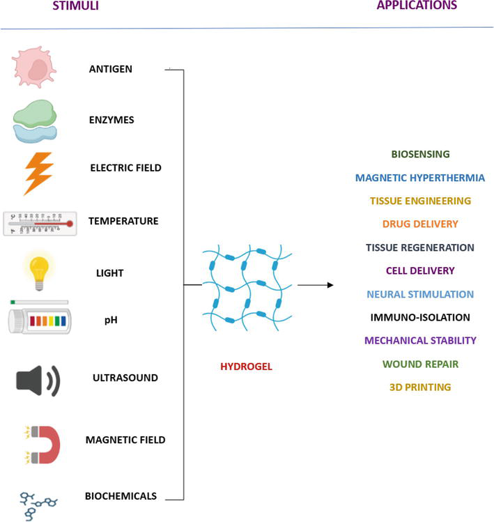
Injectable hydrogels in cardiac tissue engineering: Although patch-based and cell sheet technologies have received a lot of research attention and show promising outcomes in cardiac tissue engineering, they call for a more intrusive surgical procedure. Currently, injectable hydrogels are of attention in cardiac tissue engineering as they are the least invasive and potential therapeutic agent that provides mechanical strength to the injured tissue [66]. Clinical trial results reveal that acellular alginate-based injectable hydrogels show considerable preservation of left ventricular indices and left ventricular ejection fraction [67] in patients with myocardial infarction. Extensive study is required in the domain of injectable hydrogels to make the therapeutic strategy stable (Figure 1).
Figure 1.
Smart hydrogel: Stimuli and applications.
Cardiac tissue regeneration: Studies on cardiac tissue regeneration are hindered by the complexity of cardiac cells [68]. Nevertheless, hydrogel-based studies have allowed researchers to get around some of these challenges. Cardiomyocytes derived from pluripotent stem cells were seeded into a collagen-fibrin hydrogel blend for the regeneration of cardiac tissue. It not only showed successful regeneration capacity but also showed that the regeneration of cardiac tissue was greatly influenced by the amount of cell seeding as well as the collagen-fibrin content [69].
5. Factors affecting surface activity of the hydrogel
A hydrogel’s practicality is greatly influenced by its mechanical behavior. Reduced mechanical resistance greatly hinders its optimal activity in biomedical applications, including tissue engineering. To obtain desired mechanical properties, synthetic polymers are preferred over natural polymers due to the ability to tailor them in accordance with their application. Crystalline, chemically [70, 71] crosslinked polymers can provide better mechanical stability due to their regularly arranged structure and easier to control nature. Moreover, the synthesis and use of chemically crosslinked polymer is pH independent, making its application easier [72].
Electrical conductivity of the polymer is another crucial factor required in specific applications. The use of electroactive biomaterials as a scaffold for tissue regeneration is typically considered. Recent studies have demonstrated how controlling cell adhesion, proliferation, movement, cell death, and differentiation with electroactive biomaterials can improve the regeneration of the heart, nerve, and bone [73]. Hydrogels utilized as substrates for electroactive tissue engineering must have conductivity levels that are comparable to those of biological tissues, the lack of which will potentially affect its application. There are several ways by which the conductance of a scaffolding material can be increased, such as
Boosting the ionic conductance of the aqueous phase: Preparing or swelling the hydrogel in an ion-rich aqueous solution will increase the ionic conductivity of the aqueous phase [74].
Inclusion of a conducting material in the hydrogel network: Through the dispersion of conductive nanomaterials or other conductive materials, such as carbon fibers, in the hydrogel framework, the integration of conductive materials attempts to produce a continuous network of electronic conduction throughout the hydrogel.
Employing a conducting polymeric unit in the gel matrix: Organic macromolecules having inherent electrical conductivity are known as conducting polymers. The conducting polymers polypyrrole (PPy), polyaniline (PANI), and poly-(3,4-ethylenedioxythiophene) (PEDOT) are frequently utilized to make such hydrogels [75].
Hydrogel is used for drug delivery owing to its qualities, including minimal cytotoxicity, biodegradability, and biocompatibility. Despite the fact that it has many benefits, it also has certain drawbacks. On contacting the drug-releasing medium, drug-carrying hydrogels typically exhibit a sudden release of drugs because of the presence of substantial proportion of water [76]. The incorporation of weakly soluble drugs, which are swiftly released through diffusion, is another issue with hydrogel’s hydrophilic nature. These issues are effectively dealt with the incorporation of the drug into a nanomaterial, which is further encapsulated into a hydrogel matrix [37, 38].
Hydrophobic therapeutical drugs have restricted loading quantity and uniformity in hydrogel matrices, and the affinity of hydrogels toward hydrophobic compounds is limited.
Natural hydrogels have poor mechanical properties, low reproducibility, and high production cost.
During delivery of the drug, hydrogels have complete dependence on swellability, diffusability of water, high chance of spontaneous rupture of drug during initial stages of hydrogel swelling, slow responsive time of stimuli-sensitive hydrogels with nonspecific drug release, fast dissolution, and uncontrollable porosity.
Hydrogels cannot be used in healing wet wounds as they cannot absorb large amounts of fluids.
Hydrogels are biodegradable but they sometimes release toxic exudates, which may not be able to be excreted via kidney and remain accumulated in the body.
Due to poor mechanical strength and low stability, it’s difficult for hydrogels to store active substances.
Restriction in spatial manipulations of hydrogel, hence unable to attain full control on the organization and interaction among the cells, thus overall, there is no assurance on tissue morphogenesis.
Hydrogels differ from other forms of biomaterials in that they have a high-water content, a controlled swelling behavior, simple to handle, and are biocompatible, all of which make them desirable for biomedical applications. Hydrogels can respond to a variety of stimuli, including heat, pH, light, and chemical stimuli, based on their chemical structure and crosslink network, which can satisfy a variety of application needs. We provided detailed examples of both natural hydrogels made from polysaccharides and polypeptides as well as synthetic hydrogels, as well as different trends in their applications in various biomedical fields. It is important to conduct further research to develop materials with high mechanical strength, quick and effective self-healing, and a variety of biological activities for various specific limitations in biomedical applications. With extensive research trials and computational analysis, the employment of hydrogels can be effectively extended for various translational studies.
References
1.Shmeis R, MA. Nanotechnology in Wastewater Treatment, Comprehensive Analytical Chemistry. Netherlands: Elsevier; 2022. pp. 105-134
3.Jo H, Yoon M, Gajendiran M, Kim K. Recent strategies in fabrication of gradient hydrogels for tissue engineering applications. Macromolecular Bioscience. 2020;20(3):1-11
4.Calori IR, Braga G, Carvalho C, Bi H, Tedesco AC. Polymer scaffolds as drug delivery systems. European Polymer Journal. 2020;129:109621
5.Jabbari E. Hydrogels for cell delivery. Gels. 2018;4(3):58
6.Borges C, Colaço R, Serro AP. Poly (vinyl alcohol)-based hydrogels for joint prosthesis. Annals of Medicine. 2019;51(sup1):105
7.Catoira MC, Fusaro L, Di Francesco D, Ramella M, Boccafoschi F. Overview of natural hydrogels for regenerative medicine applications. Journal of Materials Science: Materials in Medicine. 2019;30:1-10
8.Soni V, Pandey V, Asati S, Gour V, Tekade RK. Biodegradable block copolymers and their applications for drug delivery. In: Basic Fundamentals of Drug Delivery. United States: Academic Press; 2019. pp. 401-447. DOI: 10.1016/b978-0-12-817909-3.00011-x
9.Tang G, Zhou B, Li F, Wang W, Liu Y, Wang X, et al. Advances of naturally derived and synthetic hydrogels for intervertebral disk regeneration. Frontiers in Bioengineering and Biotechnology. 2020b;8:745
10.Tang J, Katashima T, Li X, Mitsukami Y, Yokoyama Y, Sakumichi N, et al. Swelling behaviors of hydrogels with alternating neutral/highly charged sequences. Macromolecules. 2020a;53(19):8244-8254
11.Yu T, Wang H, Zhang Y, Wang X, Han B. The delivery of RNA-interference therapies based on engineered hydrogels for bone tissue regeneration. Frontiers in Bioengineering and Biotechnology. 2020;8:445
12.Madduma-Bandarage US, Madihally SV. Synthetic hydrogels: Synthesis, novel trends, and applications. Journal of Applied Polymer Science. 2021;138(19):50376
13.He J, Burgess DJ. Impact of biomaterials’ physical properties on cellular and molecular responses. In: Handbook of Biomaterials Biocompatibility. Duxford, England: Woodhead Publishing; 2020. pp. 69-84
14.Cui L, Yao Y, Yim EK. The effects of surface topography modification on hydrogel properties. APL Bioengineering. 2021;5(3):031509
15.Mantha S, Pillai S, Khayambashi P, Upadhyay A, Zhang Y, Tao O, et al. Smart hydrogels in tissue engineering and regenerative medicine. Materials. 2019;12(20):3323
16.Ben Ammar NE, Barbouche M, Hamzaoui AH. In: Chen Y, editor. Hydrogels Based on Natural Polymers. Cambridge, MA, USA: Elsevier; 2020. p. 15
17.Bai X, Gao M, Syed S, Zhuang J, Xu X, Zhang XQ. Bioactive hydrogels for bone regeneration. Bioactive Materials. 2018;3(4):401-417
18.Mariani E, Lisignoli G, Borzì RM, Pulsatelli L. Biomaterials: Foreign bodies or tuners for the immune response? International Journal of Molecular Sciences. 2019;20(3):636
19.Palencia M, Lerma TA, Garcés V, Mora MA, Martínez JM, Palencia SL. Chapter 9 - Eco-friendly hydrogels. In: Palencia M, Lerma TA, Garcés V, Mora MA, Martínez JM, Palencia SL, editors. Advances in Green and Sustainable Chemistry. Eco-friendly Functional Polymers: Elsevier; 2021. pp. 141-153
20.Egorikhina MN, Timofeeva LB, Linkova DD, Rubtsova YP, Bugrova ML, Charykova IN, et al. Biocompatibility study of hydrogel biopolymer scaffold with encapsulated mesenchymal stem cells. Polymers. 2023;15(6):1337
21.Sánchez-Cid P, Jiménez-Rosado M, Rubio-Valle JF, Romero A, Ostos FJ, Rafii-El-Idrissi Benhnia M, et al. Biocompatible and thermoresistant hydrogels based on collagen and chitosan. Polymers. 2022;14(2):272
22.Lan W, Xu M, Qin M, Cheng Y, Zhao Y, Huang D, et al. Physicochemical properties and biocompatibility of the bi-layer polyvinyl alcohol-based hydrogel for osteochondral tissue engineering. Materials & Design. 2021;204:109652
23.Axpe E, Chan D, Offeddu GS, Chang Y, Merida D, Hernandez HL, et al. A multiscale model for solute diffusion in hydrogels. Macromolecules. 2019;52(18):6889-6897
24.Nicolella P, Koziol MF, Löser L, Saalwächter K, Ahmadi M, Seiffert S. Defect-controlled softness, diffusive permeability, and mesh-topology of metallo-supramolecular hydrogels. Soft Matter. 2022;18(5):1071-1081
25.Nicolella P, Lauxen D, Ahmadi M, Seiffert S. Reversible hydrogels with switchable diffusive permeability. Macromolecular Chemistry and Physics. 2021;222(16):2100076
26.Vinchhi P, Rawal SU, Patel MM. Chapter 19 - Biodegradable hydrogels. In: Chappel E, editor. Developments in Biomedical Engineering and Bioelectronics, Drug Delivery Devices and Therapeutic Systems. United States: Academic Press; 2021. pp. 395-419
27.Zong H, Wang B, Li G, Yan S, Zhang K, Shou Y, et al. Biodegradable high-strength hydrogels with injectable performance based on poly (l-glutamic acid) and gellan gum. ACS Biomaterials Science & Engineering. 2020;6(8):4702-4713
28.Vázquez-González M, Willner I. Stimuli-responsive biomolecule-based hydrogels and their applications. Angewandte Chemie International Edition. 2020;59(36):15342-15377
29.Li Z, Zhou Y, Li T, Zhang J, Tian H. Stimuli-responsive hydrogels: Fabrication and biomedical applications. Viewpoints. 2022;3(2):20200112
30.Li X, Sun Q , Li Q , Kawazoe N, Chen G. Functional hydrogels with Tunable structures and properties for tissue engineering applications. Frontiers in Chemistry. 2018;6:1-20
31.Chaudhary S, Chakraborty E. Hydrogel based tissue engineering and its future applications in personalized disease modeling and regenerative therapy. Beni-Suef University Journal of Basic and Applied Sciences. 2022;11:3
32.Komatsu K, Shibata T, Shimada A, Ideno H, Nakashima K, Tabata Y, et al. Cationized gelatin hydrogels mixed with plasmid DNA induce stronger and more sustained gene expression than atelocollagen at calvarial bone defects in vivo. Journal of Biomaterials Science, Polymer Edition. 2016;27(5):419-430
33.Deev R, Plaksa I, Bozo I, Isaev A. Results of an international postmarketing surveillance study of pl-VEGF165 safety and efficacy in 210 patients with peripheral arterial disease. American Journal of Cardiovascular Drugs. 2017;17:235-242
34.Carballo-Pedrares N, Fuentes-Boquete I, Díaz-Prado S, Rey-Rico A. Hydrogel-based localized nonviral gene delivery in regenerative medicine approaches - An overview. Pharmaceutics. 2020;12(8):752
35.Chakradhar S. Treatments that made headlines in 2018. Nature Medicine. 2018;24(12):1785-1787
36.Seidlits SK, Gower RM, Shepard JA, Shea LD. Hydrogels for lentiviral gene delivery. Expert Opinion on Drug Delivery. 2013;10(4):499-509
37.Jiang Y, Krishnan N, Heo J, Fang RH, Zhang L. Nanoparticle–hydrogel superstructures for biomedical applications. Journal of Controlled Release. 2020;324:505-521
38.Jiang Y, Wang Y, Li Q , Yu C, Chu W. Natural polymer-based stimuli-responsive hydrogels. Current Medicinal Chemistry. 2020c;27(16):2631-2657
39.Nunes D, Andrade S, Ramalho MJ, Loureiro JA, Pereira MC. Polymeric nanoparticles-loaded hydrogels for biomedical applications: A systematic review on In vivo findings. Polymers (Basel). 2022;14(5):1010
40.Mellati A, Hasanzadeh E, Gholipourmalekabadi M, Enderami SE. Injectable nanocomposite hydrogels as an emerging platform for biomedical applications: A review. Materials Science and Engineering: C. 2021;131:112489
41.Sun Z, Wang X, Liu J, Wang Z, Wang W, Kong D, et al. ICG/l-arginine encapsulated PLGA nanoparticle-thermosensitive hydrogel hybrid delivery system for Cascade cancer photodynamic-NO therapy with promoted collagen depletion in tumor tissues. Molecular Pharmaceutics. 2021;18:928-939
42.Wang Y, Li Q , Zhou J-E, Tan J, Li M, Xu N, et al. A photopolymerized semi-interpenetrating polymer networks-based hydrogel incorporated with nanoparticle for local chemotherapy of Tumors. Pharmaceutical Research. 2021;38:669-680
43.Gao L, Yi M, Xing M, Li H, Zhou Y, Xu Q, et al. In situ activated mesenchymal stem cell (MSCs) by bioactive hydrogel for myocardial infarction treatment. Journal of Materials Chemistry B. 2020. DOI: 10.1039/d0tb01320j
44.Yang M, He S, Su Z, Yang Z, Liang X, Wu Y. Thermosensitive injectable chitosan/collagen/β-glycerophosphate composite hydrogels for enhancing wound healing by encapsulating mesenchymal stem cell spheroids. ACS Omega. 2020;5(33):21015-21023
45.Tony Thomas C, Ramanarayanan V, Krishnan V. Hydrogels - A versatile material in dentistry. European Journal of Molecular & Clinical Medicine (EJMCM). 2019;6(1):59-65
46.Liu L, Wu D, Tu H, Cao M, Li M, Peng L, et al. Applications of hydrogels in drug delivery for Oral and maxillofacial diseases. Gels. 2023;9(2):146
47.Huang Y, Li X, Yang L. Hydrogel encapsulation: Taking the therapy of mesenchymal stem cells and their derived Secretome to the next level. Frontiers in Bioengineering and Biotechnology. 2022b;10:1-9
48.Huang M, Huang Y, Liu H, Tang Z, Chen Y, Huang Z, et al. Hydrogels for the treatment of oral and maxillofacial diseases: Current research, challenges, and future directions. Biomaterials Science. 8 Nov 2022;10(22):6413-6446
49.Zagórska-Dziok M, Sobczak M. Hydrogel-based active substance release systems for cosmetology and dermatology application: A review. Pharmaceutics. 2020;12(5):396
50.Chen M, Wang Y, Zhang J, Peng Y, Li S, Han D, et al. Stimuli-responsive DNA-based hydrogels for biosensing applications. Journal of Nanobiotechnology. 2022;20(1):1-22
51.Zhang Y, Huang Y. Rational design of smart hydrogels for biomedical applications. Frontiers in Chemistry. 2021;8:615665
52.Zhang Y, Dong L, Liu L, Wu Z, Pan D, Liu L. Recent advances of stimuli-responsive polysaccharide hydrogels in delivery systems: A review. Journal of Agricultural and Food Chemistry. 2022;70(21):6300-6316
53.Bordbar-Khiabani A, Gasik M. Smart hydrogels for advanced drug delivery systems. International Journal of Molecular Sciences. 2022;23(7):3665
54.Zhao Q , Li C, Shum HC, Du X. Shape-adaptable biodevices for wearable and implantable applications. Lab on a Chip. 2020;20(23):4321-4341
55.Ying B, Liu X. Skin-like hydrogel devices for wearable sensing, soft robotics and beyond. Iscience. 2021;24(11):103174
56.Wang J, Wang Z, Yu J, Kahkoska AR, Buse JB, Gu Z. Glucose-responsive insulin and delivery systems: Innovation and translation. Advanced Materials. 2020;32(13):1902004
57.Roquero DM, Katz E. “Smart” alginate hydrogels in biosensing, bioactuation and biocomputing: State-of-the-art and perspectives. Sensors and Actuators Reports. 2022;4:100095
58.Herrmann A, Haag R, Schedler U. Hydrogels and their role in biosensing applications. Advanced Healthcare Materials. 2021;10(11):2100062
59.Li Z, Li Y, Chen C, Cheng Y. Magnetic-responsive hydrogels: From strategic design to biomedical applications. Journal of Controlled Release. 2021;335:541-556
60.Tay A, Sohrabi A, Poole K, Seidlits S, Di Carlo D. A 3D magnetic hyaluronic acid hydrogel for magnetomechanical neuromodulation of primary dorsal root ganglion neurons. Advanced Materials. 2018;30(29):1800927
61.Xu Y, Yin H, Chu J, Eglin D, Serra T, Docheva D. An anisotropic nanocomposite hydrogel guides aligned orientation and enhances tenogenesis of human tendon stem/progenitor cells. Biomaterials Science. 2021;9(4):1237-1245
62.Wu J, Ning P, Gao R, Feng Q , Shen Y, Zhang Y, et al. Programmable ROS-mediated cancer therapy via magneto-inductions. Advanced Science. 2020;7(12):1902933
63.Liu X, Zhang Y, Wang Y, Zhu W, Li G, Ma X, et al. Comprehensive understanding of magnetic hyperthermia for improving antitumor therapeutic efficacy. Theranostics. 2020;10(8):3793
64.Farzin A, Etesami SA, Quint J, Memic A, Tamayol A. Magnetic nanoparticles in cancer therapy and diagnosis. Advanced Healthcare Materials. 2020;9(9):1901058
65.de Vos P. Bioartificial pancreas: Challenges and progress. Principles of Tissue Engineering. 2020:665-679
66.Alonso JM, Andrade del Olmo J, Perez Gonzalez R, Saez-Martinez V. Injectable hydrogels: From laboratory to industrialization. Polymers. 2021;13(4):650
67.Peña B, Laughter M, Jett S, Rowland TJ, Taylor MR, Mestroni L, et al. Injectable hydrogels for cardiac tissue engineering. Macromolecular Bioscience. 2018;18(6):1800079
68.Hortensius RA, Lin WH, Ogle BM. Cardiac tissue engineering: A pathway for repair. In: Engineering in Medicine. United States: Academic Press; 2019. pp. 3-33
69.Kaiser NJ, Kant RJ, Minor AJ, Coulombe KL. Optimizing blended collagen-fibrin hydrogels for cardiac tissue engineering with human iPSC-derived cardiomyocytes. ACS Biomaterials Science & Engineering. 2018;5(2):887-899
70.You Y, Xing R, Zou Q , Shi F, Yan X. High-tolerance crystalline hydrogels formed from self-assembling cyclic dipeptide. Beilstein Journal of Nanotechnology. 2019;10:1894-1901
71.Revete A, Aparicio A, Cisterna BA, Revete J, Luis L, Ibarra E, et al. Advancements in the use of hydrogels for regenerative medicine: Properties and biomedical applications. International Journal of Biomaterials. 2022:3606765
72.Ye D, Chang C, Zhang L. High-strength and tough cellulose hydrogels chemically dual cross-linked by using low- and high-molecular-weight cross-linkers. Biomacromolecules. 2019;20(5):1989-1995
73.Ning C, Zhou Z, Tan G, Zhu Y, Mao C. Electroactive polymers for tissue regeneration: Developments and perspectives. Progress in Polymer Science. 2018:144-162
74.Peng Q , Chen J, Wang T, Peng X, Liu J, Wang X, et al. Recent advances in designing conductive hydrogels for flexible electronics. InfoMat. 2020;2. DOI: 10.1002/inf2.12113
75.Kougkolos G, Golzio M, Laudebat L, Valdez-Nava Z, Flahaut E. Hydrogels with electrically conductive nanomaterials for biomedical applications. Journal of Materials Chemistry B. The Royal Society of Chemistry. 2023;11(10):2036-2062
76.Ren Y, Li X, Han B, Zhao N, Mu M, Wang C, et al. Improved anti-colorectal carcinomatosis effect of tannic acid co-loaded with oxaliplatin in nanoparticles encapsulated in thermosensitive hydrogel. European Journal of Pharmaceutical Sciences. 2018;128:279-289
Written By
Ruby Varghese, Yogesh Bharat Dalvi, P. Lochana, S. Achinthya, Bhagyashri Omprakash Somani, Preetha Karnaver, Nebu George Thomas, S. Rupesh, Nibu Varghese and Jayachandran V.P.
Submitted: 03 June 2023Reviewed: 04 June 2023Published: 05 January 2024
 Open access peer-reviewed chapter