Open Access is an initiative that aims to make scientific research freely available to all. To date our community has made over 100 million downloads. It’s based on principles of collaboration, unobstructed discovery, and, most importantly, scientific progression. As PhD students, we found it difficult to access the research we needed, so we decided to create a new Open Access publisher that levels the playing field for scientists across the world. How? By making research easy to access, and puts the academic needs of the researchers before the business interests of publishers.
We are a community of more than 103,000 authors and editors from 3,291 institutions spanning 160 countries, including Nobel Prize winners and some of the world’s most-cited researchers. Publishing on IntechOpen allows authors to earn citations and find new collaborators, meaning more people see your work not only from your own field of study, but from other related fields too.
To purchase hard copies of this book, please contact the representative in India:
CBS Publishers & Distributors Pvt. Ltd.
www.cbspd.com
|
customercare@cbspd.com
Low-molecular-weight hydrogels (LMWHs) have garnered widespread focus as versatile soft materials owing to their distinctive characteristics and potential applications. LMWHs are synthesized from small molecules that, upon assembly, form entangled aggregates via different types of noncovalent interactions, such as hydrogen bonding, van der Waals forces, or π-π stacking interactions. LMWHs are characterized by their unique ability to mimic biological systems by effectively absorbing and retaining large quantities of water. Despite their poor mechanical properties, LMWHs are widely used in various medical applications due to their easy preparation, biocompatibility, and low toxicity. Smart LMWHs demonstrate responsiveness to external stimuli, such as light, temperature, enzymes, or pH, rendering them ideally adapted for various controlled drug delivery applications. LMWHs have been extensively employed in different biomedical applications, including drug delivery, tissue engineering and cell culture, wound healing, and biofabrication. In this chapter, we aim to explore the potential of LMWHs as drug-delivery vehicles for a range of medications, focusing on the different synthetic strategies, gelation processes, and drug-loading and releasing mechanisms.
Photochemistry Department, Chemical Industries Research Institute, National Research Centre, Cairo, Egypt
Ahmed M. Salama
State Key Laboratory of Chemical Resource Engineering, Beijing University of Chemical Technology, Beijing, China
Badria H.A. Al-Dhuwayin
Department of Chemistry, College of Science and Arts, Najran University, Najran, Saudi Arabia
Amal F. Seliem
Department of Chemistry, College of Science and Arts, Najran University, Najran, Saudi Arabia
*Address all correspondence to: hany.nour@daad-alumni.de
1. Introduction
Low-molecular-weight hydrogels (LMWHs) have gained significant attention across various disciplines as an important class of soft materials. These gels are composed of small organic molecules that can self-assemble in water, forming 3D network structures. The self-assembly of LMWHs into entangled networks is typically driven by various noncovalent interactions, such as hydrogen bonding, π-π stacking, or van der Waals forces. The noncovalent interactions between the molecules result in the formation of fibrous structures that become entangled and form hydrogels. LMWHs are known for their high water retention capability, which imparts them with a resemblance to biological tissues. LMWHs have been widely studied in various biomedical research areas due to their biocompatibility, tailored design, cost-effectiveness, and ease of preparation. LMWHs show great potential as a drug delivery platform, as they can encapsulate drugs and release them in a slow and sustained manner. For instance, LMWHs have been used to deliver vascular endothelial growth factor small interfering RNA (VEGF-siRNA) into human cells [1]. LMWHs derived from vitamin B have been utilized in the delivery of siRNA into cancer cell lines with high selectivity [2]. LMWHs have also been exploited for the delivery of proteins [3, 4].
In addition to their use in drug delivery, LMWHs have exhibited significant potential in several medicinal applications. LMWHs with inherent antibacterial properties have been widely explored [5, 6, 7]. Adams et al. reported the gelation of a naphthalene-based dipeptide, which was triggered by the oxidation of dopamine, with the generation of reactive oxygen species (ROS). The hydrogel thus formed demonstrated potent antibacterial activity against S. aureus [8]. The naturally occurring glycyrrhizic acid has been used as an injectable hydrogelator with intrinsic antibacterial activity against S. aureus [9]. Kumar et al. described the gelation of a peptide LMWH with antibacterial potential against S. aureus [10]. LMWHs that incorporate L-proline or D-phenylalanine along with thiophene were reported to show potential suppression of MRSA [11]. Diimidazolium-based LMWHs have been shown to inhibit different pathogens, such as K. rhizophila, B. subtilis, and E. coli [12]. LMWHs have been evaluated as versatile materials for wound healing applications. LMWHs, when applied to wounds, can function as a physical barrier that prevents fluid loss and offers a moist environment that promotes recovery. An illustration of this is a hydrogel prepared from cytidine, boric acid, and silver nitrate, which has been utilized for wound healing. It has been shown to have antibacterial effects against S. aureus, E. coli, and P. aeruginosa pathogens and has also been found to promote wound healing in mice [13, 14]. LMWHs have shown remarkable success in the development of potent anticancer therapeutics [15]. Marlow et al. described the synthesis of LMWHs incorporated with 5-fluoro-benzothiazole, which exhibited high inhibition of breast and ovarian cancers [16]. Amphiphilic phenylalanine gelators, when mixed with Au(III) ions in water, have been shown to generate ROS that inhibit mammalian cancer cells [17]. The use of LMWHs in tissue engineering and 3D cell cultures has undergone remarkable progress [18]. Lampe et al. demonstrated the synthesis of pentapeptide hydrogels as the supportive matrix for the cell culture of oligodendrocyte progenitor cells [19]. The oligodendrocyte progenitor cells encapsulated in the hydrogel matrix exhibited proliferation and extension, indicating the potential utility of LMWHs in tissue engineering applications. LMWHs have also been employed in biofabrication and cell adhesion applications [20, 21, 22].
This chapter aimed to provide a comprehensive understanding of the design, synthesis, and applications of LMWHs in drug delivery. We discussed the various synthetic methods utilized for the fabrication of LMWHs and highlighted the challenges of using LMWHs as drug carriers along with possible solutions. In addition, we reviewed the different drug cargos that have been utilized in drug delivery purposes incorporating LMWHs. These drugs included anticancer, anti-inflammatory, antibacterial agents, and vitamins, to name a few. The versatility and potential of LMWHs as drug-delivery vehicles in various therapeutic areas were emphasized. Furthermore, we provided insights into the mechanism of drug release from LMWHs. We also discussed the influence of the chemical structure of LMWHs on drug delivery efficiency, highlighting in particular, the importance of their functionalization.
LMWHs can form by self-assembly of small molecular weight molecules into 3D network structures capable of immobilizing water. The gelation process can be optimized by testing different parameters and observing their influences on gelation. These parameters include, but are not limited to, the concentration of the gelator, the temperature of gelation, the pH of the solution, and the addition of an external agent to promote gelation. Optimizing these parameters can aid in fine-tuning the conditions required for the organic molecules to dissolve freely in water upon heating and form a clear solution. Upon cooling, the solution can turn into a gel, which can be studied to explore the factors that promote and reinforce the gelation. The inclusion of functional motifs, or groups capable of establishing hydrogen bonding or π-π stacking, into the structural backbone of the gelator is also crucial for promoting the gelation process [23]. Therefore, by testing and optimizing these parameters, the gelation process of LMWHs can be improved and tailored for specific applications.
3. LMWHs in drug delivery: Opportunities and challenges
LMWHs possess several advantages that make them excellent candidates for various drug delivery applications, such as biocompatibility, ease of preparation, structural customization, stimuli-responsiveness, versatility, and controlled drug release. When drugs are loaded into the hydrogel matrix, they gain extra stability against degradation, oxidation, and other adverse effects, leading to a longer shelf life. Additionally, controlled drug release is a crucial benefit of using drug-loaded LMWHs, as it reduces dosing frequency. LMWHs can be tailored to deliver drugs to specific areas, which is especially useful in the case of anticancer medications, as it helps avoid harming healthy cells. Injecting anti-inflammatory and analgesic drugs into sites of injury or inflammation can also provide prolonged pain relief. Moreover, the high water content of LMWHs makes them ideal for encapsulating various drugs. The synthesis of LMWHs is often straightforward and cost-effective when compared to other drug delivery systems.
Despite their intriguing properties, using LMWHs for drug delivery may have some critical limitations. For example, some LMWHs may have weak mechanical strength, a limited capacity for drug loading, and insufficient drug release, which could influence their therapeutic effectiveness. In addition, some LMWHs may not be suitable for encapsulating drugs with poor solubility or achieving targeted drug delivery. Furthermore, certain LMWHs may have low stability for in vivo studies, and their efficiency may be affected by changes in pH or temperature, making them less reliable.
To improve the mechanical strength of hydrogels co-assembly can be utilized. For instance, acylhydrazone-based hydrogels incorporating bipyridinium units have been found to form highly stable hydrogels through donor-acceptor charge transfer interactions with electron-rich naphthyl moieties [24, 25]. The addition of metal ions can also enhance the mechanical properties and thermal stability of LMWHs [26]. The design of LMWHs can greatly impact their loading capacity, pH and temperature stability, targeted delivery ability, in vivo performance, and drug solubility. Therefore, it is important to customize the design of LMWHs to achieve the desired properties for specific applications.
Drug loading into the hydrogel matrix can be accomplished using physical entrapment or chemical modification of the structure of the hydrogelator, with the former relying on noncovalent interactions and the latter utilizing covalent binding. Nevertheless, both methods have notable drawbacks. Certain drugs may not establish strong enough interactions with the hydrogelator, which can result in rapid drug release, while others may form overly strong interactions, leading to very slow drug release. Chemical modification of the gelators to integrate drugs into their structural framework can lead to a decrease in the drug’s effectiveness after its release from the hydrogel structure. For example, the incorporation of ciprofloxacin into the structure of a peptide gelator resulted in a reduction of its antibacterial activity [27]. Therefore, it is highly important to further investigate the process of linking drugs into the backbone structure of the gelators as a drug delivery approach. Additionally, it is crucial to ensure controlled and sustained drug release, especially for physically entrapped drugs, in order to maintain the drugs’ efficacy and optimize therapeutic outcomes.
Another important challenge facing drug delivery using LMWHs is the limited drug-loading capacity. This obstacle can be overcome by modifying the hydrogel structure, such as by incorporating functional motifs or groups that reinforce the drug’s affinity for the hydrogelator. Moreover, optimizing drug concentration can also impact the drug-loading capacity. The drug loading can also be improved by increasing the porosity of the gelator. In addition, considering the pre-loading or post-loading of drugs into the hydrogel matrix is a critical factor in achieving a high loading capacity. These strategies are essential to developing effective drug delivery systems with higher drug loading capacities.
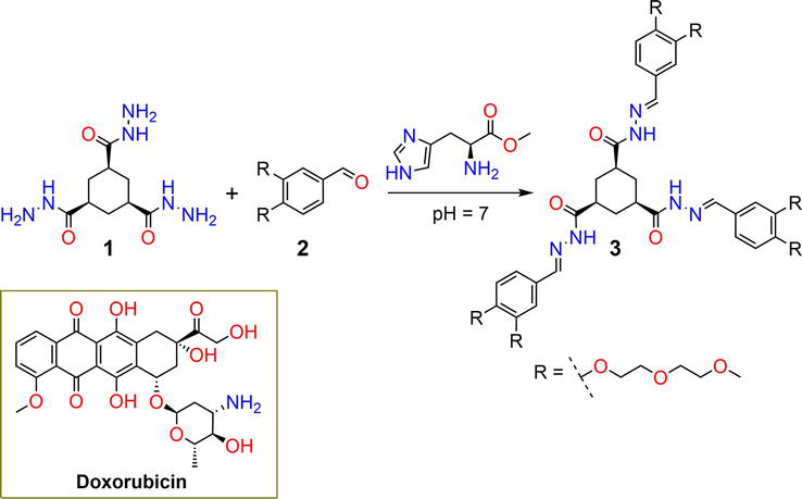
Hydrogel 3 was prepared from the condensation of tricarbohydrazide 1 with 3,4-disubstituted-benzaldehyde 2, with the formation of acylhydrazone linkages as shown in Figure 1 [28]. Augmented hydrogen bonding of the acylhydrazone functional motifs prompted gelation of derivative 3 [29]. L-Histidine methyl ester catalyzed the reaction as a biocompatible organocatalyst at a neutral pH of 7. The anticancer drug doxorubicin (DOX) was loaded into the hydrogel matrix either covalently or noncovalently based on the sequence of the reactants mixing. The drug release was accomplished in a buffer solution and peaked at pH 5 due to the hydrolysis of acylhydrazone bonds.
Figure 1.
Synthetic pathway of hydrogelator 3 and the chemical structure of the drug molecule [28, 29].
The biocompatibility of the gel was evaluated in vitro on the breast cancer cell line (MCF-7) using the 3-(4,5-dimethyl-2-thiazolyl)-2,5-diphenyl-2-tetrazolium bromide (MTT) assay. The MCF-7 cancer cell line treated with the hydrogel-DOX matrix exhibited a 50–70% reduction in cell viability over time, indicating a slow release of DOX.
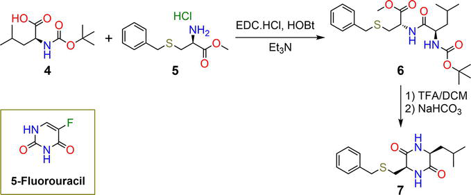
Banerji et al. reported the synthesis of a 2,5-diketopiperazine peptide 7, which gelates at a physiological pH of 7.46 at 37°C and self-assembles in water into nanofibrillar structures [30]. Cyclic peptides have a variety of intriguing features, such as biocompatibility, resistance to degradation, and rigidity. Such interesting properties enrich their biomedical applications. Hydrogelator 7 was prepared by reacting N-(tert-butoxycarbonyl)-L-leucine 4 with S-benzyl-L-cysteine methyl ester 5 in the presence of 1-ethyl-3-(3-dimethylaminopropyl)carbodiimide (EDC) and hydroxybenzotriazole (HOBt) to give compound 6, as illustrated in Figure 2. Then, derivative 6 was hydrolyzed using trifluoroacetic acid (TFA) to afford the cyclized product 7. Compound 7 forms a stable hydrogel at a critical gelation concentration (CGC) of 0.05% w/v. The backbone structure of compound 7 incorporates two amide functionalities, a thiobenzyl group, and an isobutyl chain, which promotes hydrogelation via augmented intermolecular hydrogen bonding, π-π stacking, and hydrophobic interactions. Hydrogelator 7 was employed as a drug delivery carrier for the anticancer drug 5-fluorouracil. The encapsulation efficiency of the hydrogelators 7 was reported to be 94.46 ± 1.04%. Drug release occurred by treating the drug-loaded hydrogel with a phosphate-buffered saline solution at a pH of 7.46, which resulted in 90% release. Interestingly, the drug-loaded hydrogel showed higher anticancer activity against the HCT116 human colon cancer cell with an IC50 of 6.7 ± 1.2 μM when compared to 5-fluorouracil (IC50 = 32.23 ± 3.4 μM), possibly due to the slow release of the drug [30].
Figure 2.
Synthetic pathway of hydrogelators 7 and the chemical structure of the drug molecule [30].
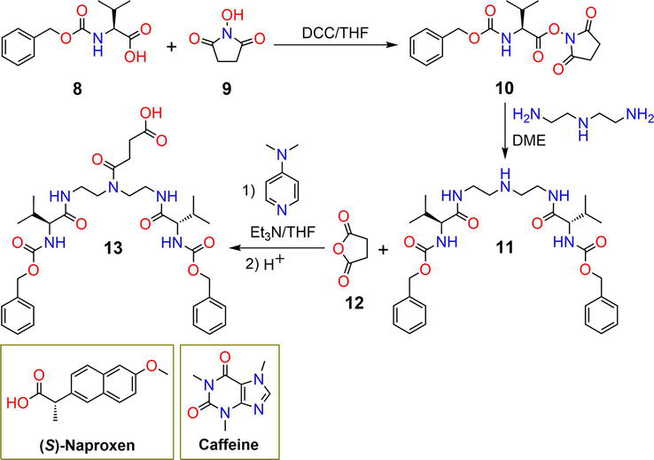
Luis et al. described the synthesis of pseudopeptide hydrogelator 13 with a C2-symmetrical structure (Figure 3). Gelator 13 functions as a drug delivery carrier for (S)-naproxen and caffeine. The reaction of protected N-Cbz-L-valine 8 with N-hydroxysuccinimide 9 in the presence of the dicyclohexylcarbodiimide (DCC) coupling agent yields compound 10 [31]. Derivative 10 reacted with a diamine in dimethoxyethane (DME) to afford 11.
Figure 3.
Synthetic pathway of hydrogel 13 and the chemical structures of the drug molecules [31, 32].
Compound 11 reacted with succinic anhydride in an alkaline medium to give the carboxylic-functionalized derivative 13 [32]. Hydrogelation of compound 13 was conducted in an acidic condition with a reported CGC of 1 mg/mL. Gelator 13 showed dual thermoresponsive and pH-responsive sol-to-gel transitions. The drug-loaded hydrogels were evaluated for transdermal drug release in animals, which revealed slow and sustained pH-dependent drug-releasing potential.
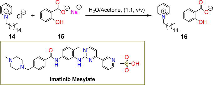
Ionic hydrogels are a readily available and cost-effective class of materials that have been widely explored as significant drug delivery systems. Cetylpyridinium salicylate ionogel 16 was prepared from the reaction of cetylpyridinium chloride 14 with sodium salicylate 15 in a mixture of H2O and acetone (Figure 4) [33]. Cetylpyridinium salicylate 16 gelates in water after standing at 25°C for 12 hours in the form of long fibers [34]. The CGC of hydrogel 16 was reported to be 4.7% w/v. The ability of gelator 16 to assemble into fibrous aggregates is mainly driven by noncovalent hydrogen bonding and hydrophobic interactions. Hydrogelator 16 exhibited high potential to encapsulate the anticancer drug imatinib mesylate. The drug-loaded ionogel exhibited 53.17, 88.30, and 94.17% releasing at pH 10, 7.4, and 5, respectively, at a typical temperature of 37°C.
Figure 4.
Synthetic pathway of ionogel 16 and the chemical structure of the drug molecule [33, 34].
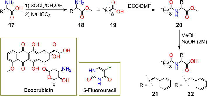
DuttKonar et al. reported the preparation of chiral hydrogelators 21 and 22, which serve as drug delivery carriers for the anticancer drugs 5-fluorouracil and DOX. As shown in Figure 5, phenylalanine 17 is esterified with thionyl chloride in methanol to give compound 18; this product, along with decanoic acid 19 and the coupling agent DCC, reacted to yield 20. The basic hydrolysis of 20 afforded derivatives 21 and 22.
Figure 5.
Synthetic pathway of hydrogels 21 and 22 and the structures of the drug molecules [35].
Compounds 21 and 22 underwent self-assembly, resulting in the formation of entangled fibrous aggregates facilitated by hydrogen bonding, hydrophobic, and π-π stacking interactions [35]. The CGCs of hydrogels 21 and 22 were found to be 0.01% w/v, classifying them as supergelators. The hydrogelators exhibited great biocompatibility when incubated with cancer cell lines, as determined by the MTT assay, and showed high stability against proteolytic degradation upon incubation with the proteolytic enzyme, proteinase K. Hydrogel nanoparticles of 21 and 22 were utilized as vehicles for drug delivery of 5-fluorouracil and DOX. Following a 45-hour duration, the release percentages of 5-fluorouracil were determined to be 43.67% and 41.23% for 21 and 22, respectively. In the instance of DOX, the release potential of nanoparticle hydrogelators 21 and 22 after 80 hours was 50.57% and 48.64%, respectively.
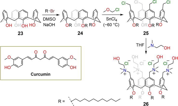
Functionalized calixarene 26 was prepared following the synthetic strategy illustrated in Figure 6 [36]. Alkylation of calixarene 23 with 1-bromododecane yields the corresponding tetraalkoxy-calixarene 24.
Figure 6.
Synthetic pathway of functionalized calixarene derivative 26 and the chemical structure of curcumin [36, 37].
The tetrachloromethyl-O-dodecyl calix[4]arene 25 is formed by the reaction of 24 with chloro(methoxy)methane in the presence of catalytic SnCl4 at −60°C. The reaction of compound 25 with N,N-dimethylaminoethanol gives derivative 26. Consoli et al. reported the gelation of the polyphenolic curcumin with the cationic choline-calix[4]arene 26, which has several medicinal applications [37]. Alkoxy chains are attached to the lower rim of calixarene, while the upper rim is functionalized with choline moieties. The calixarene-curcumin hydrogel forms by simple mixing of 26 with curcumin in a phosphate-buffered saline solution at physiological pH of 7.4. The incorporation of curcumin into the gel matrix has resulted in a remarkable enhancement of its solubility, as well as a considerable improvement in its stability against photodegradation. The calixarene-curcumin hydrogel is assembled in the form of nanosphical aggregates. Hydrogelation was assumed to occur via hydrogen bonding and ion-dipole interactions, forming 3D network structures. The curcumin showed a slow-releasing potential of 0.30 ± 0.02% per hour from the hydrogel matrix.
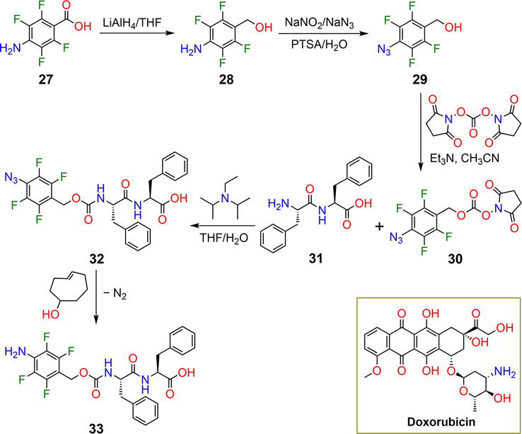
Gamble et al. described the synthesis of a diphenylalanine-based hydrogel 32 that contains fluorinated-benzyl azide and carbamate moieties [38]. Compound 28 was obtained by reducing the carboxylic group of compound 27 using lithium aluminum hydride in THF. This was followed by the conversion of compound 28 to its corresponding azido derivative 29 through a reaction with sodium azide, as shown in Figure 7. The reaction of azide derivative 29 with N,N′-disuccinimidyl carbonate in an alkaline medium gave derivative 30. Hydrogelator 32 was then synthesized by reacting compounds 30 with L-phenylalanyl-L-phenylalanine 31 in a solvent mixture of THF: H2O in the presence of Hünig’s base. Compound 32 forms a hydrogel in a mixture of H2O: DMSO (5% DMSO) at a pH of 3.7. The CGC of hydrogelator 32 was reported to be 0.02–0.1 wt%.
Figure 7.
Synthetic pathway of hydrogelator 32 and the chemical structure of the drug molecule [38].
Hydrogelator 32 assembles into entangled fibrous network structures with the aid of π-π stacking interactions. After 4 hours at 37°C, the hydrogel experienced a gel-to-sol transition by treating with 5 mM of the biorthogonal reagent cyclooct-4-enol to afford dissolution product 33 [38]. The evaluation of the drug delivery capability of hydrogelator 32 was undertaken by encapsulating it with DOX at 0.1 wt%. The drug-releasing potential was accomplished by treating the drug-loaded hydrogel with 1 mM of the biorthogonal reagent cyclooct-4-enol. The anticancer drug DOX was released from the gel matrix (89%) after 24 hours of incubation at 37°C.
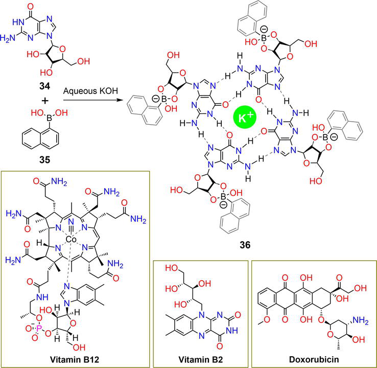
The reaction of the nucleoside guanosine 34 with 1-naphthaleneboronic acid 35 in an aqueous KOH solution forms the G-quadruplex hydrogel 36 (Figure 8) [39]. Hydrogelator 36 self-assembles through augmented interactions such as hydrogen bonding, π-π stacking, cationic, and ion-dipole interactions. In addition to the guanosine moiety, hydrogelator 36 incorporates pH-responsive boronate ester rings. The hydrogel forms nanofibers at physiological pH (pH 7.4), making it an ideal drug delivery system. The hydrogelator was found to be non-toxic and biocompatible at high doses when tested against HeLa, MCF-7 breast cancer, and HEK293 embryonic kidney cell lines [39]. The hydrogel’s drug delivery potential was assessed using vitamin B12, vitamin B2, and the anticancer drug DOX, which were separately encapsulated within the hydrogel matrix during the preparation process. The release of the vitamin or drugs was achieved at a physiological pH of 7.4 and a temperature of 37°C.
Figure 8.
Synthetic pathway of hydrogelator 36 and the structures of the drug molecules [39].
The hydrogel matrices released 30% and 60% of vitamin B12 and vitamin B2 after 40 hours, respectively, and the complete release of all entrapped vitamins was observed after 94 hours. At an acidic pH of 4.8, vitamin B12 and vitamin B2 were released in amounts of 82% and 75%, respectively. At pH 7.4 and 4.8, the DOX drug achieved 40% and 76% release, respectively. The pH-responsive behavior of the hydrogel was attributed to the breakage of hydrogen bonding interactions and boronate ester bonds.
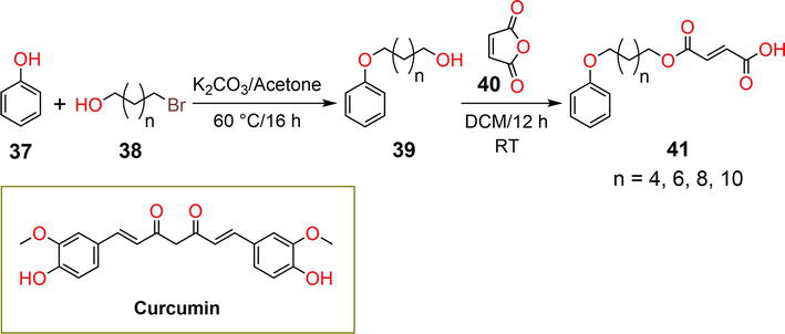
Nayak et al. described the synthesis of amphiphilic compound 41 as presented in Figure 9 [40]. Alkylation of phenol 37 with bromo-alcohol 38 in an alkaline medium yielded alkylated derivative 39. Compound 39 was reacted with maleic anhydride 40 in dichloromethane (DCM) to form derivative 41. Hydrogels were formed from compounds with (n = 8) and (n = 10) at CGCs of 1.6% and 1.3% w/v, respectively, by dissolving the compounds in phosphate buffer solutions at varied pH. Compound 41 incorporates phenoxy, carboxylic, ester, and alkyl groups that were engaged in gelation via π-π stacking, hydrogen bonding, and van der Waals interactions. Curcumin was incorporated into the hydrogel matrix by mixing it with the hydrogelator (n = 10) at the CGC in a phosphate buffer solution (pH = 8). Drug release occurred viaenzymatic- and pH-triggered mechanisms. In the former, the drug-loaded hydrogel was treated with a solution of lipozyme, which cleaved the functional ester group, transforming the hydrogel into a sol state. In the latter, acidic treatment (pH = 3.5) of the drug-loaded hydrogel mediated persistent drug release.
Figure 9.
Synthetic pathway of hydrogelator 41 and the chemical structure of curcumin [40].
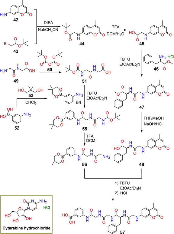
Huang et al. reported the synthesis of the photoresponsive hydrogelator 57via the synthetic steps demonstrated in Figure 10 [41]. Amino-chromenone 42 reacted with bromoacetate 43 in acetonitrile under basic conditions to produce compound 44. Derivative 44 was hydrolyzed under acidic conditions with TFA in a mixture of DCM and H2O to yield carboxylic acid-functionalized derivative 45. Coupling 45 with D-phenylalanine methyl ester hydrochloride 46 using 2-(1H-benzotriazole-1-yl)-1,1,3,3-tetramethylaminium tetrafluoroborate (TBTU) yielded the corresponding ester 47, which was hydrolyzed to afford derivative 48, with a free carboxylic group. The protection of the glycylglycine amino group using di-tert-butyl dicarbonate 50 yielded the (tert-butoxycarbonyl)glycylglycine 51. The coupling reaction of derivative 51 with the protected boronic acid 54 afforded 55, which on hydrolysis gave 56. The coupling reaction of derivatives 48 and 56 with subsequent hydrolysis yielded gelator 57. Coumarin-based derivative 57 formed a hydrogel in a 1: 2 mixture of polyethylene glycol 200 (PEG200) and water at a CGC of 2.7 mg/mL. The hydrogel assembled into entangled spiral-nanofibers with the aid of intermolecular hydrogen bonding and π-π stacking interactions. Hydrogelator 57 was found to exhibit a gel-to-sol transition upon exposure to UV irradiation at a wavelength of 365 nm, which was attributed to the breaking of the C-N bond. This property was exploited to utilize compound 57 as a drug carrier for the antineoplastic drug cytarabine hydrochloride. Thus, upon exposure to UV irradiation at 365 nm, the drug was released in a sustained and slow manner in a phosphate buffer with a pH range of 2.48–8.5. The rate of drug release was further enhanced by employing a laser as the irradiation source.
Figure 10.
Synthetic pathway of hydrogelator 57 and the structure of the drug molecule [41].
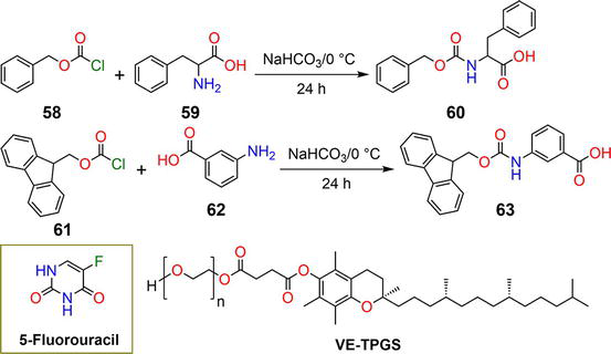
DuttKonar et al. reported a simple synthetic method of hydrogelators 60 and 63 [42]. Benzyl carbonochloridate 58 reacted with phenylalanine 59 in a basic medium to give gelator 60. Similarly, the reaction of (9H-fluoren-9-yl)methyl carbonochloridate 61 with 3-aminobenzoic acid 62, using the same conditions, resulted in the formation of hydrogelator 63, as demonstrated in Figure 11.
Figure 11.
Synthetic pathway of hydrogelators 60 and 63 and the chemical structure of the drug molecule [42].
Gelators 60 and 63 were utilized to construct fibrous network aggregates via hydrogen bonding and π-π stacking interactions. These network structures were formed at low CGCs of 0.02% and 0.05% w/v, respectively. Furthermore, when derivatives 60and 63 were mixed with Vitamin E-TPGS in light paraffin oil, they formed nanoparticles. These nanoparticles were used as drug delivery systems for the anticancer drug 5-fluorouracil. The nanoparticles of compounds 60 and 63 exhibited high entrapment efficiencies of 77.57% and 84.52%, respectively. Drug release was assessed by immersing the drug-loaded nanoparticles in a phosphate buffer at a pH of 7.4. The drug release was pH-dependent, with hydrogelator 60 exhibiting a faster release rate than hydrogelator 63. After 3 hours of immersion, hydrogelator 60 released 50% of the drug, whereas hydrogelator 63 released the same amount after 4 hours.
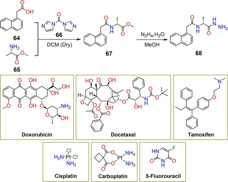
Bajaj et al. reported the synthesis of gelator 68 which incorporates naphthyl, amide, and hydrazide moieties as demonstrated in Figure 12. The synthetic method includes the reaction of 2-(naphthalen-1-yl)acetic acid 64 with methyl L-alaninate 65 in dry DCM in the presence of 1,1′-carbonyldiimidazole 66 to afford methyl (2-(naphthalen-1-yl)acetyl)-D-alaninate 67. The reaction of ester 67 with hydrazine monohydrate in methanol yields hydrazide 68 [43]. The hydrogelation of compound 68 is mainly facilitated by intermolecular hydrogen bonding of the hydrophilic and π-π stacking of the hydrophobic constituents, potentially leading to the formation of nanofiber aggregates at a CGC of 1.0% w/v [43]. Hydrogelator 68 was employed as a drug delivery carrier for a series of anticancer medications (Figure 12). The drugs were added to saturated aqueous solutions of gelator 68 on hot, with maximum encapsulation efficiency achieved by 5-fluorouracil. The drug-loading potential of hydrogelator 68 is highly attributed to its ability to establish stable noncovalent interactions with the drug molecules.
Figure 12.
Synthetic pathway of hydrogelator 68 and the structures of the drug molecules [43, 44].
Cisplatin and carboplatin showed minimum loading efficiency despite their relatively low molecular weights due to the absence of π-π stacking interactions and disruption of hydrogen bonding of the gelator by the amino groups [44]. The hydrazide group existing in the structural framework of gelator 68 could interact with DOX, resulting in high entrapment potential; however, this might lead to undesired structural modulation of the drug [44]. The release of the drugs was achieved at 37°C and a physiological pH. The drug-loaded hydrogels exhibited a release of 60–80%, except for cisplatin and carboplatin, which could be ascribed to the strong, established hydrogen bonding interactions with the hydrogelator.
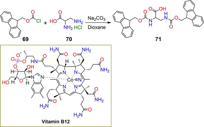
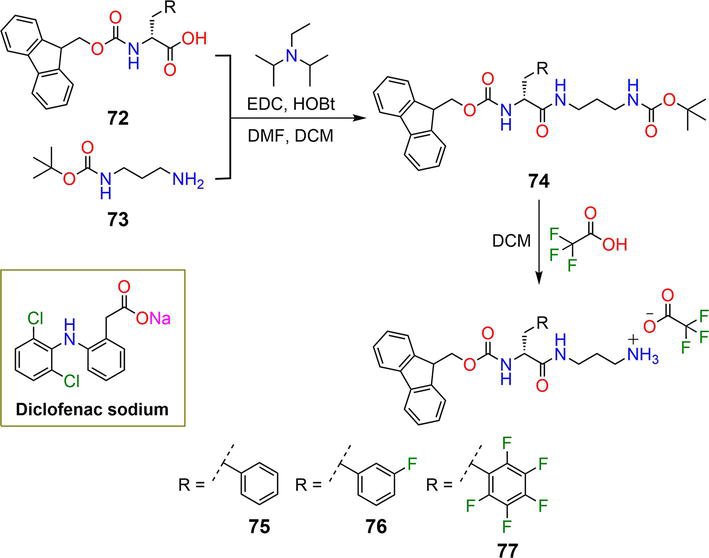
Compound 71, incorporating two fluorenyl moieties, was prepared by reacting (9H-fluoren-9-yl)methyl carbonochloridate 69 with 2,3-diaminopropanoic acid hydrochloride 70 in a basic medium as shown in Figure 13 [45]. Shanmugam et al. demonstrated the ability of compound 71 to form stable hydrogel networks [46]. Compound 71 featured carbamate and fluorenyl groups, which facilitate the formation of intermolecular hydrogen bonding and π-π stacking interactions. The gelation of compound 71 occurred by dissolving it in DMSO with the successive addition of buffer solutions at varying pH values (pH = 4.9, 7.4, and 9.1) to form entangled nanofibers. The CGCs at pH values of 4.9, 7.4, and 9.1 were reported to 0.3, 1.0, and 1.3 wt%, respectively. The drug encapsulation and release potential of hydrogel 71 was assessed at pH 7.4 using vitamin B12. The hydrogel prepared at a gelation concentration of 1.5 wt% was diluted with the phosphate buffer containing vitamin B12. The gel released 50% of vitamin B12 in a sustained and slow fashion after 24 hours of incubation. Hydrogelator 71 revealed cell viability and proliferation against the human fibroblasts (3T3) cell line utilizing the MTT assay, indicating its biocompatibility. Nilsson et al. reported the preparation of compounds 75–77 containing fluorenyl moieties (Figure 14) [47]. Compound 72 reacted with the protected tert-butyl(3-aminopropyl)carbamate 73 in DCM: DMF in the presence of EDC, HOBt, and Hünig’s base to afford 74. Acidic hydrolysis of compound 74 with TFA in DCM yielded derivatives 75–77. Gelation took place by treating heated aqueous solutions of 75–77 with NaCl at varied concentrations (2.5 mM, 5 mM, and 10 mM). The addition of NaCl boosts gelation by decreasing the repulsive effect of the cationic (−NH3+) group. Compounds 75–77 assemble into nanofibers at or below a concentration of 10 mM [48].
Figure 13.
Synthetic pathway of hydrogelator 71 and the chemical structure of the drug molecule [45, 46].
Figure 14.
Synthetic pathway of hydrogelators 75–77 and the structure of the drug molecule [47, 48].
Gelators 75–77 developed at or above 30 mM exhibited outstanding stability, rendering them appealing candidates for drug delivery applications. Gelators 75–77, as well as an equimolar co-assembly of compounds 75 and 77, were studied in vitro and in vivo for their ability to serve as drug delivery vehicles for the anti-inflammatory drug diclofenac. Diclofenac was dissolved in water and mixed with 75–77 or 75:77, with the subsequent addition of NaCl to promote hydrogelation.
The drug-loaded mixtures were incubated with a phosphate-buffered saline solution at a pH of 7 and a temperature of 37°C. All the hydrogels displayed slow and sustained drug release over the course of time in the order 75:77 > 75 > 76 > 77. When the drug-loaded gel 77 was injected into mice with localized induced injuries, the drug release was slow and sustained [48].
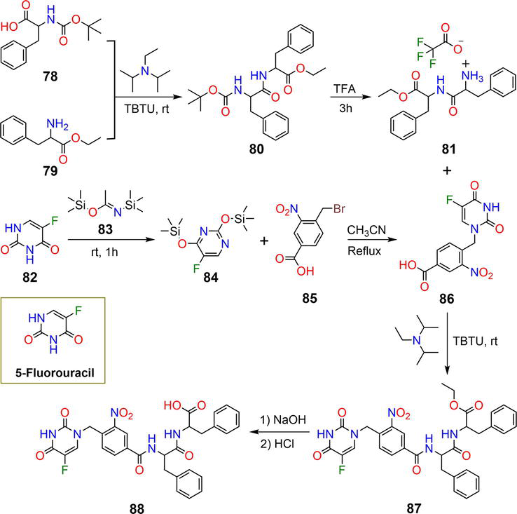
Drug loading can also be achieved by linking drug molecules to the gelators, while release can be triggered using appropriate stimuli. Kundu et al. described the synthesis of hydrogelator 87, tethered to the anticancer drug 5-fluorouracil (Figure 15) [49]. The (tert-butoxycarbonyl)phenylalanine 78 reacted with ethyl phenylalaninate 79 in the presence of the coupling agent TBTU and Hünig’s base at room temperature to yield 80, which on hydrolysis gave 81.
Figure 15.
Synthetic pathway of hydrogelator 88 and the structure of the drug molecule [49].
The reaction of the anticancer drug 5-fluorouracil 82 with the protecting agent N,O-bis(trimethylsilyl)acetamide 83 affords derivative 84, which reacted with 4-(bromomethyl)-3-nitrobenzoic acid 85 in acetonitrile under reflux to give 86. The coupling of compound 81 with 86 forms the peptide-containing drug 87, which on hydrolysis affords gelator 88. The CGC of gelator 88 was determined to be 2 wt%. The surface morphology of hydrogelator 88 revealed the formation of entangled fibrils. The irradiation of gelator 88 using UV light at 365 nm induced 26% liberation the drug after 40 minutes [49]. The biocompatibility of hydrogelator 88 was investigated using the HeLa cell lines and the MTT assay, which revealed cell viability up to 111 μg.
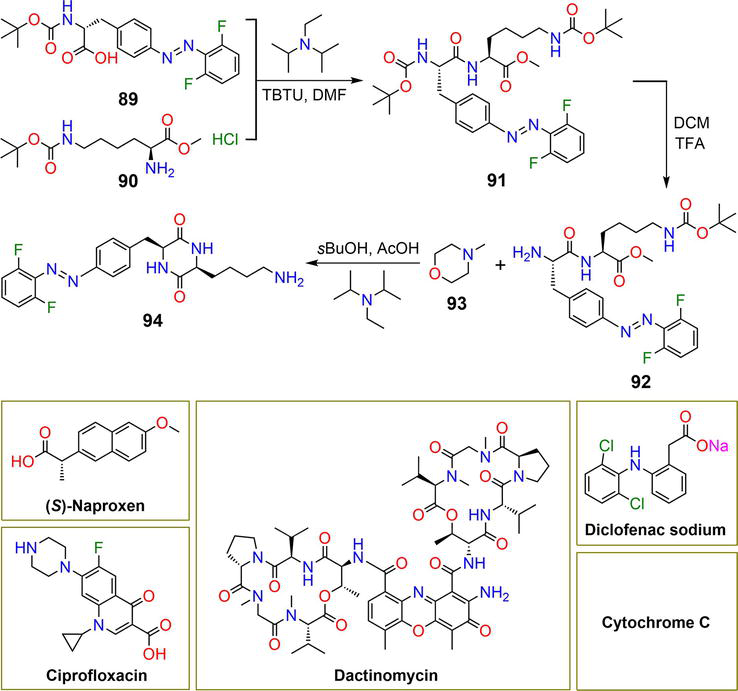
Pianowski et al. reported the synthesis of hydrogelator 94, which incorporates a photoswitchable azo functional group (−N=N−) as indicated in Figure 16 [27]. The reaction of derivative 89 with N-Boc-L-lysine methyl ester hydrochloride 90 in DMF in the presence of TBTU and N-ethyl-N-isopropylpropan-2-amine gave 91. Acidic hydrolysis of derivative 91 using TFA in DCM gave 92. The reaction of 92 with N-methylmorpholine 93 in a solvent mixture of 2-butanol: AcOH and in the presence of Hünig’s base afforded gelator 94 in the trans-form.
Figure 16.
Synthetic pathway of hydrogelator 94 and the structures of the drug molecules [27].
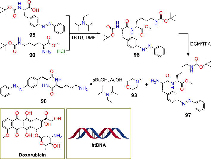
Compound 94 gelates in a phosphate-buffered saline solution (pH 7.4) in the form of fibril structures with a CGC of 3.0 wt%. The hydrogelation is triggered by hydrogen bonding of the amide functional motifs and π-π stacking interactions of the aryl moieties. The hydrogel underwent gel-to-sol transition upon exposure to green light at 523 nm after 3 hours of irradiation [27]. Several pharmaceutical molecules and a protein were encapsulated within the hydrogel matrix during fabrication, including (S)-naproxen, ciprofloxacin, dactinomycin, diclofenac, and cytochrome C. The cargo release was accomplished upon irradiation of the drug or protein-loaded hydrogel with a green light at 523 nm. The half maximal effective concentration (EC50) of hydrogelator 94 against the HeLa cell lines using the MTT assay was >500 μM, indicating lower toxicity on the human cell lines [27]. In another related study, the photoresponsive hydrogelator 98 was designed as a drug delivery carrier for the anticancer drug doxorubicin (Figure 17) [50]. The reaction of compound 95 with N-Boc-L-lysine methyl ester hydrochloride 90 in DMF in the presence of TBTU and Hünig’s base featured the azo-derivative 96.
Figure 17.
Synthetic pathway of hydrogelator 97 and the structure of the drug molecule [50].
Hydrolysis of compound 96 with TFA in DCM afforded compound 97, which, on reaction with N-methylmorpholine 93 in 2-butanol: AcOH and Hünig’s base yielded the corresponding derivative 98. Similar to fluorinated hydrogel 94, hydrogelator 98 is assembled into fibers by hydrogen bonding and π-π stacking interactions. The gel-to-sol transition was triggered by irradiation with UV light at 365 nm, while the sol-to-gel transition was achieved by irradiation with blue light at 460 nm. The irradiation of hydrogelator 98 with UV light at 365 nm disrupted intermolecular noncovalent interactions with transformation to the liquid cis-form. The CGC of gelator 98 was determined to be 1.0% [50]. The mechanical properties of hydrogelator 98 were improved by conducting gelation in aqueous NaCl solution. Hydrogelator 98 was utilized as a delivery system of DOX and herring testis DNA (htDNA). The cargos were encapsulated with the hydrogel during the synthesis at 1.5% gel concentration. After irradiating the DOX- or htDNA-loaded hydrogel with UV radiation at 365 nm for 30 minutes, 70% and 40% of DOX and htDNA were released, respectively.
In summary, Table 1 presents an overview of the hydrogelators discussed in this chapter, along with their CGCs, the substances that can be trapped within the hydrogel matrix, and the conditions under which drug release occurs.
LMWHs have emerged as promising materials with potential applications in various research areas. The unique properties of LMWHs, such as cost-effective preparation, easy functionalization, biocompatibility, low toxicity, and ability to respond to external stimuli, make them ideal candidates for different biomedical applications. LMWHs have shown great potential as drug delivery vehicles for a variety of drugs. However, there are several obstacles that should be addressed in order to fully exploit their promise in drug delivery applications. One of the major drawbacks of LMWHs is their weak mechanical strength, which can be tuned by modifying the structure of hydrogelators, such as incorporating more functional motifs, to promote and strengthen noncovalent interactions. Co-assembly can also reinforce the mechanical properties of LMWHs. The stability of LMWHs under physiological conditions or in response to external stimuli is a crucial factor that should be considered during the development of hydrogels intended for drug delivery applications. LMWHs possess limited drug-loading capacity and, in some circumstances, lack targeted drug delivery. Therefore, the development of more sophisticated strategies for improving the drug-loading potential of LMWHs is an urgent necessity. Moreover, drug release from LMWHs may also be inefficient, leading to suboptimal therapeutic effects. Thus, careful design of the structure of the hydrogelators and selection of the triggers are crucial for achieving optimal drug release. Finally, sustained and slow releases of drugs are two vital aspects to be accomplished for efficient drug delivery using LMWHs.
We hope that this chapter will spark further investigations into the synthetic approaches of LMWHs and lead to the discovery of novel gelators with potential applications in drug delivery.
1.Patil SP, Jeong HS, Kim BH. A low-molecular-weight supramolecular hydrogel of riboflavin bolaamphiphile for VEGF-siRNA delivery. Chemical Communications. 2012;48(71):8901-8903
2.Patil SP, Kim S-H, Jadhav JR, Lee J-H, Jeon EM, Kim K-T, et al. Cancer-specific gene silencing through therapeutic siRNA delivery with B vitamin-based nanoassembled low-molecular-weight hydrogelators. Bioconjugate Chemistry. 2014;25:1517-1525
3.Sedghiniya S, Soleimannejad J, Blake AJ. A low molecular weight Zr(IV) metallogel for protein delivery. Materials Today Communication. 2021;27:102448
4.Jagrosse ML, Agredo P, Abraham BL, Toriki ES, Nilsson BL. Supramolecular phenylalanine-derived hydrogels for the sustained release of functional proteins. ACS Biomaterials Science & Engineering. 2023;9(2):784-796
5.McCloskey AP, Lee M, Megaw J, McEvoy J, Coulter SM, Pentlavalli S, et al. Investigating the in vivo antimicrobial activity of a self-assembling peptide hydrogel using a galleria mellonella infection model. ACS Omega. 2019;4:2584-2589
6.Reddy SM, Shanmugam G, Duraipandy N, Kiran MS, Mandal AB. An additional fluorenylmethoxycarbonyl (Fmoc) moiety in di-Fmoc-functionalized L-lysine induces pH-controlled ambidextrous gelation with significant advantages. Soft Matter. 2015;11(41):8126-8140
7.Bernet A, Behr M, Schmidt H-W. Supramolecular hydrogels based on antimycobacterial amphiphiles. Soft Matter. 2012;8:4873-4876
8.Cross ER, Coulter SM, Fuentes-Caparros AM, McAulay K, Schweins R, Laverty G, et al. Tuning the antimicrobial activity of low molecular weight hydrogels using dopamine autoxidation. Chemical Communications. 2020;56(58):8135-8138
9.Zhao X, Zhang H, Gao Y, Lin Y, Hu J. A simple injectable moldable hydrogel assembled from natural glycyrrhizic acid with inherent antibacterial activity. ACS Applied Bio Materials. 2020;3(1):648-653
10.Aldilla VR, Chen R, Martin AD, Marjo CE, Rich AM, Black DS, et al. Anthranilamide-based short peptides self-assembled hydrogels as antibacterial agents. Scientific Reports. 2020;10(1):770
11.Zhao Q , Zhao Y, Lu Z, Tang Y. Amino acid-modified conjugated oligomer self-assembly hydrogel for efficient capture and specific killing of antibiotic-resistant bacteria. ACS Applied Materials & Interfaces. 2019;11(18):16320-16327
12.Rizzo C, Arrigo R, Dintcheva NT, Gallo G, Giannici F, Noto R, et al. Supramolecular hydro- and ionogels: A study of their properties and antibacterial activity. Chemistry - A European Journal. 2017;23(64):16297-16311
13.Tang Q , Plank TN, Zhu T, Yu H, Ge Z, Li Q , et al. Self-assembly of metallo-nucleoside hydrogels for injectable materials that promote wound closure. ACS Applied Materials & Interfaces. 2019;11(22):19743-19750
14.Wei Q , Chang Y, Ma G, Zhang W, Wang Q , Hu Z. One-pot preparation of double network hydrogels via enzyme-mediated polymerization and post-self-assembly for wound healing. Journal of Materials Chemistry B. 2019;7(40):6195-6201
15.Liang C, Zhang L, Zhao W, Xu L, Chen Y, Long J, et al. Supramolecular nanofibers of drug-peptide amphiphile and affibody suppress HER2+ tumor growth. Advanced Healthcare Materials. 2018;7(22):e1800899
16.Citossi F, Smith T, Lee JB, Segal J, Gershkovich P, Stocks MJ, et al. Self-assembling benzothiazole-based gelators: A mechanistic understanding of in vitro bioactivation and gelation. Molecular Pharmaceutics. 2018;15(4):1578-1586
17.Bhagat SD, Srivastav A. Amphiphilic phenylalanine derivatives that temporally generate reactive oxygen species from water in the presence of Au(III) ions. Biomaterials Science. 2020;8(17):4750-4756
18.Zanna N, Focaroli S, Merlettini A, Gentilucci L, Teti G, Falconi M, et al. Thixotropic peptide-based physical hydrogels applied to three-dimensional cell culture. ACS Omega. 2017;2(5):2374-2381
19.Tang JD, Mura C, Lampe KJ. Stimuli-responsive, pentapeptide, nanofiber hydrogel for tissue engineering. Journal of the American Chemical Society. 2019;141(12):4886-4899
20.Suga T, Osada S, Narita T, Oishi Y, Kodama H. Promotion of cell adhesion by low-molecular-weight hydrogel by Lys based amphiphile. Materials Science and Engineering: C. 2015;47:345-350
21.Talloj SKM, Mohammed M, Lin H-C. Construction of self-assembled nanostructure-based tetraphenylethylene dipeptides: Supramolecular nanobelts as biomimetic hydrogels for cell adhesion and proliferation. Journal of Materials Chemistry B. 2020;8(33):7483-7493
22.Dessane B, Smirani R, Bougueon G, Kauss T, Ribot E, Devillard R, et al. Nucleotide lipid-based hydrogel as a new biomaterial ink for biofabrication. Scientific Reports. 2020;10(1):2850
23.Chu NT, Chakravarthy RD, Shih NC, Lin YH, Liu YC, Lin JH, et al. Fluorescent supramolecular hydrogels self-assembled from tetraphenylethene (TPE)/single amino acid conjugates. RSC Advances. 2018;8(37):20922-20927
24.Nour HF, El Malah T, Khattab TA, Olson MA. Template-assisted hydrogelation of a dynamic covalent polyviologen-based supramolecular architecture via donor-acceptor interactions. Materials Today Chemistry. 2020;17:100289
25.Colquhoun C, Draper ER, Eden EG, Cattoz BN, Morris KL, Chen L, et al. The effect of self-sorting and co-assembly on the mechanical properties of low molecular weight hydrogels. Nanoscale. 2014;6(22):13719-13725
26.Basak S, Singh I, Banerjee A, Kraatz H-B. Amino acid-based amphiphilic hydrogels: Metal ion induced tuning of mechanical and thermal stability. RSC Advances. 2017;7:14461-14465
27.Karcher J, Pianowski ZL. Photocontrol of drug release from supramolecular hydrogels with green light. Chemistry - A European Journal. 2018;24(45):11605-11610
28.Noteborn WEM, Vittala SK, Torredemer MB, Maity C, Versluis F, Eelkema R, et al. Switching the mode of drug release from a reaction-coupled low-molecular-weight gelator system by altering its reaction pathway. Biomacromolecules. 2023;24(1):377-386
29.Poolman JM, Boekhoven J, Besselink A, Olive AG, van Esch JH, Eelkema R. Variable gelation time and stiffness of low-molecular-weight hydrogels through catalytic control over self-assembly. Nature Protocols. 2014;9(4):977-988
30.Ghosh S, Nag S, Saha KD, Banerji B. S-benzyl cysteine based cyclic dipeptide super hydrogelator: Enhancing efficacy of an anticancer drug via sustainable release. Journal of Peptide Science. 2022;28(8):e3403
31.Wadhavane PD, Gorla L, Ferrer A, Altava B, Burguete MI, Izquierdo MÁ, et al. Coordination behaviour of new open chain and macrocyclic peptidomimetic compounds with copper(ii). RSC Advances. 2015;5:72579-72589
32.Valls A, Isabel Burguete MK, Kuret L, Altava B, Luis SV. Open chain pseudopeptides as hydrogelators with reversible and dynamic responsiveness to pH, temperature and sonication as vehicles for controlled drug delivery. Journal of Molecular Liquids. 2022;348:118051
33.Bica K, Rijksen C, Nieuwenhuyzen M, Rogers RD. In search of pure liquid salt forms of aspirin: Ionic liquid approaches with acetylsalicylic acid and salicylic acid. Physical Chemistry Chemical Physics. 2010;12(8):2011-2017
34.Kuddushi M, Patel NK, Rajput S, El Seoud OA, Mata JP, Malek NI. Temperature-responsive low molecular weight ionic liquid based gelator: An approach to fabricate an anti-cancer drug-loaded hybrid ionogel. ChemSystemsChem. 2020;2(5):e1900053
35.Mehra RR, Basu A, Christman RM, Harjit J, Mishra AK, Tiwari AK, et al. Mechanoresponsive, proteolytically stable and biocompatible supergelators from ultra short enantiomeric peptides with sustained drug release propensity. New Journal of Chemistry. 2020;44:6346-6354
36.Rodik RV, Anthony A-S, Kalchenko VI, Mélya Y, Klymchenko AS. Cationic amphiphilic calixarenes to compact DNA into small nanoparticles for gene delivery. New Journal of Chemistry. 2015;39:1654-1664
37.Granata G, Petralia S, Forte G, Conoci S, Consoli GML. Injectable supramolecular nanohydrogel from a micellar self-assembling calix[4]arene derivative and curcumin for a sustained drug release. Materials Science and Engineering: C. 2020;111:110842
38.Dadhwal S, Fairhall JM, Hook S, Gamble AB. Tetrafluoroaryl azide as an N-terminal capping group for click-to-dissolve diphenylalanine hydrogels. RSC Advances. 2020;10(16):9234-9244
39.Ghosh T, Biswas A, Gavel PK, Das AK. Engineered dynamic boronate ester-mediated self-healable biocompatible G-quadruplex hydrogels for sustained release of vitamins. Langmuir. 2020;36(6):1574-1584
40.Kumar BA, Nayak RR. Supramolecular phenoxy-alkyl maleate-based hydrogels and their enzyme/pH-responsive curcumin release. New Journal of Chemistry. 2019;43:5559-5567
41.Liu Q , Wang H, Li G, Liu M, Ding J, Huang X, et al. A photocleavable low molecular weight hydrogel for light-triggered drug delivery. Chinese Chemical Letters. 2019;30:485-488
42.Tiwari P, Rajagopalan R, Moin M, Soni R, Trivedi P, DuttKonar A. Can self-assembled hydrogels composed of aromatic amino acid derivatives function as drug delivery carriers? New Journal of Chemistry. 2017;41:308-315
43.Singh M, Kundu S, Reddy M, Sreekanth V, Motiani RK, Sengupta S, et al. Injectable small molecule hydrogel as a potential nanocarrier for localized and sustained in vivo delivery of doxorubicin. Nanoscale. 2014;6:12849-12855
44.Gupta S, Singh M, Reddy M. A, Yavvari PS, Srivastava a, Bajaj a, interactions governing the entrapment of anticancer drugs by low-molecular-weight hydrogelator for drug delivery applications. RSC Advances. 2016;6:19751-19757
45.Nishikawa T, Urabe D, Isobe M. Syntheses of N-acylisoxazolidine derivatives, related to a partial structure found in zetekitoxin AB, a golden frog poison. Heterocycles. 2009;79:379-385
46.Arokianathan JF, Ramya KA, Janeena A, Deshpande AP, Ayyadurai N, Leemarose A, et al. Non-proteinogenic amino acid based supramolecular hydrogel material for enhanced cell proliferation. Colloids and Surfaces B: Biointerfaces. 2020;185:110581
47.Rajbhandary A, Raymond DM, Nilsson BL. Self-assembly, hydrogelation, and nanotube formation by cation-modified phenylalanine derivatives. Langmuir. 2017;33(23):5803-5813
48.Raymond DM, Abraham BL, Fujita T, Watrous MJ, Toriki ES, Takano T, et al. Low molecular weight supramolecular hydrogels for sustained and localized in vivo drug delivery. ACS Applied Bio Materials. 2019;2(5):2116-2124
49.Das S, Horo H, Goswami U, Kundu LM. Synthesis of a peptide conjugated 5-fluorouracil gelator prodrug for photo-controlled release of the antitumor agent. ChemistrySelect. 2019;4(22):6778-6783
50.Pianowski ZL, Karcher J, Schneider K. Photoresponsive self-healing supramolecular hydrogels for light-induced release of DNA and doxorubicin. Chemical Communications. 2016;52(15):3143-3146
Written By
Hany F. Nour, Ahmed M. Salama, Badria H.A. Al-Dhuwayin and Amal F. Seliem
Submitted: 30 May 2023Reviewed: 01 June 2023Published: 12 July 2023
 Open access peer-reviewed chapter