Open Access is an initiative that aims to make scientific research freely available to all. To date our community has made over 100 million downloads. It’s based on principles of collaboration, unobstructed discovery, and, most importantly, scientific progression. As PhD students, we found it difficult to access the research we needed, so we decided to create a new Open Access publisher that levels the playing field for scientists across the world. How? By making research easy to access, and puts the academic needs of the researchers before the business interests of publishers.
We are a community of more than 103,000 authors and editors from 3,291 institutions spanning 160 countries, including Nobel Prize winners and some of the world’s most-cited researchers. Publishing on IntechOpen allows authors to earn citations and find new collaborators, meaning more people see your work not only from your own field of study, but from other related fields too.
To purchase hard copies of this book, please contact the representative in India:
CBS Publishers & Distributors Pvt. Ltd.
www.cbspd.com
|
customercare@cbspd.com
This book chapter give an overview of natural and synthetic polymeric moieties consumed for developing hydrogels and their types. Different properties of nanogels are the advancement of hydrogels characterized by nano-size range, stimuli-responsive swelling, and release. Stimuli responsiveness is imparted by the presence of a suitable monomer. A number of polymerization approaches are presented in the literature that are employed to prepare such networks. These systems are elastic, rubbery, nontoxic, and biocompatible and offer prolonged release of the drugs without chances of dose dumping. These types of networks have potential pharmaceutical, agricultural, food, and biotechnological applications in terms of controlled, prolonged, and targeted drug delivery, solubility enhancements, stimuli-dependent intelligent drug delivery, such as contact lenses, wound healing, etc. In the current chapter, we have tried to introduce hydrogels and microgels, their different types, the variety of polymers used to develop such carrier systems, approaches to develop such drug delivery systems, and their utilization in various sectors in addition to the pharmaceutical sector.
Department of Pharmacy, University of Chakwal, Pakistan
Hira Ijaz
Department of Pharmaceutical Sciences, Pak-Austria Fachhochschule: Institute of Applied Sciences and Technology, Haripur, Pakistan
Rai Muhammad Sarfraz
College of Pharmacy, University of Sargodha, Sargodha, Pakistan
Nadiah Zafar
Faculty of Pharmacy, Department of Pharmaceutics, University Teknologi MARA, Malaysia
Muhammad Zaman
Faculty of Pharmacy, University of Central Punjab, Lahore, Pakistan
Mariya Azam
Department of Pathology, Niazi Medical and Dental College, Sargodha, Pakistan
*Address all correspondence to: asif.mahmood@uoc.edu.pk
1. Introduction
Drug delivery encompasses a variety of techniques, formulations, and systems for delivering therapeutic moieties toward desired areas within the body to attain therapeutic outcomes effectively [1]. Conventional dosage forms are more likely required to be frequently administered in high doses, thereby resulting in certain side effects and patient noncompliance. To resolve such issues, controlled drug delivery systems with minimal side effects have been introduced that control the release of drugs in a number of ways [2]. By developing such delivery systems, the bioavailability of active agents can be potentiated through rate controlled delivery approach [3].
The term hydrogel has been used repeatedly in the literature. The first cross-linked polymeric network material was developed in 1960 [4]. It was equipped with properties of hydrogels, i.e., mainly having attraction toward water. Polyhydroxy ethyl methacrylate (PHEMA) hydrogel is massively employed in the preparation of contact lenses. These soft materials are also consumed by the patients for therapeutic purposes [5].
A new polymeric material was developed that was responsive to a number of environmental stimuli like pH, temperature, and concentration of different solutes in a solvent system. These stimuli play an important role in the swelling transition behavior of the networks and, subsequently, the release of therapeutic agents [6].
The third generation of hydrogels offered further advances in the field of polymeric materials. It has focused on stereo-complex materials, their interactions, and cross-linking through physical interactions [2]. Yana and his partners worked on collagen and shark cartilage tissues to develop a novel dressing that effectively decreased the healing burn [7]. Natural and synthetic hydrogels are used extensively in tissue engineering. Both of these hydrogels have variable potential applications in the control of thrombosis, drug delivery, biosensor coating, and cell transplant. Furthermore, the properties of hydrogels, like water-loving, biocompatibility, and their response to stimuli, established a great interest of researchers and scientists to introduce smart hydrogels [8].
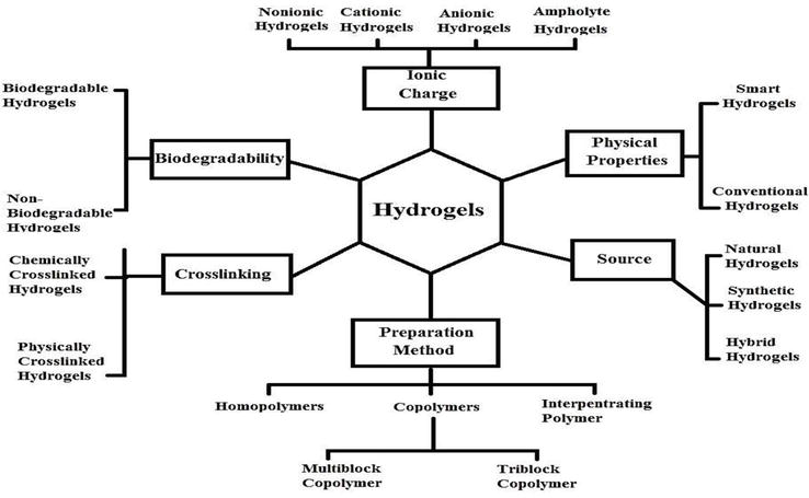
Hydrogels are 3D water-loving carrier systems that have the ability to imbibe plenty of aqueous media or biological solvents without showing any transition in their structure [9]. These carrier systems, at their optimal swelling, show resemblance with body tissues because of their delicate and elastic consistency [10]. Hydrogels being intelligent carrier systems show several advantages like absence of active ingredient exposure in acidic environment, targeted drug delivery, and prolonged release of incorporated drugs [11]. A number of bonding types are involved development of chemically cross-linked hydrogels like ionic or covalent [5]. Presence of hydrophilic functional groups in developed polymer structure like –OH, –COOH, –CONH2, –CONH, and SO3H under suitable pH conditions offer absorption of a large amount of water by hydrogels and is helpful in the encapsulation of hydrophilic drugs [12]. Hydrogels are available in numerous forms, so classification may be based on various factors including method of preparation, source, biodegradation, response to certain stimuli, cross-linking, physical properties, etc. [13]. The detailed classification of hydrogels is summarized in Figure 1.
Figure 1.
Classification of polymeric hydrogels.
Hydrogels offer a variety of pharmaceutical, medical, and clinical applications like tissue regeneration, diagnostic applications, mucoadhesive potentials, etc. Hydrogels in literature are formulated in different shapes like nano and micro-sized particles, hydrogel membranes, beads, matrics, etc. [7, 8, 9, 10, 11], depending upon the required use.
Hydrogels are high molecular weight network systems primarily consisting of hydrophilic polymers holding excess physiological fluid or water in polymer chains. Due to higher cross-linking reaction within polymers a grafted network structure is developed that prevent dissolution of hydrogels [14]. These type of network systems, when fabricated to be influenced by certain external stimuli such as temperature, pH, enzyme, light, electric and magnetic fields, etc., are known as stimuli-responsive hydrogels [15]. On exposure to these environmental stimuli they exhibit response by undergoing volume or shape transitions [16, 17].
2.1 pH-responsive hydrogels
Hydrogels sensitive to pH are being developed extensively. There are various factors that control the swelling and de-swelling in hydrogels like ionic charge, extent of ionization, pka/pkb values, pH of medium, type of the monomer, concentration of polymer, and hydrophilicity of the developed networks [6]. At higher pH, weakly acidic functional groups present in anionic hydrogels like -COOH group undergo ionization resulting in swelling of the network. Similarly at lower pH, cationic hydrogels show swelling behavior due to ionization of basic functional groups like amines [18, 19, 20].
2.1.1 Mechanism of swelling in pH-responsive hydrogels
Swelling in hydrogels is caused by variations in osmotic pressure created within the network on exposure to physiological fluids that is controlled by the presence of charge on polymer, its hydrophilicity, and counter ions within the network system. Swelling of hydrogels proceeds in three steps [21]. Firstly, water penetrates into the hydrogel by diffusion, causing the hydration of polymeric chains. As a result, the relaxation of the polymeric chains promotes swelling in the grafted network [19]. Primary-bound water induces swelling due to its interaction with polar groups in the hydrogel, while secondary-bound water interacts with available hydrophobic groups. An osmotic driving force is created that promotes diffusion of additional water called as free water into the hydrogel resulting them attaining equilibrium swelling. Flory and Rehner’s theory describe this swelling response in terms of elasticity of polymeric chains and their compatibility with water molecules [22].
2.1.2 Mechanism of drug release in pH-sensitive Hydrogels
Active agents loaded or entrapped in hydrogels are released by different ways like diffusion-based, swelling-based, and chemically controlled approaches.
2.1.2.1 Diffusion-based controlled release
Cross-linked network structure of hydrogels composed of mesh-like geometry within the polymeric chains involved in the retention of small particles and liquids. The release of therapeutic agents through these hydrogels is described by size of mesh or pores [23]. Diffusion process is dominated in grafted network systems having mesh size greater than the size of drug [8]. In this case, size of pores has no significant impact on diffusion as smaller particles can pass easily through the cross-linked network. When the pores size in network becomes equal to the size of active moiety, then drug mainly diffuses through steric hindrance effect [21]. By using increased proportions of polymers or cross-linker contents in hydrogels pores size can be decreased. Due to reduced mesh size a greater frictional drag is exerted on drug by polymeric chains resulting in greater path length for drug transportation. In this case, diffusion is approximated by certain theoretical principles, and the net result is prolonged drug release by slow diffusion [24].
2.1.2.2 Swelling-based controlled release
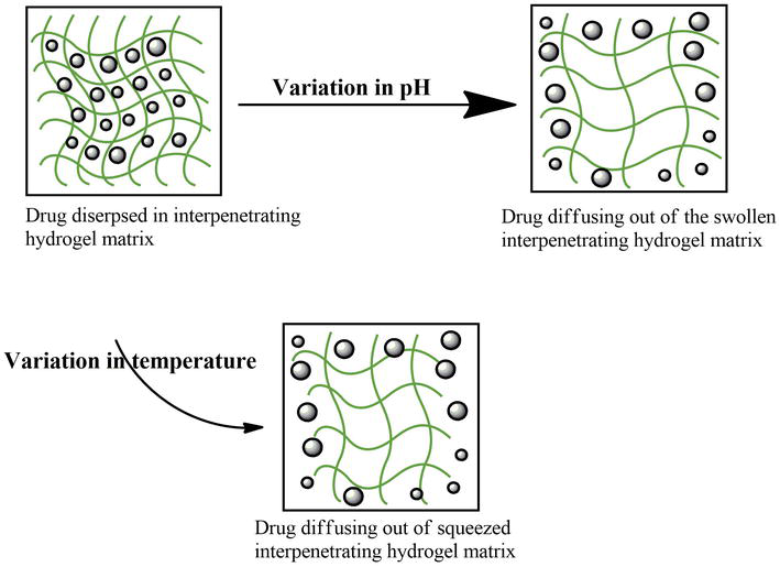
Therapeutic agents can be released from hydrogels by another approach that is controlled by swelling mechanism [25]. Hydrogels swelling involves the absorption of physiological, biological, or buffer fluids that results in the enlargement of mesh size. Numerous factors can influence the swelling response like pH, temperature, light, ionic strength, electric field, glucose, etc. An important approach for cancer and oral drug delivery systems is pH mediated swelling [8]. By employing such technique certain pH responsive monomers and polymers are copolymerized to develop hydrogels that show negligible swelling in acidic pH of stomach, causing protection of entrapped drugs [26]. While at intestinal pH, these grafted networks show appreciable swelling attributing to drug release by swelling and diffusion modes. Numerous polymers having pendent basic or acidic functional groups are employed to develop such stimuli-responsive hydrogels. Furthermore, therapeutic moieties can be guided to targeted areas having solid tumors (more acidic environment) by using pH-responsive release systems (Figure 2) [27].
Figure 2.
Drug release mechanism in pH and temperature-responsive hydrogels.
2.1.2.3 Chemically controlled release
Another strategy of drug release from hydrogels involves enzymatic or hydrolytic degradation of chains and follows a chemically controlled method [28]. By adopting this technique, release of drugs from cross-linked structures is kinetically controlled or reaction–diffusion controlled [29]. In kinetically controlled method, chemical reaction within hydrogel contents causes polymer decomposition by cleavage of its chains. In this case, there is no significant diffusion process involved. While, diffusion-controlled method results in drug release by both mechanisms involving polymer decomposition and followed by drug diffusion [30].
Hydrogels are declared biocompatible due to their high water content. These offer unique physical properties as their elasticity resembles the living tissues, due to which a lot of researchers have employed them for drug delivery applications. Structurally, these are highly porous, and their porosity can be tuned by the extent/degree of cross-linking in gel-matrix. Porosity is vital as it impacts on swelling, loading of drugs and release drug at desired rate depending upon the diffusion coefficient of the micro as well as macro molecules [28]. Hydrogels do not trigger or identified as foreign particles by body’s own immune system due to the difference in interfacial tension between the surface of hydrogel and body fluids leading to minimized adsorption of protein and cell adhesion. Hydrogels are advantageous owing to ease of preparation. Hydrogels are capable of molding themselves according to the shape of surface applied [29]. Therefore, they are deformable. Nonirritant due to their soft and rubbery/elastic nature. Biodegradable hydrogels can be designed via implication of suitable hydrolytic, enzymatic, and environmental factors (e.g., pH, temp, or electric field).
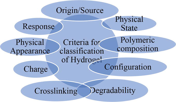
These can be classified based on various criteria as follows (Figure 3) [31]:
Origin-based classification
According to their source, hydrogels are categorized as:
Natural
Synthetic
Semi-synthetic
Based on physical state
Solid hydrogels
Semi-solid hydrogels
Liquid hydrogels
Polymeric composition-Based Classification
Homopolymeric hydrogels
Co-polymeric hydrogels
Interpenetrating multipolymer hydrogels
Homopolymeric hydrogels are formed by single monomer species, depending upon the nature of polymer and method of polymerization.
Co-polymeric hydrogels are made up of two or more monomer species. Among these, one is hydrophilic. They have arranged randomly, block-wise or alternatively along the polymeric chain network [32].
Interpenetrating multipolymer hydrogels are networks of two or more cross-linked polymers. One of which is natural, and the other is synthetic arranged in a network [33].
Configuration-based classification
On the basis of their physical and chemical configuration, there are four types:
Cross-linking may be chemical or physical depending upon the nature of cross-linked junctions; thus, hydrogels are classified into physically or chemically cross-linked hydrogels [34].
Chemically cross-linked hydrogels are made up of stable bonds.
Physically cross-linked hydrogels have unstable junctions which are formed either by polymer chain entanglements or as a result of physical interactions, hydrogen bonds, or hydrophobic interactions.
Based on physical appearance
Solid hydrogels
Semi-solid hydrogels
Liquid hydrogels
Charge-based classification
Cross-linked chains possess electric charges and classified accordingly.
Nonionic (neutral)
Amphoteric (composed of both acidic and basic groups)
Ionic (anionic/cationic)
Zwitterionic (having both anionic or cationic groups in each structural unit) [35]
Based on response to environmental stimuli
Hydrogels have the ability to respond to various chemical and physical stimuli [36, 37] and are classified into the followings:
Physically responsive hydrogels
Chemically responsive hydrogels
Biochemically responsive hydrogels
Figure 3.
Classification of hydrogels.
Physical stimuli may include temperature, light, electric or magnetic field, pressure, and sound. Chemical stimuli are pH, ionic strength, solvent composition, and molecular species. Biochemical stimuli include antigens, ligands, and enzymes [38].
Hydrogels get responsive to any kind of stimuli by means of volume alterations in their structure. In dry state, polymeric network of hydrogel is tightly bound and looks compacted, but as it comes in contact with fluid of suitable pH value, water molecules get diffused into its structure, and the polymer chains start separating, leading to subsequent swelling as a result of repulsion among ionized functional groups [31]. The extent of swelling depends upon three major factors:
Ionic constituents of hydrogels
Increase in the ionic content of hydrogels would result in higher electrostatic repulsion between ionized groups; thereby, elevation of swelling ratio occurs.
Cross-linking content of hydrogels
It controls the swelling of hydrogel system. Highly cross-linked network shows delayed swelling and vice versa.
Hydrophilic constituents
As hydrophilicity of hydrogel contents increases, there is more interaction between water and hydrophilic groups and ultimate increase in swelling [36].
Once swelling starts, it continues from surface to interior core of hydrogel matrix until equilibrium is achieved, which is indicated by optimum swelling. At peak swelling stage, polymeric chains maintain a greater distance, and the solvent absorbed creates a pressure on polymeric chains, which is compensated by the presence of a cross-linker [37, 39]. Through diffusion, solvent and solute gets in and out of the hydrogel structure. The theory for swelling of hydrogel, which narrates kinetic properties for transport of solvent molecule into hydrogel system, can be demonstrated as follows:
t=a2D
Where,
t = time required for swelling.
a = length of hydrogel.
D = diffusion coefficient
D=K+4μ/3f
K = bulk modulus.
f = friction between polymer and bulk molecule.
μ = shear modulus.
Thus, the length of hydrogel has a direct relation with time of swelling thus the response of hydrogel to any stimuli is controlled by increasing or decreasing length of hydrogel. There are numerous strategies by which swelling of hydrogel can be enhanced, e.g., increasing the size of hydrogel by graft polymerization. This technique is beneficial for any material which de-swells promptly as the graft polymerization increases the sensitivity of hydrogel, and the resultant hydrogel responds much more rapidly by diffusion of solvent [40].
Hydrogels being unique carrier systems have certain limitations along with different advantages.
Hydrogels offer poor tensile strength leading to early breakdown and hence dislocation from target sites.
More significantly, drug delivery issues may arise, like heterogeneous drug loading, as in the case of hydrophobic drugs, which can affect drug release from the hydrogel network.
Greater pore size promotes the uptake of larger water contents and hence faster drug release rates.
Although hydrogels are deformable and can be used as injectable still, some require surgical implantation, which is exorbitant and demands sterile conditions for implantation [41, 42].
Constituents used in hydrogels are mentioned in Table 1.
Monomer: Single units used in network fabrication by polymerization.
Initiator: In order to kick-start the polymerization reaction.
Cross-linking agents: Cross-links polymeric chains for network fabrication to resist disintegration and dissolution of IPN itself.
7.1 Drug loading and drug release
Drug loading in IPNs:
Generally, two methods are followed for drug loading in IPNs; preloading and postloading [43].
Preloading: Drug loading in this method is carried out during the preparation of polymeric networks. Preweighed amount of medication (active pharmaceutical ingredient) is solubilized in the most suitable solvent, and afterward, this drug solution is added to the final polymerization mixture or solution. Another approach is to blend medication solution with a monomer solution, and other solutions are added separately. After the completion of polymerization, API will remain entrapped within the network framework.
Postloading: This method involves the formation 1% solution of medication. This is done by dissolving 1 g of medication (drug) in the most suitable solvent, and then the volume is made up to 100 ml. Dried and preweighed hydrogel discs are dipped in drug solution for predefined intervals until and unless maximum swelling occurs. Loaded IPNs are dried at normal room temperature. Sometimes, drying is accomplished by using an oven for a particular time period and temperature. In this way, dried and drug-loaded IPNs are obtained.
Drug release in IPNs:
Generally, three methods are being followed for drug release in IPNs [37];
7.2 Swelling- controlled release
When water or biological fluids comes in contact with the dried or dehydrated IPNs it imbibes into the voids in between the polymeric chains. Thus, the drug, after its dissolution, is released in solution form from the networks.
7.3 Diffusion-controlled release
Drug release from IPNs is done by following the diffusion law (Fick’s diffusion law), i.e., the drug is released according to the concentration gradient [23].
7.4 Chemically controlled release
Chemical reactions like enzymatic reaction and hydrolysis cause cleavage of polymeric chains resulting in drug release. Drug release from IPNs is studied by performing dissolution studies, and then it is validated through different kinetic models like zero-order, first order, Higuchi, Korsmeyer–Peppas, and Hixson Crowell models to find out the drug release kinetics in terms of best-fit model and mechanism of drug release from regression coefficient and value of exponent n, respectively [39].
Numerous polymers from natural and synthetic sources are being used for development of hydrogels. Polymers obtained from natural sources have excellent characteristics like biocompatibility, biodegradability, nontoxic nature, and economical, but hydrogels developed by these polymers show poor durability [40, 43]. While synthetic polymers are expensive and nonbiodegradable, and their hydrogels are good in mechanical strength. So these limitations can be overcome by blending the natural polymers with synthetic ones to develop more stable hydrogel networks [41, 42]. Natural polymers can be categorized as plant-based and animal-based polymers that have been extensively employed for various applications. Gums obtained from plants have gained considerable interest in drug release applications owing to their biocompatibility, low cost, and easy accessibility [44]. Detail of some natural and synthetic polymers that are being employed for hydrogel network formation given in the proceeding sections.
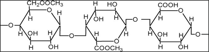
8.1 Pectin
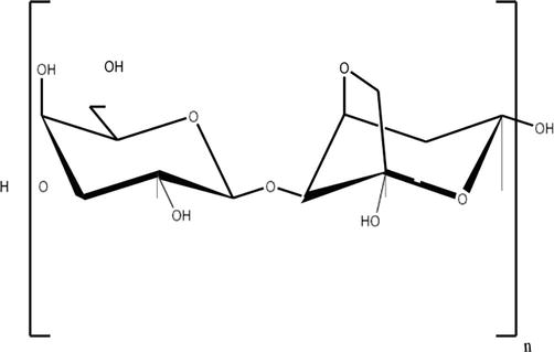
Pectin is a carbohydrate-based polymer having a natural origin. It is prepared by extraction process on a commercial scale; from different types of citrus plants like apples, guavas, strawberries, oranges, and grapes under slightly acidic conditions [44]. High methoxyl pectin and low methoxyl pectin are two groups in which pectin can be divided. In the food industry, it is employed as a gelling agent as well as a stabilizer. Pectin has already been investigated for the development of targeted delivery of drugs and other biomedical applications [45]. Rapid degradation of pectin occurs by colonic microorganisms and thus making it a possible carrier for colon-targeted drug delivery because of its exceptional biocompatible and biodegradable nature [46]. Nowadays, scientists have been working on the fabrication of pectin-based preparations such as polymeric hydrogels, tablets, microspheres, films, and nanoparticles [47].
Structurally, pectin is a polysaccharide. It consists of repetitive galacturonic acid units joined together through ɑ-1,4-glucoside bonds, as shown in Figure 4 [48]. Pectin has linear as well as a branched structure, thus making it a complex molecule. Methylation and esterification both contribute to the gelling ability of pectin. The degree of esterification not only contributes to the gelling potential of pectin but also affects solubility profile of pectin. Pectin forms aggregates in cold water but solubilizes in slightly warm water under continuous stirring [49].
Figure 4.
Chemical structure of pectin.
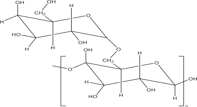
8.2 Natrosol or hydroxyethyl cellulose (HEC)
Natrosol is a polymer that is derived from cellulose. It is a water-soluble and nonionic derivative of cellulose. The basic structure is shown in Figure 5. It has a chain structure in which anhydrous glucose units are attached to each other by β-1,4-linkage giving rise to three free hydroxyl groups in each unit. These free hydroxyl groups make it a suitable polymer for hydrogel fabrication [50].
Figure 5.
Chemical structure of natrosol.
It is marketed in two grades; R-grade and B-grade. The R-grade of Natrosol facilitates preparation of solution in water without formation of lumps. Natrosol has been used in various pharmaceutical preparations because of its emulsifying, water-retaining, thickening, gelling, and stabilizing properties. It is most widely consumed in ophthalmic and topical preparations to facilitate drug delivery. It is also used in household products like lubricants, detergents, and cosmetics attributed to its water-soluble and nonionic nature [51]. It is soluble in hot water as well as cold water. Its viscous nature has indirect relation with increasing temperature can be reduced by increasing temperature. This property also makes it a useful ingredient for making gels at lower temperatures [52].
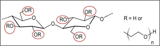
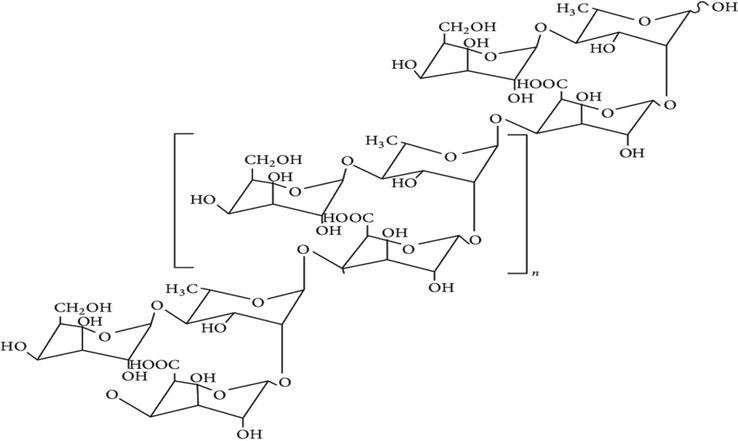
8.3 Tamarind gum
Tamarind gum is a polysaccharide, a natural polymer obtained by extraction of Tamarindus indica seeds. Its structure consists of β-(1,4)-d-glucan attached with pendant chains of α-(1,4)-d-xylopyranose and [β-d-galactopyranosyl-(1,2)-α-d-xylopyranosyl] linked to glucose units [53]. It is extracted by employing enzymatic and chemical-based extraction methods. In chemical method, ground seeds are soaked in water to prepare a slurry, which is then added to boiling distilled water with continuous stirring. The resultant mixture is filtered and poured in twice its volume of ethanol to get the required precipitates [54]. These precipitates are dried to prepare the gum. In enzymatic-based extraction, the protease enzyme is allowed to act on tamarind seed powder after mixing it with ethanol. Mixture is then centrifuged, and the supernatant collected is poured into ethanol to obtain precipitates of tamarind gum. These are dried after separation for further use [55].
It contains abundant hydroxyl groups. It is transformed into mucilage while contacting with water. This can be used in the pharmaceutical sector as an excipient because of its compatibility with living tissues, nontoxic nature, and bioadhesive potentials (Figure 6) [56].
Figure 6.
Structural formula of tamarind seed polysaccharide.
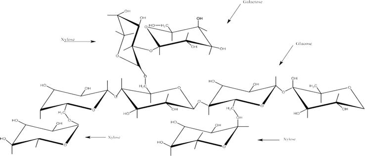
8.4 Fenugreek
Fenugreek seeds gum is obtained by extraction of seeds endosperm of fenugreek (Trigonella foenum-graecum L.) [57]. High content of gum is present in fenugreek seeds that are converted into thick and viscous mass on exposure to water and biological fluids. Main constituents of gum are galactomannans which are polysaccharides in which single d-galactose units are linked to main β-(1–4)-d-mannan backbone through α-(1-6)-glycosides bond [58, 59]. The gum is extracted by soaking the fenugreek seeds in distilled water (1:10 w/v) at 40°C for 4 hours with occasional shaking. After that thick mixture is filtered through muslin cloth, and ethanol is added to the filtrate (1:1 v/v). Resulting precipitates of gum are kept in oven at 40–45°C till drying. Fenugreek seed gum has numerous applications in food industry and pharmaceutical formulations as binding, suspending, gelling agent, etc. (Figure 7) [60].
Figure 7.
Structural formula of fenugreek seed gum.
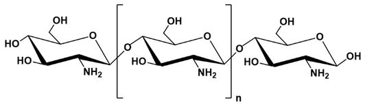
8.5 Chitosan
Chitosan is commercially developed by deacetylation of chitin and is mainly comprised of d-glucosamine and N-acetyl-d-glucosamine units having β-(1-4)-linkages [61]. It is available as high, medium, and low molecular-weight chitosan. This polymer is cationic, contains nitrogen in its chemical structure, and has ability to form polyelectrolyte complexes [62]. It becomes water soluble by forming carboxylate salts like acetate, citrate, formate, malate, glycolate, pyruvate, lactate, and ascorbate. These properties make it different from other polysaccharides [63]. Chitosan is widely employed in biotechnology and biomedical areas owing to its stable, nontoxic, and biodegradable characteristics [64]. Various controlled-release formulations have been developed by chitosan keeping in view its ideal properties. Furthermore, it is largely employed for the development of hydrogels in different forms including microparticles, microspheres, beads, sponges, composites, tablets, etc. (Figure 8) [65].
Figure 8.
Chemical structure of chitosan.
8.6 Agarose
Agarose is a neutral polysaccharide found in certain seaweeds [66]. Its chemical structure consists of agarbiose units in which 3,6-anhydro-α-l-galactopyranose are linked to C-1 and C-3 positions of β-d-galactopyranose. Agarose dissolves in boiling water and gels on lowering the temperature (30–40°C) [67]. The gelation mechanism of this polymer involves self-assembly of its units along with molecular cross-linking and conformational change [68]. Different factors affecting this mechanism are rate of agitation, temperature, concentration of polymer, co-solutes, and solvent properties [69]. Mechanism of gel formation is not fully understood; however, it can be explained regarding liquid–liquid phase separation. Hydrogels developed by using pure agarose are transparent, hard, elastic, and thermoreversible (Figure 9) [51].
Figure 9.
Structural formula of agarose.
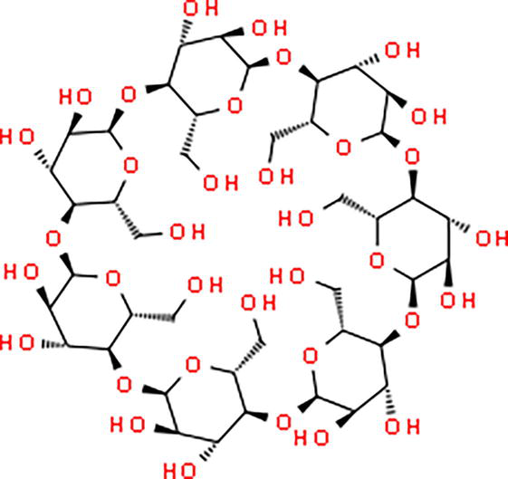
8.7 Cyclodextrins
Cyclodextrins (CDs) are very familiar oligosaccharides containing 6 to 7 glucose units that are linked together via α-1, 4 glucosidic linkages. Cyclodextrins are stable at lower pH regions like stomach and intestine, but these are fermented at higher pH regions, i.e., colon. These have nest, like geometry with inner hydrophobic lining and outer hydrophilic end. Microflora of colon is capable of degrading and completely digest within the colon; hence these polymers can be employed for drugs to be deliver in colon. The structural formula is shown in Figure 10.
Figure 10.
Structural representation of cyclodextrin.
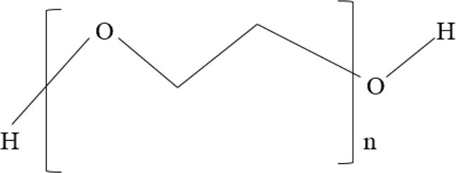
8.8 Polyethylene Glycol 8000
Polyethylene Glycol 8000 is a polymer that can be prepared by combining ethylene oxide with water. PEG occurs as white or off-white flakes that are waxy in nature with a faint and sweet odor [70]. This form is a result of condensation reaction between ethylene oxide and water under pressure in the presence of a catalyst. It is a highly water-soluble polymer that normally does not cause any harm to skin and is also stable in nature [71]. They are capable of promoting dissolution and systemic availability of numerous less water-soluble drugs. Different grades of PEG are generally used in wide range of pharmaceutical formulations (Figure 11) [72].
Figure 11.
Structural formula of polyethylene glycol.
8.9 Okra gum
Okra is one of the traditional plant and a multipurpose crop with botanical name Hibiscus esculentus belongs to the family Malvaceae. It is an annual plant cultivated in tropical and subtropical areas of the world [73]. It can grow on all soil types and is the most tolerant plant. It is superfluous in nutritional content. Different parts of the plant are extensively used in traditional medicines as antidiabetic, diuretic, and anticancer agents [74]. It has wide applications in the pharmaceutical and food industries. Okra is abundant in bioactive compounds like flavonoids, polysaccharides, polyphenols, caffeine, and pectin. The whole plant is edible and is used for food, nonfood, and medical applications.
As compared to synthetic polymers, natural polymers offer good biocompatibility and biodegradability. However, natural polymers yield less mechanical strength in polymeric formulations [75]. Okra gum is chemically inert, nonirritant, biodegradable, biocompatible, and eco-friendly. It is economical and widely cultivated crop. It is comprised of l-rhamnose, d-galactose, and l-galacturonic acid in its composition (Figure 12) [76].
Figure 12.
Chemical structure of okra gum polysaccharide.
In pharmaceutical industry, although its role has not been explored enough as hydrogel preparations, it is evaluated as an economical and effective binding agent in solid dosage forms [45, 47, 48], in forming controlled release matrices [49, 50], for preparation of sustained release beads [51], microspheres [52, 53], in mucoadhesive nasal gel [54] and in many other formulations as a sustained release polymer.
8.10 Xanthan gum
It is obtained from sucrose or glucose by the process of fermentation by Xanthomonas campestric. It is soluble in both hot and cold water. Xanthan gum has stability over a pH ranging from 4 to 10. It is used in cosmetic, topical formulation and as a stabilizer in food industry [77].
8.11 Hyaluronic acid
It is also known as hyaluronate and hyaluronan. Hyaluronic acid is present in the tissues of animals. It is composed of N-acetyl-d-glucosamine and d-glucuronic acid, which are bonded together by β-(1-3) link [78]. It is used for ophthalmic, parenteral, and nasal preparation owing to its nonimmunogenicity and biocompatibility [79].
8.12 Gelatin
It is a natural polymer that has aqueous solubility. Collagen is hydrolyzed to obtain gelatin, where collagen is present in the bone and connective tissues of animals. Gelatin is insoluble in alcohol, whereas it is soluble in a hot mixture of water and glycerin. It has wide applications in protein and drug delivery, cell culture, and gene delivery [80, 81].
8.13 Polyethylene glycol
It has aqueous solubility. Hydrogels based on PEG are also known as intelligent or smart hydrogels due to their stimuli dependence. The stimuli can be either chemical (specific ion or pH) or physical (radiation, light, pressure, solvent, or temperature). PEG base hydrogels are widely used for the controlled release of therapeutic agents [82, 83].
8.14 Polyvinyl alcohol
It is an aqueous soluble polymer. Hydrogels based on polyvinyl alcohol are prepared by freeze and thawing process. Their water retention capacity is high; therefore, they are used in wound dressing. PVA is also used in cartilage reconstitution and contact lenses [84, 85].
8.15 Polyvinyl pyrolidone
It has high solubility in polar solvents as well as in water. It has good elasticity, and the production cost is less. PVP is combined with other different polymers such as CMC to enhance biocompatibility and mechanical properties [86, 87].
8.16 Pluronic acid F127
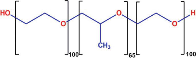
Pluronic acid F127 is composed of two blocks, a water-loving polyethylene oxide (PEO) and water-hating polypropylene oxide (PPO). These polymer blocks are arranged in a basic tri-block structure of repeating units, i.e., A-B-A. Molecular weight of F127 is approximately 12,600 Da. At temperature above lower critical solution temperature, i.e., 30°C, F127 loses solubility of its PPO block, leading to micelle development, and solution is converted into gel [88]. Pluronic acid serves as an important base for thermo-sensitive hydrogels. For localized malignancy treatment, intra-tumoral or peri-tumoreal infusion of gel formed by pluronic acid F127 leads to the development of ‘depot’ at the point of administration that gradually and constantly releases medicinal agents to the malignant and neighboring tissues [89]. Utilizing a topical or injectable gel for physical targeting has added preferences over passive or other actively targeted treatments that possess ability to supply medicinal agent to the whole tumor irrespective of vascular status, thus giving precise dosing of the drug without causing systemic toxicity [90]. Pluronic Acid F127 has a very vital job among in situ gelling frameworks due to its sol–gel transition character, a solution of poloxamer in water at a low temperature converts into polymeric gel at a higher temperature. Due to this capability, PF127 has confirmed its use in medical and therapeutic fields like in fertility management cases, pain management, and delivery of drugs at a controlled rate via eyes or rectal route (Figure 13) [91].
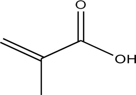
Methacrylic acid (MAA) is a colorless organic liquid and has a strong pungent odor. It is widely employed as a monomer due to its excellent pH-responsive characteristics [92]. Hydrogels developed from this monomer exhibit appreciable swelling in basic media, while this effect is negligible in acidic media. This effect is contributed by ionizable carboxylic groups from MAA structure, creating repulsion within the network (Figure 14) [93].
Figure 14.
Structural formula of MAA.
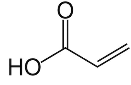
9.2 Acrylic acid
It is an organic compound and a simplest form of unsaturated carboxylic acid. It also contains a vinyl group, which is directly attached to a carboxylic acid. It is a colorless liquid with a characteristic acrid odor and a tart smell. It is miscible with aqueous media and weakly acidic compounds. It is highly corrosive and irritant to skin. It is also known as 2-propenoic acid. In literature, it is widely used as an initiator in various hydrogel preparations (Figure 15) [94, 95].
Figure 15.
Structural representation of AA.
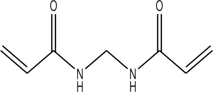
9.3 N,N′-methylenebisacrylamide
A bi-functional monomer, N,N′-methylenebisacrylamide (MBA) consists of two double bonds in its structure. It is majorly used in polymerization reactions due to its cross-linking properties [44]. It is a hydrophilic, cross-linking agent used during polymerization reactions. It has the ability to form a network structure instead of making linear chains, thus assisting in maintaining the firmness of prepared gel. It has diverse applications as a monomer and cross-linker in various polymerization reactions. Chemically, it contains two unsaturated bonds and have been employed in the preparation of various pharmaceutical dosage forms (Figure 16) [96].
A number of methods have been quoted in literature to prepare polymeric networks. These include ionic gelation, spray drying, dispersion photopolymerization, suspension cross-linking technique, ionotropic gelation method, free radical polymerization, membrane emulsification technique, free radical precipitation polymerization, Michael addition reaction, and inverse emulsion polymerization techniques depending upon the objective of the study.
11. Applications
11.1 Biomedical applications
The hydrogels exculpate oneself comparably to anthropoid organs in reply to climate state such as change in pH, change in temperature, electric field, and enzymes. They are cast off in pharmaceuticals lodge, diagnostic gadgets, bone implants, embolus, and computerized grasp [17, 46, 97]. They are also employed in urinary catheter by intercepting them from germs attack on the surface [98]. Kampa [99] successfully used the hydrogel electrodes in the diagnosis and treatment of body tissue [18].
11.2 Biotechnology applications
Hydrogels are also giving an important role in various mechanisms. They are used in the refining of various gadgets and centralizing the moist forum [19]. Murphy et al. [100] have worked on the use of hydrogels in the treatment of wounds. They have worked on the development of new technology containing hydrogel-based bioprinting. This technology was used for faster healing of wounds. They find out that hyaluronic acid-based hydrogels are best for bio printing applications [101, 102].
11.3 Pharmaceutical applications
There are various petitions for hydrogels in pharmaceutical factories. They are mostly used for the intended site distribution of drugs, sustained delivery of drugs, controlled release of medicine, and reducing the frequency of drugs [21]. Peppas et al. have studied hydrogels to find a suitable way to cure a number of diseases. This study showed that hydrogels are water-loving and can store bulk amounts of water and medicaments. These medicaments can be made to release as desired in response to different stimuli. The release of drugs from hydrogel can be controlled [103].
11.4 Separation technology
Currently water has been polluted broadly by the factories’ consumption containing various categories of pigments from textile factories, pharmaceuticals, nonmedical, and paper industries. Hydrogels play a vital character in the separation of dyes by acting as adsorbents [22, 23].
11.5 Agriculture industry
In the cultivation part, hydrogels give a dominant part by assuming control availability of supplements to crops and plants from loaded hydrogels [25]. The advancement of a cellulose-based biodegradable superabsorbent hydrogel has been studied for the streamlining of water assets in agriculture. The sorption capacity of the proposed hydrogel was rightly evaluated, with explicit respect to two factors that may assume a key job in the soil condition: ionic strength and pH. A fundamental assessment of the hydrogel’s potential to store water was performed by utilizing the hydrogel in greenhouses to develop tomatoes. The soil and water retention curve was noted. The results demonstrated that the material permitted productive storage and spontaneous supply of water to the soil and the plant roots [26].
11.6 Contact lenses
Hydrogels are used to create contact lenses that envelop the enormous mass of the essential ingredients needed to be used in different situations. There are some requirements to make it safe during use for a hydrogel material that is used as a contact trifocal. Such essential elements contain amounts of water, oxygen pores, and great focus point. So as to construct the water tranquil of hydrogel and to acquire additional bulging effects, different kinds of monomers can be used. These include dihydroxy methacrylates, acrylamides, and MAA [27, 28].
11.7 Food packaging
Some analysts are working on hydrogels as a part of nourishment construction by preventing them from degradation [30]. It is a promising and rising idea, as the greater part of the biopolymer-based hydrogels should be biodegradable, they can be considered as elective environment friendly packaging materials. Polyvinyl Pyrollidone (PVP) and Carboxymethyl Cellulose (CMC)-based hydrogel was prepared under controlled environmental condition. He prepared different solutions containing PVP and CMC in different concentrations. These concentrations were 20:80, 10:90, 50:50, 80:20, and 90:10. The results showed that CMC-PVP hydrogels of 80:20 ratio was observed useful in packaging of food [32].
In this book chapter, different types of hydrogel, along with properties as well as medical applications were discussed. However, there are different mechanisms involved in formation of hydrogel. Physically cross-linked nexus employed as substrate for tissue engineering with various medical applications. Drug release from nexus is attributed to swelling. However, stimuli-responsive nexus is gaining much importance. Temperature and pH aid in a controlled release from the hydrogel nexus. Moreover, they have excellent water absorption, soft architecture and are biocompatible.
References
1.Mignani S, El Kazzouli S, Bousmina M, Majoral JP. Expand classical drug administration ways by emerging routes using dendrimer drug delivery systems: a concise overview. Advanced Drug Delivery Reviews. 2013;65(10):1316-1330
2.Kaur G, Arora M, Kumar MR. Oral drug delivery technologies—a decade of developments. Journal of Pharmacology and Experimental Therapeutics. 2019;370(3):529-543
3.Campbell S, Smeets N. Drug delivery: polymers in the development of controlled release systems. In: Functional Polymers. Cham: Springer; 2019. pp. 719-747
4.Hardenia A, Maheshwari N, Hardenia SS, Dwivedi SK, Maheshwari R, Tekade RK. Scientific rationale for designing controlled drug delivery systems. In: Tekade RK, editor. Basic Fundamentals of Drug Delivery. Cambridge, Massachusetts: Academic Press; 2019. pp. 1-28
5.Park K. Controlled drug delivery systems: past forward and future back. Journal of Controlled Release. 2014;190:3-8
6.El-Hamshary H et al. Evaluation of clay-ionene nanocomposite carriers for controlled drug delivery: synthesis, in vitro drug release, and kinetics. Materials Chemistry and Physics. 2019;225:122-132
7.Karna S, Chaturvedi S, Agrawal V, Alim M. Formulation approaches for sustained release dosage forms: a review. Asian Journal of Pharmaceutical and Clinical Research. 2015;8(5):46-53
8.Rodríguez-Rodríguez R, Espinosa-Andrews H, Velasquillo-Martínez C, García-Carvajal ZY. Composite hydrogels based on gelatin, chitosan and polyvinyl alcohol to biomedical applications: a review. International Journal of Polymeric Materials and Polymeric Biomaterials. 2020;69(1):1-20
9.Patel GC, Joshi SA. Targeting aspects of hydrogels in drug delivery. In: Grumezescu AM, editor. Biomedical Applications of Nanoparticles. Norwich, NY: William Andrew Publishing; 2019. pp. 195-231
10.Jagtapa SD, Mohite AM, Tayadeb S, Supriya S. Synthesis of hydrogels and their applications in control fertilizer release. Journal of Emerging Technologies and Innovative Research. 2019;6:457-468
11.Shafiq K, Mahmood A, Salem-Bekhit MM, Sarfraz RM, Algarni AS, Taha EI, et al. Development and Optimization of Tamarind Gum-β-Cyclodextrin-g-Poly (Methacrylate) pH-Responsive Hydrogels for Sustained Delivery of Acyclovir. Pharmaceuticals. 8 Dec 2022;15(12):1527
12.Gupta P, Vermani K, Garg S. Hydrogels: from controlled release to pH-responsive drug delivery. Drug Discovery Today. 2002;7(10):569-579
13.Elsayed MM. Hydrogel preparation technologies: relevance kinetics, thermodynamics and scaling up aspects. Journal of Polymers and the Environment. 2019;27(4):871-891
14.Trotta F, Mele A, editors. Nanosponges: Synthesis and Applications. Hoboken, New Jersey: John Wiley & Sons; 2019
15.Nayak AK, Hasnain MS. Plant Polysaccharides-Based Multiple-Unit Systems for Oral Drug Delivery. Singapore: Springer; 2019
16.Bandawane A, Saudagar R. A review on novel drug delivery system: a recent trend. Journal of Drug Delivery and Therapeutics. 2019;9(3):517-521
17.Chirani N, Yahia LH, Gritsch L, Motta FL, Chirani S, Farè S. History and applications of hydrogels. Journal of Biomedical Science. 2015;4(02):1-23
19.Shivashankar M, Mandal BK. A review on interpenetrating polymer network. International Journal of Pharmacy and Pharmaceutical Sciences. 2012;4(5):1-7
20.Qadri MF, Malviya R, Sharma PK. Biomedical applications of interpenetrating polymer network system. Open Pharmaceutical Sciences Journal. 2015;2(1):21-30
21.Soman A, Mathew F, Chacko AJ, Alias M, Poosan GV. Interpenetrating polymer network (Ipn)-hydrogels. The Pharma Innovations. 2014;3(8, Part A):59-66
22.Li Y, Wang C, Zhang W, Yin Y, Rao Q. Preparation and characterization of PAM/SA tough hydrogels reinforced by IPN technique based on covalent/ionic crosslinking. Journal of Applied Polymer Science. 2015;132(4):1-7
23.Panahi Y, Gharekhani A, Hamishehkar H, Zakeri-Milani P, Gharekhani H. Stomach-specific drug delivery of clarithromycin using a semi interpenetrating polymeric network hydrogel made of montmorillonite and chitosan: synthesis, characterization and in vitro drug release study. Advanced pharmaceutical bulletin. 2019;9(1):159
24.Wang JJ, Liu F. UV-curing of simultaneous interpenetrating network silicone hydrogels with hydrophilic surface. Polymer Bulletin. 2012;69:85-97
25.Tang Q, Yu JR, Chen L, Zhu J, Hu ZM. Porous silicone hydrogel interpenetrating polymer networks prepared using a template method for biomedical use. Polymer International. 2011;60(7):1136-1141
26.Dragan ES. Design and applications of interpenetrating polymer network hydrogels. A review. Chemical Engineering Journal. 2014;243:572-590
27.Vaghani SS, Patel MM. pH-sensitive hydrogels based on semi-interpenetrating network (semi-IPN) of chitosan and polyvinyl pyrrolidone for clarithromycin release. Drug Development and Industrial Pharmacy. 2011;37(10):1160-1169
28.Bhardwaj V, Harit G, Kumar S. Interpenetrating polymer network (IPN): novel approach in drug delivery. International Journal of Drug Development and Research. 2012;4(3):41-54
30.Jayaramudu T, Varaprasad K, Sadiku ER, Amalraj J. Temperature-sensitive semi-IPN composite hydrogels for antibacterial applications. Colloids and Surfaces A: Physicochemical and Engineering Aspects. 2019;572:307-316
31.Malikmammadov E, Hasirci N. Dual-and multistimuli-responsive polymers for biomedical applications. In: Aguilar MS, Román JS, editors. Smart Polymers and Their Applications. Sawston, Cambridge: Woodhead Publishing; 2019. pp. 255-278
32.Lohani A, Singh G, Bhattacharya SS, Verma A. Interpenetrating polymer networks as innovative drug delivery systems. Journal of Drug Delivery. 2014;2014:1-11
33.Amiji MM, inventor; Northeastern University Boston, assignee. Drug delivery using pH-sensitive semi-interpenetrating network hydrogels. United States patent US 5,904,927. 1999
34.Mukhopadhyay P, Kundu PP. Stimuli-responsive polymers for oral insulin delivery. In: Makhlouf ASH, Abu-Thabit NY, editors. Stimuli Responsive Polymeric Nanocarriers for Drug Delivery Applications. Sawston, Cambridge: Woodhead Publishing; 2019. pp. 525-546
35.Sano J, Habaue S. Synthesis of interpenetrating polymer networks using silicone polymer with silanol residue. Reactive and Functional Polymers. 2019;137:21-26
36.Hoque J, Sangaj N, Varghese S. Stimuli-responsive supramolecular hydrogels and their applications in regenerative medicine. Macromolecular Bioscience. 2019;19(1):1800259
37.Wang T et al. Biocompatible, injectable anionic hydrogels based on poly (oligo ethylene glycol monoacrylate-co-acrylic acid) for protein delivery. Advanced Therapeutics. 2019;2(9):1900092
38.Fujiyoshi T, Carrez O, Imizcoz M, Zornoza A, Isasi JR. Interpenetrated polymer networks of poly (β-cyclodextrin) and polyvinylpyrrolidone with synergistic and selective sorption capacities. Carbohydrate Polymers. 2019;219:105-112
39.Deen GR, Loh XJ. Stimuli-responsive cationic hydrogels in drug delivery applications. Gels. 2018;4(1):13
40.Olad A, Pourkhiyabi M, Gharekhani H, Doustdar F. Semi-IPN superabsorbent nanocomposite based on sodium alginate and montmorillonite: reaction parameters and swelling characteristics. Carbohydrate Polymers. 2018;190:295-306
41.Rudhrabatla VP, Jalababu R, Rao KK, Reddy KS. Fabrication and characterisation of curcumin loaded pH dependent sodium alginate-g-poly (acryloyl phenylalanine)-cl-ethylene glycol vinyl ether-co-hydroxyethyl acrylate hydrogels and their in-vitro, in-vivo and toxicological evaluation studies. Journal of Drug Delivery Science and Technology. 2019;51:438-453
42.Hajikhani M, Khanghahi MM, Shahrousvand M, Mohammadi-Rovshandeh J, Babaei A, Khademi SM. Intelligent superabsorbents based on a xanthan gum/poly (acrylic acid) semi-interpenetrating polymer network for application in drug delivery systems. International Journal of Biological Macromolecules. 2019;139:509-520
43.Jalababu R, Veni SS, Reddy KS. Synthesis and characterization of dual responsive sodium alginate-g-acryloyl phenylalanine-poly N-isopropyl acrylamide smart hydrogels for the controlled release of anticancer drug. Journal of Drug Delivery Science and Technology. 2018;44:190-204
44.Kim SJ, Lee CK, Kim IY, Hyeok An K, Kim SI. Preparation and characterizations of interpenetrating polymer network hydrogels of poly (ethylene oxide) and poly (methyl methacrylate). Journal of Applied Polymer Science. 2003;89(1):258-262
45.Willats WG, Knox JP, Mikkelsen JD. Pectin: new insights into an old polymer are starting to gel. Trends in Food Science & Technology. 2006;17(3):97-104
46.Cui L, Jia J, Guo Y, Liu Y, Zhu P. Preparation and characterization of IPN hydrogels composed of chitosan and gelatin cross-linked by genipin. Carbohydrate Polymers. 2014;99:31-38
47.Sriamornsak P. Application of pectin in oral drug delivery. Expert Opinion on Drug Delivery. 2011;8(8):1009-1023
48.Fawzy A, Toghan A, Alqarni N, Morad M, Zaki ME, Sanad MM, et al. Experimental and computational exploration of chitin, pectin, and amylopectin polymers as efficient eco-friendly corrosion inhibitors for mild steel in an acidic environment. Kinetic, thermodynamic, and mechanistic aspects. Polymers. 2023;15(4):1-33
49.Singh A, Suri A. Synergistic kinetic hydrate inhibition of pectin, PVP, and PVCap with monoethylene glycol. Energy & Fuels. 2023;37(6):4524-4543
50.Ijaz H, Tulain UR. Development of interpenetrating polymeric network for controlled drug delivery and its evaluation. International Journal of Polymeric Materials and Polymeric Biomaterials. 2019;68(18):1099-1107
51.Zafar N, Mahmood A, Sarfraz RM, Ijaz H, Ashraf MU, Mehr S. Facile synthesis of β-cyclodextrin-cyclophosphamide complex-loaded hydrogel for controlled release drug delivery. Polymer Bulletin. 2022:1-33. DOI: 10.1007/s00289-022-04567-7
52.Abdel-Halim ES. Chemical modification of cellulose extracted from sugarcane bagasse: preparation of hydroxyethyl cellulose. Arabian Journal of Chemistry. 2014;7(3):362-371
53.Nayak AK, Pal D. Functionalization of Tamarind Gum for Drug Delivery. In: Thakur V, Thakur M, editors. Functional Biopolymers. Springer, Cham: Springer Series on Polymer and Composite Materials; 2018
55.Dey S, Nandy BC, De JN, Hasnain MS, Nayak AK. Tamarind gum in drug delivery applications. In: Hasnain MS, Nayak AK, editors. Natural Polysaccharides in Drug Delivery and Biomedical Applications. Cambridge, Massachusetts: Academic Press; 2019. pp. 285-306
56.Malviya R, Sundram S, Fuloria S, Subramaniyan V, Sathasivam KV, Azad AK, et al. Evaluation and characterization of tamarind gum polysaccharide: the biopolymer. Polymers. 2021;13(18):3023
57.Nayak AK, Pal D, Pradhan J, Hasnain MS. Fenugreek seed mucilage-alginate mucoadhesive beads of metformin HCl: design, optimization and evaluation. International Journal of Biological Macromolecules. 2013;54:144-154
58.Gopinath R, Manoharan S, Likhith M, Bhuvan PG, Kailash R, Akash M, et al. Augmentation of soil-moisture retention with natural hydrogel extracted from fenugreek seed husk for loamy soil with real-time monitoring using FC-28 sensors. ECS Transactions. 2022;107(1):1139
59.Sharma P, Laddha H, Agarwal M, Gupta R. Selective and effective adsorption of malachite green and methylene blue on a non-toxic, biodegradable, and reusable fenugreek galactomannan gum coupled MnO2 mesoporous hydrogel. Microporous and Mesoporous Materials. 2022;338:111982
60.Avachat AM, Dash RR, Shrotriya SN. Recent investigations of plant based natural gums, mucilages and resins in novel drug delivery systems. Indian Journal of Pharmaceutical Education and Research. 2011;45(1):86-99
61.Ijaz H, Tulain UR, Minhas MU, Mahmood A, Sarfraz RM, Erum A, et al. Design and in vitro evaluation of pH-sensitive crosslinked chitosan-grafted acrylic acid copolymer (CS-co-AA) for targeted drug delivery. International Journal of Polymeric Materials and Polymeric Biomaterials. 2022;71(5):336-348
62.Malatani RT, Bilal S, Mahmood A, Sarfraz RM, Zafar N, Ijaz H, et al. Development of tofacitinib loaded pH-responsive chitosan/mucin based hydrogel microparticles: in-vitro characterization and toxicological screening. Gels. 2023;9(3):187
63.Qureshi J, Iqbal FM, Hussain F, Ijaz H, Aamir MF, Afzal M. Fabrication and evaluation of Olopatadine hydrochloride ocular inserts. Journal of Plastic Film & Sheeting. 2020 Jul;36(3):243-259
64.Mahmood A, Mahmood A, Sarfraz RM, Ijaz H, Zafar N, Ashraf MU. Hydrogel-based intelligent delivery system for controlled release of diloxanide furoate. Polymer Bulletin. 2022;1:1-37
65.Batool N, Mahmood A, Sarfraz RM, Ijaz H, Zafar N, Hussain Z. Formulation and evaluation of interpenetrating polymeric network for controlled drug delivery. Drug Development and Industrial Pharmacy. 2021;47(6):931-946
66.López-Marcial GR, Zeng AY, Osuna C, Dennis J, García JM, O’Connell GD. Agarose-based hydrogels as suitable bioprinting materials for tissue engineering. ACS Biomaterials Science & Engineering. 2018;4(10):3610-3616
67.Salati MA, Khazai J, Tahmuri AM, Samadi A, Taghizadeh A, Taghizadeh M, et al. Agarose-based biomaterials: opportunities and challenges in cartilage tissue engineering. Polymers. 2020;12(5):1150
68.Hur J, Im K, Kim SW, Kim J, Chung DY, Kim TH, et al. Polypyrrole/agarose-based electronically conductive and reversibly restorable hydrogel. ACS Nano. 2014;8(10):10066-10076
69.Martínez-Sanz M, Ström A, Lopez-Sanchez P, Knutsen SH, Ballance S, Zobel HK, et al. Advanced structural characterisation of agar-based hydrogels: Rheological and small angle scattering studies. Carbohydrate Polymers. 2020;236:115655
70.Sarfraz RM, Ahmad M, Mahmood A, Ijaz H. Development, in vitro and in vivo evaluation of pH responsive β-CD-comethacrylic acid-crosslinked polymeric microparticulate system for solubility enhancement of rosuvastatin calcium. Polymer-Plastics Technology and Engineering. 2018;57(12):1175-1187
71.Huang Z, Wu Q, Liu S, Liu T, Zhang B. A novel biodegradable β-cyclodextrin-based hydrogel for the removal of heavy metal ions. Carbohydrate Polymers. 2013;97(2):496-501
72.Daoud-Mahammed S, Grossiord JL, Bergua T, Amiel C, Couvreur P, Gref R. Self-assembling cyclodextrin based hydrogels for the sustained delivery of hydrophobic drugs. Journal of Biomedical Materials Research Part A. 2008;86(3):736-748
73.Tan H, DeFail AJ, Rubin JP, Chu CR, Marra KG. Novel multiarm PEG-based hydrogels for tissue engineering. Journal of Biomedical Materials Research Part A: An Official Journal of The Society for Biomaterials, The Japanese Society for Biomaterials, and The Australian Society for Biomaterials and the Korean Society for Biomaterials. 2010;92(3):979-987
74.Weber LM, He J, Bradley B, Haskins K, Anseth KS. PEG-based hydrogels as an in vitro encapsulation platform for testing controlled β-cell microenvironments. Acta Biomaterialia. 2006;2(1):1-8
75.Sagle AC, Ju H, Freeman BD, Sharma MM. PEG-based hydrogel membrane coatings. Polymer. 2009;50(3):756-766
76.Shaw GS, Uvanesh K, Gautham SN, Singh V, Pramanik K, Banerjee I, et al. Development and characterization of gelatin-tamarind gum/carboxymethyl tamarind gum based phase-separated hydrogels: A comparative study. Designed Monomers and Polymers. 2015;18(5):434-450
77.Pushpamalar J, Meganathan P, Tan HL, Dahlan NA, Ooi LT, Neerooa BN, et al. Development of a polysaccharide-based hydrogel drug delivery system (DDS): an update. Gels. 2021;7(4):153
78.Sennakesavan G, Mostakhdemin M, Dkhar LK, Seyfoddin A, Fatihhi SJ. Acrylic acid/acrylamide based hydrogels and its properties - a review. Polymer Degradation and Stability. 2020;180:109308
79.Tang Y, Hu M, Tang F, Huang R, Wang H, Wu D, et al. Easily-injectable shear-thinning hydrogel provides long-lasting submucosal barrier for gastrointestinal endoscopic surgery. Bioactive Materials. 2022;15:44-52
80.Ijaz H, Tulain UR, Qureshi J. Formulation and in vitro evaluation of pH-sensitive cross-linked xanthan gum-grafted acrylic acid copolymer for controlled delivery of perindopril erbumine (PE). Polymer-Plastics Technology and Engineering. 2018;57(5):459-470
81.Gao Z, Golland B, Tronci G, Thornton PD. A redox-responsive hyaluronic acid-based hydrogel for chronic wound management. Journal of Materials Chemistry B. 2019;7(47):7494-7501
82.Park YD, Tirelli N, Hubbell JA. Photopolymerized hyaluronic acid-based hydrogels and interpenetrating networks. Biomaterials. 2003;24(6):893-900
83.Jaipan P, Nguyen A, Narayan RJ. Gelatin-based hydrogels for biomedical applications. MRS Communications. 2017;7(3):416-426
84.Kang JI, Park KM. Advances in gelatin-based hydrogels for wound management. Journal of Materials Chemistry B. 2021;9(6):1503-1520
85.Zheng Y, Andreopoulos FM, Micic M, Huo Q, Pham SM, Leblanc RM. A novel photoscissile poly (ethylene glycol)-Based Hydrogel. Advanced Functional Materials. 2001;11(1):37-40
86.Park Y, Lutolf MP, Hubbell JA, Hunziker EB, Wong M. Bovine primary chondrocyte culture in synthetic matrix metalloproteinase-sensitive poly (ethylene glycol)-based hydrogels as a scaffold for cartilage repair. Tissue Engineering. 2004;10(3–4):515-522
87.Kumar A, Han SS. PVA-based hydrogels for tissue engineering: a review. International Journal of Polymeric Materials and Polymeric Biomaterials. 2017;66(4):159-182
88.Husain MS, Gupta A, Alashwal BY, Sharma S. Synthesis of PVA/PVP based hydrogel for biomedical applications: a review. Energy Sources, Part A: Recovery, Utilization, and Environmental Effects. 2018;40(20):2388-2393
89.Roy N, Saha N, Kitano T, Saha P. Biodegradation of PVP–CMC hydrogel film: A useful food packaging material. Carbohydrate Polymers. 2012;89(2):346-353
90.Doan P, Nhi TT, Nguyen DT, Nguyen BT, Nguyen TP, Tran NQ. Multifunctional injectable pluronic-cystamine-alginate-based hydrogel as a novel cellular delivery system towards tissue regeneration. International Journal of Biological Macromolecules. 2021;185:592-603
91.Xiong XY, Tam KC, Gan LH. Polymeric nanostructures for drug delivery applications based on Pluronic copolymer systems. Journal of Nanoscience and Nanotechnology. 2006;6(9–10):2638-2650
92.Engel L, Berkh O, Adesanya K, Shklovsky J, Vanderleyden E, Dubruel P, et al. Actuation of a novel pluronic-based hydrogel: Electromechanical response and the role of applied current. Sensors and Actuators B: Chemical. 2014;191:650-658
93.Ijaz H, Tulain UR, Azam F, Qureshi J. Thiolation of arabinoxylan and its application in the fabrication of pH-sensitive thiolated arabinoxylan grafted acrylic acid copolymer. Drug Development and Industrial Pharmacy. 2019;45(5):754-766
94.Murphy N, Lynch MA. Activation of the P 2 X 7 receptor induces migration of glial cells by inducing cathepsin B degradation of tissue inhibitor of metalloproteinase 1. Journal of Neurochemistry. 2012;123(5):761-770
95.Peppas NA, Bures P, Leobandung WS, Ichikawa H. Hydrogels in pharmaceutical formulations. European Journal of Pharmaceutics and Biopharmaceutics. 2000;50(1):27-46
96.Lin F, Lu X, Wang Z, Lu Q, Lin G, Huang B, et al. In situ polymerization approach to cellulose–polyacrylamide interpenetrating network hydrogel with high strength and pH-responsive properties. Cellulose. 2019;26:1825-1839
98.Bera R, Dey A, Chakrabarty D. Synthesis and characterization of acrylic acid-2-hydroxyethyl methacrylate IPN hydrogels. RSC Advances. 2015;5(93):75870-75880
100.Murphy SV, Skardal A, Atala A. Evaluation of hydrogels for bio‐printing applications. Journal of Biomedical Materials Research Part A. Jan 2013;101(1):272-284
101.Zhang N, Shen Y, Li X, Cai S, Liu M. Synthesis and characterization of thermo-and pH-sensitive poly (vinyl alcohol)/poly (N, N-diethylacrylamide-co-itaconic acid) semi-IPN hydrogels. Biomedical Materials. 2012;7(3):035014
102.Mohamed RR, Seoudi RS, Sabaa MW. Synthesis and characterization of antibacterial semi-interpenetrating carboxymethyl chitosan/poly (acrylonitrile) hydrogels. Cellulose. 2012;19:947-958
103.Sun XF, Zeng Q, Wang H, Hao Y. Preparation and swelling behavior of pH/temperature responsive semi-IPN hydrogel based on carboxymethyl xylan and poly (N-isopropyl acrylamide). Cellulose. 2019;26:1909-1922
104.Lee DS, Kang JI, Hwang BH, Park KM. Interpenetrating polymer network hydrogels of gelatin and poly (ethylene glycol) as an engineered 3D tumor microenvironment. Macromolecular Research. 2019;27:205-211
105.Das S, Subuddhi U. Controlled delivery of ibuprofen from poly (vinyl alcohol)− poly (ethylene glycol) interpenetrating polymeric network hydrogels. Journal of Pharmaceutical Analysis. 2019;9(2):108-116
106.Hennink WE, Franssen O. U.S. Patent No. 6,303,148. Washington, DC: U.S. Patent and Trademark Office. 2001
107.Hennink WE, van Dijk-Wolthuis WNE. U.S. Patent No. 6,497,903. Washington, DC: U.S. Patent and Trademark Office. 2002
108.Hubbell JA, Kieras MT, Ron ES, Rowe SC. U.S. Patent No. 6,703,037. Washington, DC: U.S. Patent and Trademark Office. 2004
109.Hubbell JA, Pathak CP, Sawhney AS, Desai NP, & Hill-West JL. U.S. Patent No. 6,060,582. Washington, DC: U.S. Patent and Trademark Office. 2000
110.Hennink WE, van Dijk-Wolthuis WNE. U.S. Patent No. 7,060,296. 2006
111.Jennings Jr RN, Yang B, Protter AA, Wang YCJ. U.S. Patent No. 6,331,309. Washington, DC: U.S. Patent and Trademark Office. 2001
112.Winkler RA. U.S. Patent No. 6,200,257. Washington, DC: U.S. Patent and Trademark Office. 2001
113.Chow SL, Wong D. U.S. Patent Application No. 13/032,453. 2011
114.Lim TH, Park JB, Lee JW. U.S. Patent No. 8,940,311. Washington, DC: U.S. Patent and Trademark Office. 2015
115.Sukuru K. U.S. Patent No. 8,333,989. Washington, DC: U.S. Patent and Trademark Office. 2012
116.Chow SL, Wong D, Garcia D. U.S. Patent Application No. 11/772,017. 2008
117.Seliktar D, Oss-Ronen L. U.S. Patent No. 9,492,376. Washington, DC: U.S. Patent and Trademark Office. 2016
118.Jani D, Salamone J, Hu Z, Xia E, Borazjani R, Ammon D. U.S. Patent Application No. 10/319,132. 2004
119.Rubinstein A, Radai R, Friedman M, Tirosh B, Baluom M, & Nassar T. U.S. Patent No. 6,692,766. Washington, DC: U.S. Patent and Trademark Office. 2004
120.Shalaby SW. U.S. Patent No. 6,413,539. Washington, DC: U.S. Patent and Trademark Office. 2002
 Open access peer-reviewed chapter