Open Access is an initiative that aims to make scientific research freely available to all. To date our community has made over 100 million downloads. It’s based on principles of collaboration, unobstructed discovery, and, most importantly, scientific progression. As PhD students, we found it difficult to access the research we needed, so we decided to create a new Open Access publisher that levels the playing field for scientists across the world. How? By making research easy to access, and puts the academic needs of the researchers before the business interests of publishers.
We are a community of more than 103,000 authors and editors from 3,291 institutions spanning 160 countries, including Nobel Prize winners and some of the world’s most-cited researchers. Publishing on IntechOpen allows authors to earn citations and find new collaborators, meaning more people see your work not only from your own field of study, but from other related fields too.
To purchase hard copies of this book, please contact the representative in India:
CBS Publishers & Distributors Pvt. Ltd.
www.cbspd.com
|
customercare@cbspd.com
Nowadays, nanoparticles are used in a variety of biological applications where they enhance treatments and laboratory tests. Due to their distinctive properties and minor adverse effects, nanoparticles are being used more specifically for medication delivery, not only in the treatment of cancer but also for other diseases. Magnetic nanoparticles like SPION (superparamagnetic Iron Oxide nanoparticles) are regarded to be the most viable in the midst of these materials. SPION are frequently used in biomedical applications due to their low cost and lack of toxicity. Within the developing field of nanomedicine, superparamagnetic iron oxide nanoparticles (SPION) are basic technological classes that have been widely studied for cancer imaging and treatment. Additionally, SPION employ super paramagnets, which seem to be beneficial for focusing on particular tumor areas within a body. For instance, the superparamagnetic abilities of magnetite (Fe3O4), which are frequently utilized in delivery of drug, diagnosis and therapy. SPION was envisioned as a tool for the “golden therapeutic era” since it minimized cellular absorption by macrophages, targeted cancer cells preferentially while sparing healthy cells, monitored cancer cells before and after therapy, and controlled drug release. In order to give a concise overview of SPION, there will be focus on their biomedical applications includes hyperthermia (HT), magnetic resonance imaging (MRI), magnetic drug targeting (MDT), gene delivery as well as nanomedicine.
Department of Chemistry and Chemical Science, School of Physical and Material Sciences, Central University of Himachal Pradesh, Dharamshala, India
Purnima Justa
Department of Chemistry and Chemical Science, School of Physical and Material Sciences, Central University of Himachal Pradesh, Dharamshala, India
Hemant Kumar
Department of Chemistry, Ramjas College, University of Delhi, Delhi, India
Deepshikha
Department of Chemistry and Chemical Science, School of Physical and Material Sciences, Central University of Himachal Pradesh, Dharamshala, India
Krishna
Department of Chemistry and Chemical Science, School of Physical and Material Sciences, Central University of Himachal Pradesh, Dharamshala, India
Balaram Pani
Department of Chemistry, Bhaskaracharya College of Applied Sciences, University of Delhi, Delhi, India
Pramod Kumar*
Department of Chemistry and Chemical Science, School of Physical and Material Sciences, Central University of Himachal Pradesh, Dharamshala, India
*Address all correspondence to: pramodgang03@gmail.com
1. Introduction
SPION (Super magnetic iron oxide nanoparticles) are the nanoparticles with a functionalized shell surrounding an iron oxide core. These are iron oxide crystals {magnetite (Fe3O4) or maghemite (γ-Fe2O3)} with a shell that can be altered to improve stability in aqueous conditions and to alter the biochemical properties for a wide range of applications in biomedical sectors [1, 2]. The scientific community has recently become very interested in SPION due to their intriguing potential diagnostic and therapeutic applications. They have wide range of biomedical applications such as MRI, dual modality MRI/computed tomography (CT), magnetic fluid hyperthermia (MFH), biosensors, drug delivery as well as bio- separation [3]. SPION are a unique contrast agent for magnetic resonance imaging and a fascinating family of tracers for nuclear medicine due to their modifiable surface and core properties. A contrast agent can accelerate the relaxation rate of water, which is known as relaxation rates, since MRI evaluates the change in magnetic moment of water protons after applying radiofrequency (RF) pulses. In essence, after being excited by RF pulses, protons return to their equilibrium state, which is longitudinal T1 relaxation. Transfer (T2) relaxation is the exchange of the spin angular momentum between the protons (Figure 1).
Figure 1.
Showing physiochemical properties of SPIONs [4].
SPION exhibit contrast enhancing behavior, engage with nearby water molecules and speed up the rate at which water protons relax. The T1 relaxation times are sped up by the contrast agents. The regions where SPION are taken up typically have a lower MR a signal intensity, which causes those regions to appear darker in MRI. Transfer (T2) relaxation is the exchange of protons’ spin angular momentum. SPION act as a contrast agent, interact with nearby water molecules, and speed up the rate at which water protons relax. The T1 relaxation and/or T2 relaxation times are sped up by the contrast agents. The areas where SPION are taken up typically have a lower MR signal intensity, which causes those areas to appear darker in MRI [5, 6, 7, 8]. The current chapter comprehensively reviews the biomedical applications of SPION in theranostic area (Figure 2).
2. Synthesis and biomedical applications of SPIONS
2.1 Synthesis methods
The morphology as well as composition both affects the magnetic properties of the Iron oxide nanoparticles. In order to ensure control over the particle’s size, shape, size distribution as well as crystallinity, the method of synthesis must be carefully chosen. SPION can be created by using as variety of techniques, including chemical, physical and biosynthetic ones [10, 11, 12, 13].
2.1.1 Co-precipitation
The co-precipitation technique, one of the most straightforward and effective synthesis methods, relies on the addition of a weak or strong base to precipitate Fe2+ and Fe3+ aqueous salt solutions simultaneously. This process is primarily used to synthesize SPION commercially. Several artificial parameters affect the size, shape and the composition of iron oxide nanoparticles, such as Fe2+/Fe3+ ratio, temperature, pH, and salt type (chloride, nitrate, sulphate, perchlorate), as well as the kind of base employed (NaOH, NH4OH, Na2CO3). This technique is one of the most economical ways to make SPION with the right magnetic properties in high degree of polydispersity and little crystallinity. A number of modified versions of this technique have been developed to address these drawbacks [14, 15, 16]. These for example, include in vivo co-precipitation in a carboxyl-functionalized polymer matrix, assistance from ultrasound, the use of alkanolamines as base, the preparation of Fe3O4 nanoparticles under a static magnetic field and at the end co-precipitation of FeCl3.6H2O, FeSO4.7H2O, and Gd(NO3)3 aqueous solutions is done by addition of NaOH [17, 18, 19, 20].
2.1.2 Microemulsion
Microemulsion systems are the isotropic dispersion of two immiscible liquids that are thermodynamically stable. Essentially, there are two types of microemulsions: oil-in-water (o/w; normal micelles) and water-in-oil (w/o; reversed micelles). Usually, the dispersed phase acts as a nano/micro-reactor, offering a constrained setting for the initiation and controlled development of nano- and microparticles. Micellar microemulsion systems are created using a variety of amphiphilic surfactants, such as dioctyl sodium dodecyl sulphate (DSS), cetyltrimethylammonium bromide (CTAB), sodium dodecyl sulphate (SDS), and polyethoxylates (such as Tween-20 and -80) [14]. The main benefit of using microemulsion methodology to prepare SPION is the ability to control the nanoparticle size by varying the size of the micelles. Furthermore, a rise in the particle’s polydispersity is seen, which is likely caused by the relatively homogenous size of micelles. The typical low and constrained reaction temperature, which leads to SPION with poor crystallinity and low yields, is a drawback of microemulsion synthesis. The crystallinity of the particles produced by this method can be increased by thermal annealing the synthesized iron oxide or maintaining the micelle structure at high reaction temperatures [12, 21, 22].
2.1.3 Thermal decomposition
By thermally decomposing organoiron precursors in high-boiling-point organic solvents with stabilizing surfactants, SPION with excellent size and shape control, narrow size distribution, and good crystallinity can be produced [23, 24]. Oleic acid, oleylamine, fatty acids, and hexadecylamine are examples of amphiphilic surfactants that enable fine-tuning of the nucleation and growth kinetics of the nanoparticles. The presence of the surfactant in the reaction mixture and the high reaction temperatures produce samples with excellent size dispersion and crystallinity. However, because this method involves the synthesis of toxic chemicals like iron pentacarbonyl, chloroform, and hexane, it is not particularly eco-friendly. Furthermore, a further surface modification step is required to produce water-dispersible and biocompatible nanoparticles that are helpful for biomedical applications because the surface of the magnetic nanoparticles has a hydrophobic coating. Control over morphology and nanoparticle size when using the thermal decomposition method to create SPION is highly dependent on reaction time, reaction temperature, and the ratio of precursor to surfactant [25].
2.1.4 Sol: gel
The sol-gel method, which is frequently used for the production of silica-coated SPION, is based on the condensation and hydrolysis of tetraethyl orthosilicate (TEOS) in ethanol and 30% aqueous H2O2 with Fe3+ solutions to create colloidal sols. A 3D iron oxide network is then created by gelling the sol through a chemical reaction or solvent removal. After drying and solvent removal, the formed gel needs to be crushed in order to obtain iron oxide nanoparticles. When surfactant is added before gelation, the system’s free energy is reduced, which causes the formation of nanoscale iron oxides without the development of a 3D network. A simple method for producing high yields of relatively large and monodisperse nanoparticles in ambient conditions is the sol-gel synthesis technique. But because the sol-gel method is used at room temperature, additional heating is required to produce the desired crystalline structures. The technique also produces contaminated by-products, necessitating post-treatment purification. The parameters that affect the SPION gel’s structure and properties are temperature, pH, the solvent being used, and the concentration of salt precursors. TEOS and ammonia concentrations are typically used to adjust the silica shell’s thickness (Table 1) [26, 27, 43].
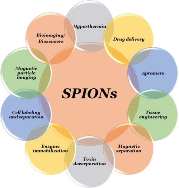
As SPION are mainly used due to their main features such as their size, surface charge, shape, surface stability and biocompatibility. There are many uses of SPION in biomedicine, which are listed below: MRI, drug delivery, hyperthermia, cell labeling and separation, aptamers, biosensors etc. (Figure 3) [30].
Figure 3.
Showing SPIONs application in biomedicine [30].
3.1 Drug delivery
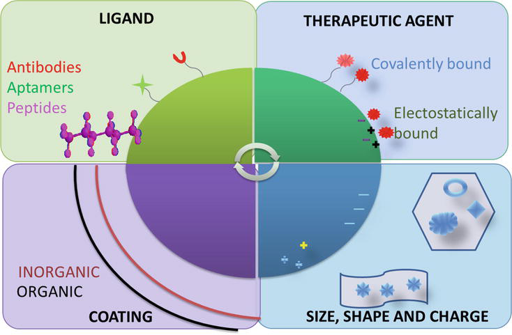
The Transmission of a remedial agent to a recommended site in the body is known as drug delivery. Drug delivery to directed disease is a critical step in the treatment of a variety of conditions, including cardiac disease, microbial attack and cancer. For targeted drug delivery of many diseases different kinds of magnetic nanoparticles are used. For example, in detection lesions in the liver tumor at a 23 mm level. SPION have been used by clinical imaging. For the treatment of MCF-5 breast cancer, drug administration and MRI monitoring have also been studied [39, 44, 45]. In the whole body, chemotherapy causes serious damage, so it is important to focus selectively on cancer cells to create targeted therapies. For higher doses of drugs, the targeting strategy should also increase the effectiveness of drugs without being limited by the harm to healthy tissue. For the SPION-based drug delivery system to be effective, many important criteria must be considered.in order to link targeting units, the carrier should provide a functional group and also provide the delivery system with suitable hydrophily, so that it can easily disperse into aqueous environment (Table 2) [8].
Functional molecules
Structure of DDS
Application
Results
References
Methotrexate
via a self-assembled monolayer PEG it immobilized on the surface of the iron oxide nanoparticles
Through MRI follow-up drug administration to brain tumors
High absorption of targeted nanoparticles, non-toxic methotrexate in vitro, significantly improved contrast
Nuclei of SPIONs covered with a mixture of the triblock copolymer methoxy PEG-b-poly (methacrylic acid -co-n-butyl methacrylate)-b-poly (glycerol monomethacrylate) and the folate-conjugated block copolymer folate-PEG-b-poly (glycerol monomethacrylate) loaded with doxorubicin
For the treatment of cancer of cervix
The Targeting strategy improved absorption of nanoparticles and cytotoxicity
A nanocomposite with quantum dots conjugated to the surface and an internally embedded PS matrix that is spherical, high fraction of SPIONs+PLGA+paclitaxel load
Prostate cancer imaging and targeting, medicine storage
Showing use of SPION in drug carrier and preclinical studies.
3.2 Bioimaging
In bioimaging and clinical purpose magnetic nanoparticles show great scientific interest because of their unique properties. At molecular and cellular level magnetic resonance imaging (MRI) is one of the strongest techniques used in biomedical imaging. In magnetic resonance imaging (MRI), many magnetic nanoparticles are used as a contrast medium, such as SPION, the core, magnetic gel nanoparticles and iron oxide. In these SPION demonstrate an important role in differentiating pathogenic and healthy cells. Because of the high resolution and capability of 3D imaging MRI provides information about soft tissue as well as for the detection of intra-tumor cancerous tissue. SPION and magnetic nanoparticles of iron oxide have aroused major interest in the administration of cancer drugs over the last decades [39].
3.3 Cell labeling and separation
There is numerous SPION cell labelling techniques that can be applied, each with a different goal in mind. There are three main categories that they fall under:
In-vitro methods (such as endocytosis, transfection agents, magnetofection, and electroporation);
In-vivo cell labelling by reticuloendothelial system (RES) through systemic application; and
receptor-mediated binding and internalization of SPION by targeted cells (e.g., targeted labelling and imaging). Stem cell tracking and monitoring for cell transplantation therapy reasons is one of the uses for cell labelling [52]. Living organisms, such as peptides, proteins, large molecules (such as cell receptors) or structural components of cell membranes (such as glycoproteins or cholesterol)—should make it possible to recognize activated cells, organs, or pathogenetic states for exact cell and tissue instruction to achieve therapeutic success (Table 3) [4].
Blood pool agent, cellular labeling, iron replacement therapy in patients with chronic kidney failure, lymph node imaging
r1 = 15 r2 = 89
N.a.
17–31
f
Ferucarbotran
SHU-555A
Carboxydextran
Liver imaging, CNS imaging, cell labeling
r1 = 9.7 r2 = 189
4
62
d
Table 3.
Showing the clinically approved iron oxide nanoparticles examples [42, 53, 54, 55].
3.4 Tissue engineering
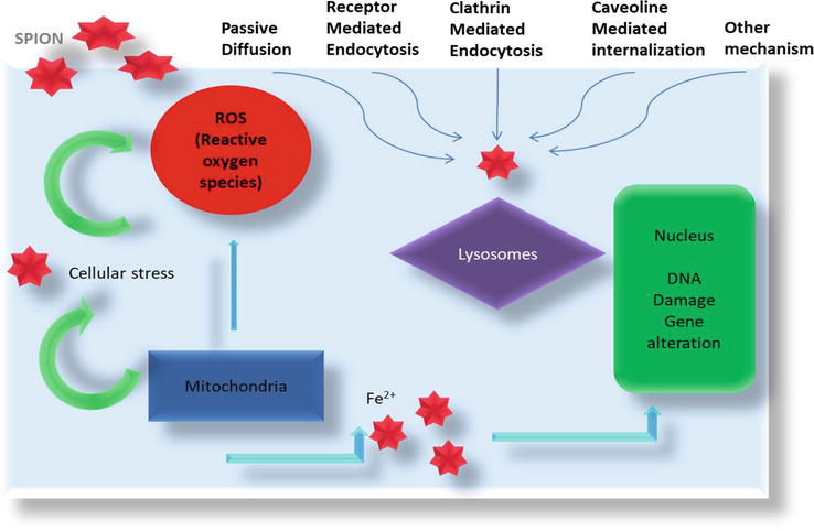
The creation of functional replacement tissues or organs using patient cells has been proposed as a therapeutic strategy. These tissues or organs can develop ex vivo (in a bioreactor) for later implantation or in the patient’s body at the site of the defect. It was suggested that nanomagnetic actuation be used as a mechanical simulator in TE and regenerative medicine. Bone TE bioreactors are created employing mechanical actuators made of magnetic nanoparticles. The applications of MNPs in bone tissue engineering can be expanded by the use of an external magnetic field to regulate their movement and operation. An oscillating external field in conjunction with MNPs inside a defect may increase cell induction and remotely send biomechanical cues to the cells to promote osteogenesis (Figure 4) [56, 57, 58].
Figure 4.
Showing mechanism involving cell damage persuade by SPION [56].
3.5 Magnetic particle imaging
SPION mainly used as to detect substance in the magnetic particle imaging (MPI). MPI was announce in 2005, novel pictorial representation method [59]. Generally, the SPION are superparamagnetic. After the action of the magnetic field is turned off, superparamagnetic particle will not remain magnetized. Due to Brownian and Neel relaxation, the magnetization direction can change even at ambient temperature when thermal stimulation occurs. High spatial sensitivity and sensitivity, along with the potential for high-quality real-time imaging, are the benefits of MPI above existing imaging approaches. The spatial bioavailability of the particles affects the MPI approach’s ability to produce high-quality images. Producing tracers with the best magnetically particle spectroscopy (MPS) efficiency is the difficulty of SPION synthesis (Figure 5) [60].
Figure 5.
Showing combination of diagnosis and therapy effect of SPION [52].
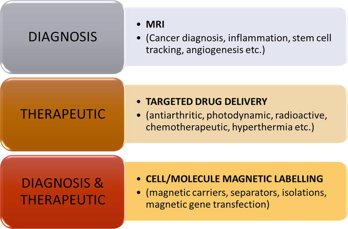
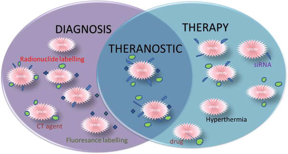
4. SPIONs: a better alternative for theranostic agent
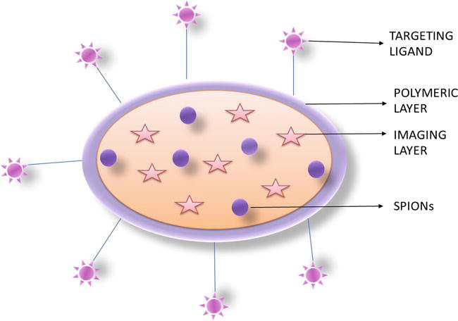
The fusion of diagnostics and therapy is known as theranostics or theragnostics, and it can be divided into three types: the simultaneous use of therapeutics and diagnostics, identification followed by therapy, and therapy followed by diagnosis. Theranostics agents are created with the intent of detecting and treating disease at an early stage, monitoring the effectiveness of the therapeutic process, and minimizing time wasted during diagnosis and treatment. Theranostics nanomedicine, also known as nano-theranostics, is based on the aggregation of diagnostic and therapeutic substances into nanocarriers for use in medicine, including liposomes, micelles, carbon nanotubes, nanoparticles, and polymer-based nanomaterials. The imaging component of nanotheranostics relies on the use of fluorescent dyes like quantum dots (for optical imaging), magnetic nanoparticles, such as SPION (for MRI), radionuclides and heavy metals, such as iodine [52]. For the treatment of the diseases, theranostic medicines are used. Theranostic nanomedicine aims to reduce systemic toxicity related to cancer treatment while also improving cancer detection and treatment effectiveness (Figure 6) [52].
Figure 6.
Shows the representation of theranostic system [61].
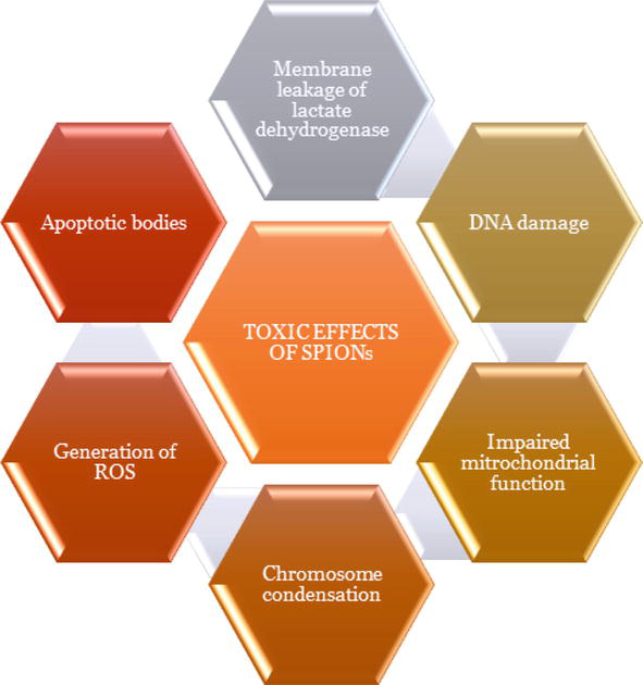
Studies on the effects of SPION on human and animal cells at the cellular level revealed that SPION can enter cells through passive diffusion as well as endocytosis and have a number of harmful effects by changing gene expression and producing oxidative radicals [62]. Toxic or cytotoxic effects from SPION with altered physicochemical properties are possible. High levels of free ferric ions in that tissue could consequently cause an imbalance in homeostasis and abnormal cellular responses like osmotic damage, cytotoxicity, epigenetic events, DNA damage, and inflammatory disorders, which could result in cancer development or significantly affect subsequent generations [63, 64, 65, 66, 67, 68, 69]. In conclusion, toxicity of SPION, despite being suspected to be low, has not yet been adequately established because human epidemiological studies are almost nonexistent, in vivo studies are scarce, and results from in vitro studies are frequently incongruent. In the field of biomedical applications, a lot of future work has still to be done. For this, the requirement is to understood the interactions and the harmful health consequences of these nanoparticles on cellular system (Figure 7) [71].
One-pot biosynthetic pathway of γ-Fe2O3 NPs using natural plant extracts as reducing and capping agents has been promoted in recent years as an easy, affordable, and environmentally friendly substitute for chemical and physical methods of producing nanoparticles. The world economic scientific community is in urgent need of simple, affordable methods to produce ultrasmall (2–20 nm) maghemite nanoparticles using naturally existing plant materials. Single phase γ-Fe2O3 NPs can be synthesized by using Ficus carica, Wedelia urticifolia, Plantago major, Pisum sativum, Citrus paradisi, Hibiscus sabdariffa, Ruellia tubercosa as plant extract that shows antioxidant as well as catalytic activity [72, 73]. Magnetite nanoparticles has also been developed using Andean blackberry leaf extract via green synthesis approach. Environmentally friendly and appealing, this straightforward, safe, and inexpensive phytosynthesis of Fe3O4 NPs can generate substantial quantities for a variety of nanotechnology applications [74].
At the molecular and cellular level, SPION are an alluring platform for cell tracking, tumor diagnosis, and drug delivery owing to their unique magnetic properties and capacity to act as theranostic agents. We have described significant innovations in superparamagnetic Iron Oxide nanoparticles for biomedical applications in this book chapter. Superparamagnetic nanoparticles have been created and synthesized using a variety of chemistry principles. These distinctive structured nanoparticles, such as magneto-core-shell nanoparticles, magneto-micelles, and magnetosomes, promise applications in biomedicine for the detection of bacteria, proteins, and cells as well as benefits for contrast agents and drug delivery. Recent developments in nanotechnology and nanomaterials have led to the development of superparamagnetic nanoparticles with tunable size, morphology, and relaxivity. High r2 relaxivity, appropriate particle diameters for long circulation, permeation, and immobilization of biomolecules, a narrow size distribution for a uniform response to an external magnetic field, biodegradability and stimuli responsiveness for controlled loading, and r2 relaxivity are key characteristics that need to be better controlled in future design and development of effective superparamagnetic nanoparticles for biomedical applications. Additionally, establishing a strong structure-pharmacokinetics relationship is likely a crucial aspect of superparamagnetic research and requires additional research evidence. Superparamagnetic nanoparticles may soon be used for disease theranostics in translational medicine once these problems are resolved.
Dr. Pramod Kumar would like to sincerely acknowledge the start-up funding (SERB-SRG/2020/000381) provided by the Central University of Himachal Pradesh, India.
core size diameter (nm) determined by laser light scattering.
Dh
hydrodynamic diameter (nm) determined by laser light scattering.
d
only limited countries available.
e
withdrawn from the market.
f
withdrawn from EU market.
RM
regenerative medicine.
TE
tissue engineering.
ROS
reactive oxygen species.
References
1.Lassenberger A, Scheberl A, Stadlbauer A, Stiglbauer A, Helbich T, Reimhult E. Individually stabilized, superparamagnetic nanoparticles with controlled shell and size leading to exceptional stealth properties and high relaxivities. ACS Applied Materials & Interfaces. 2017;9(4):3343-3353. DOI: 10.1021/acsami.6b12932
2.Uthaman S, Lee SJ, Cherukula K, Cho CS, Park IK. Polysaccharide-coated magnetic nanoparticles for imaging and gene therapy. BioMed Research International. 2015;2015:1-14. DOI: 10.1155/2015/959175
3.Sabale S, Kandesar P, Jadhav V, Komorek R, Motkuri RK, Yu XY. Recent developments in the synthesis, properties, and biomedical applications of core/shell superparamagnetic iron oxide nanoparticles with gold. Biomaterials Science. 2017;5(11):2212-2225. DOI: 10.1039/C7BM00723J
4.Almeida AF, Vinhas A, Gonçalves AI, Miranda MS, Rodrigues MT, Gomes ME. Magnetic triggers in biomedical applications–prospects for contact free cell sensing and guidance. Journal of Materials Chemistry B. 2021;9(5):1259-1271. DOI: 10.1039/d0tb02474k
5.Yoffe S, Leshuk T, Everett P, Gu F. Superparamagnetic iron oxide nanoparticles (SPIONs): Synthesis and surface modification techniques for use with MRI and other biomedical applications. Current Pharmaceutical Design. 2013;19(3):493-509. DOI: 10.2174/138161213804143707
6.Amiri H, Saeidi K, Borhani P, Manafirad A, Ghavami M, Zerbi V. Alzheimer’s disease: Pathophysiology and applications of magnetic nanoparticles as MRI theranostic agents. ACS Chemical Neuroscience. 2013;4(11):1417-1429. DOI: 10.1021/cn4001582
7.Wang Y, Ye F, Jeong EK, Sun Y, Parker DL, Lu ZR. Noninvasive visualization of pharmacokinetics, biodistribution and tumor targeting of poly [N-(2-hydroxypropyl) methacrylamide] in mice using contrast enhanced MRI. Pharmaceutical Research. 2007;24(6):1208-1216. DOI: 10.1007/s11095-007-9252-1
8.Dulińska-Litewka J, Łazarczyk A, Hałubiec P, Szafrański O, Karnas K, Karewicz A. Superparamagnetic iron oxide nanoparticles—Current and prospective medical applications. Materials. 2019;12(4):617. DOI: 10.3390/ma12040617
9.Friedrich RP, Janko C, Unterweger H, Lyer S, Alexiou C. SPIONs and magnetic hybrid materials: Synthesis, toxicology and biomedical applications. Physical Sciences Reviews. 2021:1-30. DOI: 10.1515/psr-2019-0093
10.Wu W, Wu Z, Yu T, Jiang C, Kim WS. Recent progress on magnetic iron oxide nanoparticles: Synthesis, surface functional strategies and biomedical applications. Science and Technology of Advanced Materials. 2015;16(2):023501. DOI: 10.1088/1468-6996/16/2/023501
11.Laurent S, Forge D, Port M, Roch A, Robic C, Vander Elst L, et al. Magnetic iron oxide nanoparticles: Synthesis, stabilization, vectorization, physicochemical characterizations, and biological applications. Chemical Reviews. 2008;108(6):2064-2110. DOI: 10.1021/cr068445e
12.Hasany SF, Ahmed I, Rajan J, Rehman A. Systematic review of the preparation techniques of iron oxide magnetic nanoparticles. Nanoscience and Nanotechnology. 2012;2(6):148-158. DOI: 10.5923/j.nn.20120206.01
13.Sodipo BK, Aziz AA. Recent advances in synthesis and surface modification of superparamagnetic iron oxide nanoparticles with silica. Journal of Magnetism and Magnetic Materials. 2016;416:275-291. DOI: 10.1016/j.jmmm.2016.05.019
14.Remya NS, Syama S, Sabareeswaran A, Mohanan PV. Toxicity, toxicokinetics and biodistribution of dextran stabilized iron oxide nanoparticles for biomedical applications. International Journal of Pharmaceutics. 2016;511(1):586-598. DOI: 10.1016/j.ijpharm.2016.06.119
15.Petcharoen K, Sirivat AJ. Synthesis and characterization of magnetite nanoparticles via the chemical co-precipitation method. Materials Science and Engineering: B. 2012;177(5):421-427. DOI: 10.1016/j.mseb.2012.01.003
16.Liu Y, Jia S, Wu Q , Ran J, Zhang W, Wu S. Studies of Fe3O4-chitosan nanoparticles prepared by co-precipitation under the magnetic field for lipase immobilization. Catalysis Communications. 2011;12(8):717-720. DOI: 10.1016/j.catcom.2010.12.032
17.Wu S, Sun A, Zhai F, Wang J, Xu W, Zhang Q , et al. Fe3O4 magnetic nanoparticles synthesis from tailings by ultrasonic chemical co-precipitation. Materials Letters. 2011;65(12):1882-1884. DOI: 10.1016/j.matlet.2011.03.065
18.Pereira C, Pereira AM, Fernandes C, Rocha M, Mendes R, Fernández-García MP, et al. Superparamagnetic MFe2O4 (M= Fe, Co, Mn) nanoparticles: Tuning the particle size and magnetic properties through a novel one-step coprecipitation route. Chemistry of Materials. 2012;24(8):1496-1504. DOI: 10.1021/cm300301c
19.Suh SK, Yuet K, Hwang DK, Bong KW, Doyle PS, Hatton TA. Synthesis of nonspherical superparamagnetic particles: in situ coprecipitation of magnetic nanoparticles in microgels prepared by stop-flow lithography. Journal of the American Chemical Society. 2012;134(17):7337-7343. DOI: 10.1021/ja209245v
20.Roy E, Patra S, Madhuri R, Sharma PK. Stimuli-responsive poly (N-isopropyl acrylamide)-co-tyrosine@ gadolinium: Iron oxide nanoparticle-based nanotheranostic for cancer diagnosis and treatment. Colloids and Surfaces B: Biointerfaces. 2016;142:248-258. DOI: 10.1016/j.colsurfb.2016.02.053
21.Park J, An K, Hwang Y, Park JG, Noh HJ, Kim JY, et al. Ultra-large-scale syntheses of monodisperse nanocrystals. Nature materials. 2004;3(12):891-895. DOI: 10.1038/nmat1251
22.Hufschmid R, Arami H, Ferguson RM, Gonzales M, Teeman E, Brush LN, et al. Synthesis of phase-pure and monodisperse iron oxide nanoparticles by thermal decomposition. Nanoscale. 2015;7(25):11142-11154. DOI: 10.1039/C5NR01651G
23.Sharifi I, Shokrollahi H, Amiri S. Ferrite-based magnetic nanofluids used in hyperthermia applications. Journal of Magnetism and Magnetic Materials. 2012;324(6):903-915. DOI: 10.1016/j.jmmm.2011.10.017
24.Lee Y, Lee J, Bae CJ, Park JG, Noh HJ, Park JH, et al. Large-scale synthesis of uniform and crystalline magnetite nanoparticles using reverse micelles as nanoreactors under reflux conditions. Advanced Functional Materials. 2005;15(3):503-509. DOI: 10.1002/adfm.200400187
25.Tartaj P, Serna CJ. Microemulsion-assisted synthesis of tunable superparamagnetic composites. Chemistry of Materials. 2002;14(10):4396-4402. DOI: 10.1021/cm021214d
26.Puscasu E, Sacarescu L, Lupu N, Grigoras M, Oanca G, Balasoiu M, et al. Iron oxide-silica nanocomposites yielded by chemical route and sol–gel method. Journal of Sol-Gel Science and Technology. 2016;79(3):457-465. DOI: 10.1007/s10971-016-3996-1
27.Fernandes MT, Garcia RB, Leite CA, Kawachi EY. The competing effect of ammonia in the synthesis of iron oxide/silica nanoparticles in microemulsion/sol–gel system. Colloids and Surfaces A: Physicochemical and Engineering Aspects. 2013;422:136-142. DOI: 10.1016/j.colsurfa.2013.01.025
28.Sun S, Zeng H, Robinson DB, Raoux S, Rice PM, Wang SX, et al. Monodisperse mfe2o4 (m= fe, co, mn) nanoparticles. Journal of the American Chemical Society. 2004;126(1):273-279. DOI: 10.1021/ja0380852
29.Lee YT, Woo K, Choi KS. Preparation of water-dispersible and biocompatible iron oxide nanoparticles for MRI agent. IEEE Transactions on Nanotechnology. 2008;7(2):111-114. DOI: 10.1109/TNANO.2007.909949
30.Mahmoudi M, Sant S, Wang B, Laurent S, Sen T. Superparamagnetic iron oxide nanoparticles (SPIONs): Development, surface modification and applications in chemotherapy. Advanced Drug Delivery Reviews. 2011;63(1-2):24-46. DOI: 10.1016/j.addr.2010.05.006
31.Sharapova VA, Uimin MA, Mysik AA, Ermakov AE. Heat release in magnetic nanoparticles in AC magnetic fields. The Physics of Metals and Metallography. 2010;110(1):5-12. DOI: 10.1134/S0031918X10070021
32.Buyukhatipoglu K, Morss CA. Controlled flame synthesis of αFe2O3 and Fe3O4 nanoparticles: Effect of flame configuration, flame temperature, and additive loading. Journal of Nanoparticle Research. 2010;12(4):1495-1508. DOI: 10.1007/s11051-009-9724-9
33.Kang YS, Risbud S, Rabolt JF, Stroeve P. Synthesis and characterization of nanometer-size Fe3O4 and γ-Fe2O3 particles. Chemistry of Materials. 1996;8(9):2209-2211. DOI: 10.1021/cm960157j
34.Qu S, Yang H, Ren D, Kan S, Zou G, Li D, et al. Magnetite nanoparticles prepared by precipitation from partially reduced ferric chloride aqueous solutions. Journal of Colloid and Interface Science. 1999;215(1):190-192. DOI: 10.1006/jcis.1999.6185
35.Chen D, Xu R. Hydrothermal synthesis and characterization of nanocrystalline Fe3O4 powders. Materials Research Bulletin. 1998;33(7):1015-1021. DOI: 10.1016/S0025-5408(98)00073-7
36.Arbain R, Othman M, Palaniandy S. Preparation of iron oxide nanoparticles by mechanical milling. Minerals Engineering. 2011;24(1):1-9. DOI: 10.1016/j.mineng.2010.08.025
37.Janot R, Guérard D. One-step synthesis of maghemite nanometric powders by ball-milling. Journal of Alloys and Compounds. 2002;333(1-2):302-7DOI: 10.1016/S0925-8388(01)01737-6
38.Islam MN, Jeong JR, Kim C. A facile route to sonochemical synthesis of magnetic iron oxide (Fe3O4) nanoparticles. Thin Solid Films. 2011;519(23):8277-8279. DOI: 10.1016/j.tsf.2011.03.108
39.Mirza S, Ahmad MS, Shah MI, Ateeq M. Magnetic nanoparticles: Drug delivery and bioimaging applications. In: Metal Nanoparticles for Drug Delivery and Diagnostic Applications. Elsevier; 2020:189-213. DOI: 10.1016/B978-0-12-816960-5.00011-2
40.Pinkas J, Reichlova V, Zboril R, Moravec Z, Bezdicka P, Matejkova J. Sonochemical synthesis of amorphous nanoscopic iron (III) oxide from Fe (acac) 3. Ultrasonics Sonochemistry. 2008;15(3):257-264. DOI: 10.1016/j.ultsonch.2007.03.009
41.Zhang S, Zhang Y, Wang Y, Liu S, Deng Y. Sonochemical formation of iron oxide nanoparticles in ionic liquids for magnetic liquid marble. Physical Chemistry Chemical Physics. 2012;14(15):5132-5138. DOI: 10.1039/C2CP23675C
42.Dadfar SM, Roemhild K, Drude NI, von Stillfried S, Knüchel R, Kiessling F, et al. Iron oxide nanoparticles: Diagnostic, therapeutic and theranostic applications. Advanced Drug Delivery Reviews. 2019;138:302-325. DOI: 10.1016/j.addr.2019.01.005
43.Darmawan A, Smart S, Julbe A, Diniz da Costa JC. Iron oxide silica derived from sol-gel synthesis. Materials. 2011;4(2):448-456. DOI: 10.3390/ma4020448
44.Kohler N, Sun C, Fichtenholtz A, Gunn J, Fang C, Zhang M. Methotrexate-immobilized poly (ethylene glycol) magnetic nanoparticles for MR imaging and drug delivery. Small. 2006;2(6):785-792. DOI: 10.1002/smll.200600009
45.Kohler N, Sun C, Wang J, Zhang M. Methotrexate-modified superparamagnetic nanoparticles and their intracellular uptake into human cancer cells. Langmuir. 2005;21(19):8858-8864. DOI: 10.1021/la0503451
46.Guo M, Que C, Wang C, Liu X, Yan H, Liu K. Multifunctional superparamagnetic nanocarriers with folate-mediated and pH-responsive targeting properties for anticancer drug delivery. Biomaterials. 2011;32(1):185-194. DOI: 10.1016/j.biomaterials.2010.09.077
47.Lin JJ, Chen JS, Huang SJ, Ko JH, Wang YM, Chen TL, et al. Folic acid–Pluronic F127 magnetic nanoparticle clusters for combined targeting, diagnosis, and therapy applications. Biomaterials. 2009;30(28):5114-5124. DOI: 10.1016/j.biomaterials.2009.06.004
48.Kim DK, Chang JH, Kang YJ. Efficient internalization of peptide-conjugated SPIONs in dendritic cells for tumor targeting. Journal of Nanoscience and Nanotechnology. 2012;12(7):5191-5198. DOI: 10.1166/jnn.2012.6379
49.Cho HS, Dong Z, Pauletti GM, Zhang J, Xu H, Gu H, et al. Fluorescent, superparamagnetic nanospheres for drug storage, targeting, and imaging: A multifunctional nanocarrier system for cancer diagnosis and treatment. ACS Nano. 2010;4(9):5398-5404. DOI: 10.1021/nn101000e
50.Taratula O, Garbuzenko O, Savla R, Andrew Wang Y, He H, Minko T. Multifunctional nanomedicine platform for cancer specific delivery of siRNA by superparamagnetic iron oxide nanoparticles-dendrimer complexes. Current Drug Delivery. 2011;8(1):59-69. DOI: 10.2174/156720111793663642
51.Leuschner C, Kumar CS, Hansel W, Hormes J. Targeting breast cancer cells and their metastases through luteinizing hormone releasing hormone (LHRH) receptors using magnetic nanoparticles. Journal of Biomedical Nanotechnology. 2005;1(2):229-233. DOI: 10.1166/jbn.2005.027
52.Zarepour A, Zarrabi A, Khosravi A. Spions as nano-theranostics agents. In: SPIONs as Nano-Theranostics Agents. Singapore: Springer; 2017. pp. 1-44. DOI: 10.1007/978-981-10-3563-0_1
53.Iv M, Telischak N, Feng D, Holdsworth SJ, Yeom KW, Daldrup-Link HE. Clinical applications of iron oxide nanoparticles for magnetic resonance imaging of brain tumors. Nanomedicine. 2015;10(6):993-1018. DOI: 10.2217/nnm.14.203
54.Qiao R, Yang C, Gao M. Superparamagnetic iron oxide nanoparticles: From preparations to in vivo MRI applications. Journal of Materials Chemistry. 2009;19(35):6274-6293. DOI: 10.1039/B902394A
55.Jin R, Lin B, Li D, Ai H. Superparamagnetic iron oxide nanoparticles for MR imaging and therapy: Design considerations and clinical applications. Current Opinion in Pharmacology. 2014;18:18-27. DOI: 10.1016/j.coph.2014.08.002
56.Nabavinia M, Beltran-Huarac J. Recent progress in iron oxide nanoparticles as therapeutic magnetic agents for cancer treatment and tissue engineering. ACS Applied Bio Materials. 2020;3(12):8172-8187. DOI: 10.1021/acsabm.0c00947
57.Dasari A, Xue J, Deb S. Magnetic nanoparticles in bone tissue engineering. Nanomaterials. 2022;12(5):757. DOI: 10.3390/nano12050757
58.Gobbo OL, Sjaastad K, Radomski MW, Volkov Y, Prina-Mello A. Magnetic nanoparticles in cancer theranostics. Theranostics. 2015;5(11):1249. DOI: 10.7150/thno.11544
59.Lindemann A, Lüdtke-Buzug K, Fräderich BM, Gräfe K, Pries R, Wollenberg B. Biological impact of superparamagnetic iron oxide nanoparticles for magnetic particle imaging of head and neck cancer cells. International Journal of Nanomedicine. 2014;9:5025. DOI: 10.2147/IJN.S63873
60.Zanganeh S, Aieneravaie M, Erfanzadeh M, Ho JQ , Spitler R. Magnetic particle imaging (MPI). In: Iron Oxide Nanoparticles for Biomedical Applications. Elsevier; 2018:115-133. DOI: 10.1016/B978-0-08-101925-2.00004-8
61.Bai RG, Muthoosamy K, Manickam S. Nanomedicine in theranostics. In: Nanotechnology Applications for Tissue Engineering. William Andrew Publishing; 2015:195-213. DOI: 10.1016/B978-0-323-32889-0.00012-1
62.Barhoumi L, Dewez D. Toxicity of superparamagnetic iron oxide nanoparticles on green alga Chlorella vulgaris. BioMed Research International. 2013;2013:1-11. DOI: 10.1155/2013/647974
63.Vakili-Ghartavol R, Momtazi-Borojeni AA, Vakili-Ghartavol Z, Aiyelabegan HT, Jaafari MR, Rezayat SM, et al. Toxicity assessment of superparamagnetic iron oxide nanoparticles in different tissues. Artificial Cells, Nanomedicine, and Biotechnology. 2020;48(1):443-451. DOI: 10.1080/21691401.2019.1709855
64.Kumar H, Agnihotri S, Roy I, Pani B, Kumar P. Microemulsion mediated multifunction of doxorubicin encapsulated Core–Shell iron oxide/Ormosil nanoparticles as efficient magnetically-guided delivery, bioimaging and In-vitro studies. Advanced Science, Engineering and Medicine. 2020;12(9):1166-1173. DOI: 10.1166/asem.2020.2674
65.Kumar P, Agnihotri S, Roy I. Preparation and characterization of superparamagnetic iron oxide nanoparticles for magnetically guided drug delivery. International Journal of Nanomedicine. 2018;13(T-NANO 2014 Abstracts:43. DOI: 10.2147/IJN.S125002
66.Kumar P, Agnihotri S, Roy I. Synthesis of photoactive SPIONs doped with visible light activated photosensitizer. Journal of Nanomedicine Nanotechnology. 2016;7(392):2. DOI: 10.4172/2157-7439.1000392
67.Kumar P, Agnihotri S, Roy I. Synthesis of dox drug conjugation and citric acid stabilized superparamagnetic iron-oxide nanoparticles for drug delivery. Biochemistry & Physiology. 2016;5(194):2. DOI: 10.4172/2168-9652.1000194
68.Kumar H, Kumar J, Pani B, Kumar P. Multifunctional folic acid-coated and doxorubicin encapsulated mesoporous silica nanocomposites (FA/DOX@ silica) for cancer therapeutics, bioimaging and invitro studies. Chemistry Select. 2022;7(44):e202203113. DOI: 10.1002/slct.202203113
69.Kumar H, Kumar J, Pani B, Kumar P. In-vitro and bioimaging studies of mesoporous silica nanocomposites encapsulated iron-oxide and loaded doxorubicin drug (DOX/IO@silica) as magnetically guided drug delivery system. Current Pharmaceutical Biotechnology. 2022. DOI: 10.2174/1389201023666220428084920
70.Singh N, Jenkins GJ, Asadi R, Doak SH. Potential toxicity of superparamagnetic iron oxide nanoparticles (SPION). Nano Reviews. 2010;1(1):5358. DOI: 10.3402/nano.v1i0.5358
71.Valdiglesias V, Fernández-Bertólez N, Kiliç G, Costa C, Costa S, Fraga S, et al. Are iron oxide nanoparticles safe? Current knowledge and future perspectives. Journal of Trace Elements in Medicine and Biology. 2016;38:53-63. DOI: 10.1016/j.jtemb.2016.03.017
72.Kumar B, Smita K, Galeas S, Guerrero VH, Debut A, Cumbal L. One-pot biosynthesis of maghemite (γ-Fe 2 O 3) nanoparticles in aqueous extract of ficus carica fruit and their application for antioxidant and 4-nitrophenol reduction. Waste and Biomass Valorization. 2021;12:3575-3587. DOI: 10.1007/s12649-020-01279-9
73.Kumar B. Green synthesis of gold, silver, and iron nanoparticles for the degradation of organic pollutants in wastewater. Journal of Composites Science. 2021;5(8):219. DOI: 10.3390/jcs5080219
74.Kumar B, Smita K, Cumbal L, Debut A, Galeas S, Guerrero VH. Phytosynthesis and photocatalytic activity of magnetite (Fe3O4) nanoparticles using the Andean blackberry leaf. Materials Chemistry and Physics. 2016;179:310-315. DOI: 10.1016/j.matchemphys.2016.05.045
Written By
Nancy Jaswal, Purnima Justa, Hemant Kumar, Deepshikha, Krishna, Balaram Pani and Pramod Kumar
Submitted: 16 December 2022Reviewed: 25 January 2023Published: 17 March 2023
 Open access peer-reviewed chapter