 Open access peer-reviewed chapter
Abstract
Chronic venous disease is a highly prevalent disorder. Risk factors are obesity, smoking, orthostasis, and family history. Pathophysiology encompasses changes such as venous hypertension, reflux, valve incompetencies, and calf-muscle weakness. Patients complain about skin changes, leg edema, pain, and ulcers. Possible recurrence of varicoses and surgery complications shift the focus on conservative approaches. The mainstay is compression therapy, applied by using multi-layer compression bandages or adequate compression stockings. Active exercises tackle muscle pump weakness, ankle joint restrictions, and physical activity. Aerobic exercises focus on lower limb activities (walking, cycling, aqua exercises) and are complemented by resistance exercises and muscle stretching. The gait pattern needs analysis and adaptation. Breathing exercises and manual lymphatic drainage act as a supplement. A critical factor for success is the patient’s adherence to lifestyle changes and health behavior. Therapists must motivate, guide, and educate their patients. They advise them on clothes/shoes and activities of daily life.
Keywords
- chronic venous disease
- post-thrombotic syndrome
- venous ulcer
- compression therapy
- exercise training
- breathing exercises
- manual lymphatic drainage
1. Introduction
Chronic venous disease (CVD) is a highly prevalent morbidity, affecting up to 17% in male and 40% in female gender, respectively [1]. Well-known risk factors are obesity, older age, smoking, orthostasis, and specific family history [2, 3]. CVD pathophysiology encompasses morphological and functional changes such as venous hypertension, venous reflux, valve incompetencies [1], and calf muscle weakness or failure [4]. Patients complain about skin changes, leg edema, pain, and recurrent ulcers all of which impact quality of life (QoL) negatively [5].
CVD can be staged by using the Clinical condition, Etiology, Anatomic location, Pathophysiology (CEAP) classification (C0–C6), involving clinical condition, etiology, anatomic location, and pathophysiology as denominators, ranging from no clinical signs to active ulcer [6].
Conservative (compression therapy [CT], physiotherapy) and invasive/operative treatment methods (endovenous sclerosis, valvuloplasty) [7] are available and are applied based on the disease’s severity. Possible recurrences of varicose veins after surgery and surgery complications (infection, scarring, and nerve injury) [1] shift the focus to the beforementioned conservative approaches. Various current scientific evidence hints at the successful implementation of physiotherapeutic treatment methods for CVD [8].
The mainstay of treatment is CT [9]. Multi-layer compression bandages including padding, if necessary, or compression stockings in the long-term can be used. Active exercise training strives at counteracting muscle pump weakness, ankle joint restrictions, and patients’ overall often diminished physical activity [3]. As an adjunctive component, breathing exercises and manual lymphatic drainage are applied to enhance venous backflow [8]. In this chapter, it was decided to not mention other possible therapies, such as drugs or surgery, as this would deviate from the primary intention of physiotherapeutic approaches.
A critical factor for therapy success is the patient’s therapy adherence hindered by a need to change lifestyle and health behavior as well as a perceived discomfort with the CT [10]. Patients with more advanced health literacy adhere better to their prescribed therapies. So, therapists must motivate for exercise and CT, guide, and educate their patients to change their daily life, and wear proper clothes and shoes in order to achieve long-term results. Lifestyle changes need first time, second, support and advice, and finally tailoring to the patient’s needs to succeed [3].
This chapter will give an overview of different physiotherapeutic approaches, guiding the patient through different stages of CVD disease, i.e., varicoses, chronic venous insufficiency, deep venous thrombosis, and venous ulcer by showing the latest evidence for physiotherapeutic protocols and regimes.
2. Physiology and pathophysiology
2.1 Vein and tissue physiology and pathophysiology
A total of 90% of the venous blood flow is executed in the deep venous system, lying subfascial in the musculature. The remaining 10% of blood is transported by the superficial veins. Under physiological conditions (normal vein diameter, sufficient valves, no obstacles) during a muscle contraction, the venous blood from the deep veins is promoted forward to the heart against gravity. Both systems are interconnected by the so-called communicating, i.e. perforating veins. During muscle relaxation, more blood is sucked from the superficial system on the surface into the deep system [8] and from the distal parts of the leg to the more proximal ones. This kind of microcirculation is the reason for instructing dynamic exercises, which combine both, contraction and relaxation alternatingly.
CVD pathophysiology encompasses morphological and functional changes such as venous hypertension, venous reflux, and valve incompetencies [1]. CVD is differentiated in primary venous insufficiency, characterized by a reduced elastin component in venous walls. Primary venous insufficiency is more common in women than in men. Secondary venous insufficiencies are the consequence of previous deep venous thrombosis (DVT) leading to inflammatory responses and valve destruction at the thrombus site. Inflammation further leads to increased permeability of venous walls [11]. It has been shown that ongoing inflammation is a considerable part of CVD. Inflammatory-related cells such as neutrophils, inflammation-related proteins [12], and the neutrophil-lymphocyte ratio were elevated in patients with CVD [13]. If CVD is not treated accordingly, the disease will progress to increasing morbidity and higher stages [1].
Post-thrombotic syndrome (PTS) develops with an incidence of about 25–50% in the first 2 years after DVT [14, 15]. This wide range can be explained by the thrombus length and location as well as patient characteristics (age) and other existing comorbidities. Patients develop chronic venous hypertension, which cannot be reduced with walking or exercise alone, as the primary morbidity leads to damaged venous valves and subsequently venous reflux. The vein is also constricted because of incomplete thrombus removal and inner wall scarring [15]. Besides this, a significant number of DVTs remain unrecognized and therefore undiagnosed and untreated [16]. A vicious cycle develops with venous hypertension leading to further endothelial permeability of plasma and proteins into the interstitial space [4, 17].
A considerable number of patients develop venous leg ulcer (VLU) with a total prevalence of 1.5% [18]. Superficial skin trauma (e.g., after scratching because of the itchy condition) is often the reason for VLU or its recurrence. Ulcers are highly prone to recurrence with a 60–70%-rate within 10 years [19]. Ulcers are most likely located cranially of the medial malleolus, where the Cockett perforator veins connect the superficial and the deep venous system, and a high pressure is exerted on the superficial venous system. Additional symptoms are pruritus, atrophic skin changes, atrophy blanche, hyperpigmentation from hemosiderin deposition, eczema, and lipodermatosclerosis [4, 20]. Persistent VLUs for more than 3 months, with a length of ≥10 cm or combined with obesity or advanced age have a poor prognosis in healing. For proper therapy VLUs must be differentiated from other ulcers, e.g. caused by peripheral arterial disease, diabetes mellitus, or malignancies [20]. Though not showing any vein obstruction or reflux at ultrasonography, obese patients (stage 1–3 = BMI 35—≥ 40) can develop VLUs, indicating a correlation between CVD and obesity because of increased intraabdominal pressure [21]. It was confirmed that obesity contributes to hindered venous backflow [22].
Patients complain about skin changes, leg edema, pain, itching night cramps [11], and recurrent VLUs, all of which reduce QoL gravely [5]. The more skin manifestations are present as symptoms, the higher the reported pain was in a sample of 250 patients from CEAP stages C2 to C6 [6]. Loss of work productivity, sick leaves, dependences, and limited self-esteem can follow. The economic and social burden of CVD should not be underestimated. It was calculated that the cost of treating VLU per year reaches 3 billion dollars in the USA [18]. These costs are mainly attributed to the intensive wound care requirements [15], but also to hospital admissions.
2.2 Clinical assessment
After taking a detailed patient’s history (including family history) a physical examination including inspection and palpation of both legs follows. During this examination, patients are undressed in the lower body and remain in a standing position. Visible varicose veins are documented on the body chart. Palpation or more precise infrared thermometers can determine elevated leg/foot temperature [23]. Ulcers are photographed with a measurement ruler lying nearby for later comparison. Their location is also documented on the body chart.
Poor knee alignment is another indicator for reduced venous backflow as knee recurvation pushes the veins at knee level against the upper dorsal part of the tibia. Orthopedic comorbidities, such as osteoarthritis or several foot alignment disorders (pes valgus, hallux valgus, flat foot, etc.) can afflict efficient stance phase and muscle force meanwhile and should be targeted later by e.g. insoles, foot muscle exercises, and joint reharmonization techniques. To exclude other circulatory diseases, distal pulses (
A test of possible indentation (pressing the thumb into the patient’s forefoot/lower limb tissue gives a first hint of pitting edema. This is followed by a Stemmer’s sign, where the assessor takes a skinfold between his thumb and index finger at the second toe, the forefoot, the ankle, and the lower limb. Stemmer’s sign is an easily applicable, quick, but sensitive means for edema detection [24]. Physiotherapists should measure leg and foot circumference at certain predefined points: forefoot (4 cm/8 cm above little toenail, ankle (lateral malleolus), 10/20/30 cm above ankle level (Figure 1). The figure-of-eight measurement can be used to specify ankle proportions.

Figure 1.
Foot/lower leg measurement.
For gait analysis patients are asked to walk through the room while the assessor is watching for step/stride length, foot, and joint mobility. Video apps can assist in re-watching the gait sequence, also at low speed. It should be noticed if patients are using some kind of cane, crutches, or rollator. The easiest way to assess patients’ QoL is by employing the Visual Analog Scale (0 = very low, 100 = very high). Physical activity can be assessed with e.g. the International Physical Activity Questionnaire [25, 3].
2.3 Muscle status
Lower limb muscle strength is very often reduced in CVD patients. Physiotherapists are measuring muscle strength from 0 (no muscle activity at all) to 5 (full exhibition against maximal resistance). This symptom worsens with CVD severity. Possible explanations are the inactivity/reduced physical activity level [25] in combination with obesity and pain. Physical activity is also correlated to QoL, both declining with progressing disease [26], resulting in compromised active social participation. Muscle strength measurements with an isokinetic dynamometer showed significant reductions in lower limb muscle strength of the plantar flexion muscle groups in comparison to healthy controls [27]. Muscle strength, measured with a heel-rise test capability, was reduced in more severe cases of CVD [28].
Lower limb muscle strength is connected to limited ankle range of motion (ROM) which does not allow full joint movements and therefore inhibits muscle strength enforcement at different angles and an altered gait pattern while walking. The latter leads to a reduction of bodily counter-acting of ground reaction forces in the transition from terminal stance to pre-swing. During this gait phase, the foot should actively push off the ground. The gastrocnemius muscle (M. triceps surae) is responsible for the important calf muscle pump. Additional muscles (M. flexor hallucis longus, M. flexor digitorum longus) are responsible for the so-called “sole-pump”. Impossibility or restriction (bedrest, plaster cast) of muscle pump activity, therefore, results in reduced muscle strength and venous flow.
2.4 Ankle range of motion
Ankle ROM measurement in physiotherapy practice is taken with a (digital) goniometer. More sophisticated devices such as inclinometers are available, but not commonly used in everyday practice. A normal ROM is ranged at 20/0/50.
Although adjusted for age, gender, obesity, and previous trauma, Tavares et al. documented a significant loss in ankle ROM in individuals with CVD (n = 810) of about 14° which represents a loss of 20% of the total ROM. This reduction was applied to legs with active VLUs compared to healthy legs. The authors could not detect significant differences in ROM between legs with confirmed CVD, but no ulcer and healthy legs [29]. A very recent meta-analysis was able to show that both, dorsiflexion, and plantarflexion is impaired in patients with VLU when comparing healthy subjects and patients with CVD without ulcer [30]. As more women are affected by CVD, also high-heeled shoes can be a predisposing factor for loosing full ankle ROM because of the pronounced plantarflexion during standing and walking.
ROM and muscle strength are in close connection: without full ROM the muscle is unable to develop its force at different joint angles. The calf muscle efficacy depends on the ankle joint, i.e. talocrural mobility as well as on muscular force [8]. Furthermore, without sufficient dorsiflexion during the swing phase, while walking, the calf muscles are not pre-stretched in the same way as they would have been with normal ROM. This results in a diminished muscle force capacity in the upper-next contraction while walking, that is the following transition from mid-stance to pre-swing, where the calf and plantar foot muscles should push off the ground effectively.
2.5 Assessments and staging tools
Duplex ultrasonography is an efficient means to evaluate the venous system and the gold standard for imaging CVD and confirms the diagnosis. It can provide an assessment of vein anatomy and its function. It is widely used because of its low costs, non-invasiveness, and good reliability. It serves for treatment planning as well as for evaluation [31].
The CEAP classification stages CVD into 7 different categories. It uses denominators such as clinical condition, etiology, anatomic location, and pathophysiology. Stage C0 is characterized by not visible or palpable signs of CVD, C1 by teleangiectasia or varicoses <3 mm, C2 by varicoses >3 mm, C3 by edema, C4a/b by stasis dermatitis and lipodermatosclerosis, C5 by a healed venous ulcer, and finally C6 by active ulcer [6]. Clinical signs given as cutaneous symptoms and disease severity measured with CEAP and pain are significantly correlated [6].
The Venous Clinical Severity Score (VCSS) uses pain, varicoses, edema, skin pigmentation, inflammation, induration, number of active ulcers, their duration, size, and whether CT is applied as denominators. The scale is given as a 4-point Likert description (absent = 0, mild =1, moderate = 2, severe =3). The results range from 0 to 30, the more points, the severe the CVD is [6].
The CICIQ (Chronic Lower Limb Venous Insufficiency Questionnaire) is a validated questionnaire to check health-related quality of life in CVD with several cross-cultural adaptations. Two versions are available: 14 or 20 items (4 dimensions: physical, psychological, social aspects, pain). Both versions display adequate psychometric properties and can differentiate between various levels of CVD [32].
The Villalta classification is recommended to stage and diagnose PTS. It shows better sensitivity than comparable tools. Symptoms such as pain, cramps, heaviness, paresthesia, and pruritus are self-rated by the patient. The clinical signs (pretibial edema, skin induraion, hyperpigmentation, redness, venous ectasia, pain on calf compression, and ulcer) are rated by the physician with a scale of 0–3, except ulcer (present vs. absent) [33].
2.6 Six-minute-walk test
The Six-Minute-Walk Test can depict functional capacity and treatment efficacy over time in CVD patients. This test is easy to execute for physiotherapists with only a corridor with a minimum walking length of 30 or 50 m, several plastic cones to subdivide the distance into smaller units and a watch. For safety reasons, the blood pressure, heart rate, oxygen saturation, and the patient’s level of fatigue after completing the test are also measured. In CVD patients the 6MWT distance was significantly reduced when compared with healthy controls [25]. When assessed with their preferred walking speed, CVD patients choose to walk significantly slower than healthy controls (1.25 m/sec ± 0.31 vs. 1.44 m/sec ± 0.015 [34].
3. Compression therapy
The mainstay of treatment is CT [9]. Physiological reasons for compression encompass improvement of valve function, reduction of fluid filtration into the interstitial tissue, support of insufficient muscle pump, increase of blood flow velocity, and pain reduction [35]. After endovenous thermal ablation CT could reduce pain in the first 10 days after surgery and enforce a quicker return to normal activities of daily life (ADL) [36]. During walking CT can improve venous hemodynamics by reducing venous reflux [37]. Because of a reduction of several inflammation-associated proteins CT could improve endothelial function [12]. Contraindications to be considered are peripheral arterial disease with an ankle-brachial index of <0.6, heart failure class III and IV, severe sensitivity disorders, e.g. neuropathy accompanying diabetes mellitus as well as allergy to the material used.
CT can be applied by using multi-layer compression bandages or compression stockings in the long term. Four different compression stocking classes with pressures from 18 to >47 mm Hg are prescribed in different lengths according to the patient’s symptoms and needs. They exert a graduated pressure, the highest at the ankle level, subsequently decreasing up to the knee or thigh, ensuring upward blood flow. Flat-knitted customized stockings are recommended for patients with greatly varying leg diameters or CVD in combination with lymphedema. Over-the-counter sold round-knitted stockings are prescribed in CVD.
Although CT is strongly recommended for CVD, patients’ adherence is rather poor. Given reasons are discomfort, difficulties with donning/doffing (e.g. limited mobility, finger/hand arthritis), feelings of claustrophobia, etc. Several donning aids such as handy gloves or more sophisticated devices (slippery sleeves, stocking donning butlers, or the Doff N´ Donner, a soft silicone sleeve) can easily CT use. To overcome the donning problems for elderly patients, compression wrapping systems can be an alternative [38]. It was stated that achieving a good adherence to CT is “
Even for skilled and trained medical staff, applying a thorough multi-layer compression bandage remains a challenge. To overcome the difficulties with applying the pressure warranted and needed to support venous backflow, various pressure sensor devices such as the Kikuhime are recommended for bandage practice [40]. Bandages with optic marks that indicate optimal elongation can support healthcare staff and even lay persons in reaching an effective pressure [41].
After a mean wear time of 9.66(±1.7) hours of different CT (tights, knee-high) in CEAP class 3 for 6 months, a recent study showed significant improvements in CEAP class, pain, and QoL (measured with CIVIQ-20). The authors strongly support the physicians’ task to educate patients on the importance of CT and to prescribe CT. Otherwise they “
Makedonov et al. recommend elastic compression stockings immediately after DVT, as they have the potential to prevent PTS development. It may not be necessary to apply these stockings for 2 years (which was a long-followed tradition). If a period of symptom stability is reached after just 1 year, this also may be enough. They also recommend early mobilization after DVT to diminish the risk of PTS [43]. Early mobilization also applies to patients after endovascular recanalization techniques [17] and superficial thrombophlebitis. Interestingly, patients with PTS who have a good adherence to their compression stockings showed similar scores in QoL as patients without PTS, underlining the importance of this intervention [44]. Below-knee stockings are better tolerated than above-knee stockings. The pressure should be selected at 20–40 mm Hg for patients with leg edema and discomfort after DVT [33]. After VLU healing, good adherence to CT is crucial for the prevention of a recurrence [39]. Additionally, intermittent pneumatic compression may be an option for CVD and VLU. It should not come as a stand-alone treatment in VLU but can be used to amend conventional CT [20]. Multiple chamber devices should be preferred with a pressure set at 20–70 mm Hg.
4. Exercise therapy
Active exercise training strives at counteracting muscle pump weakness, ankle joint restrictions, and patients’ overall diminished physical activity. This influences microcirculation and venous hypertension by regulating bodily fluid balance [45] and is accompanied by losing body weight. Patients can combine exercise and CT effects by wearing their stockings during exercise. PTS patients should never exercise without CT. This ensures proper heart-directed venous backflow and led to higher transcutaneous oxygen partial pressure measurements than exercise alone [18]. Different therapy approaches (aerobic, resistance, stretching) are combined for rehabilitative goals. Aerobic exercises with a focus on lower limb activities such as walking, cycling, running, aqua exercises, and swimming are complemented by lower limb resistance exercises and muscle stretching.
A systematic review scrutinized the effectiveness of therapeutic exercises for CVD patients. 11 studies with different therapy regimes were included. Aside from hemodynamic benefits (venous reflux, ejection fraction, residual volume), higher production of nitric oxide was described. Nitric oxide is a neuromodulator of venous tone which can prevent hypoxia injury [46].
The gait pattern needs to be therapeutically analyzed and changed, if necessary, for an improvement of muscle pump activity during walking. Patients need to adhere to a physiologic foot axis, that is a slight divergent line from the center of the heel to the proximal joint of the great toe. While climbing stairs, patients should not use the handrail, if possible. Furthermore, maximal muscle strength while ascending stairs can be achieved if the person puts only her/his forefoot on the next stair. This reduces the contact surface to the ground and subsequently leads to increased muscle deployment.
In PTS, exercise therapy has the capacity to reduce swelling and via an improvement of the calf muscle pump alleviate the patients’ pain and discomfort. A systematic review that scrutinized exercise therapy in PTS was only able to include one study meeting their inclusion criteria (exercise therapy, Villalta score as outcome measurement). Supervised and unsupervised exercise for 6 months (aerobic, strength, stretching) was compared with an educational session. Unfortunately, this study with only 39 participants bears a high risk of bias and failed to show significant differences. The paucity of evidence on this topic calls for further high-quality studies, as the authors could not give recommendations for exercise in PTS [15].
Mutlak et al. performed a 4-armed study (n = 80, exercise only, compression only, combination of both, control) to explore easy-to-perform dynamic exercises (10 dorsiflexions every wakening hour). After 3 months ulcers had increased in diameter in the control group and stayed equal in the compression group. Both, exercise and exercise plus CT reduced ulcer size. The reduction was more noticeable in the combined group [18].
4.1 Aerobic exercises
Each rehabilitation protocol must be tailored to patients’ capabilities and needs. Therefore, a thorough assessment, a therapy protocol, and patient education are crucial. This approach facilitates patients’ motivation and can advance healthcare literacy for improving self-care management [47]. To enhance venous backflow, dynamic muscle contractions are recommended. The actual full joint ROM should be exploited during exercise.
The easiest exercise is the venous pump which can be applied in active and bed-ridden weak patients. As longer bed rest deteriorates venous capacity [48], countermeasures are warranted to avoid DVT. Patients are lying supine, legs elevated, and are wearing their CT if prescribed any. The ankle is moved in full range plantar- and dorsiflexion for 20 to 30 repetitions in 3 series with a short break (30 secs) in between ideally every wakening hour. The toes should be involved in the movement as well because plantar veins contribute to the physiology of venous return [49]. The venous pump exercise could reduce edema significantly better in a group of CVD patients (CEAP C3–C5) in comparison to leg-elevated bed rest [50]. To enhance effectiveness, patients can use rubber bands for resistance or tight pillows to push the feet against. Pedal ergometers are small practical transportable training devices to be placed at the lower end of the bed for lying patients or on the floor in front of sitting patients (Figure 2). Patients can exercise with the pedal ergometer at different predefined resistances. Short training sessions for weak patients can start at 5–10 minutes of continuous training and progress to 20–30 minutes.
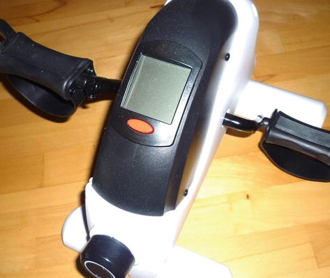
Figure 2.
Pedal ergometer.
Walking/treadmill walking or bicycle exercises are useful means to employ great parts of lower limb musculature. They are also recommendable because of their familiar movement pattern, the low need for additional exercise equipment, and their easy compatibility with everyday life. Patients should walk at a moderate velocity, that is 3–4 km/h for at least 30 minutes daily wearing trainers or other comfortable flat shoes.
4.2 Resistance training
Strength/resistance training (RT) is indicated because of the before-mentioned muscle pump weakness [4]. RT should involve different lower limb muscle groups with a focus lying on ankle plantarflexion, i.e. the gastrocnemius muscle to ensure an active calf-muscle pump. Additionally, also ankle dorsiflexion, knee flexion, and extension components need to be integrated into the training sequence. To decipher the correct number of repetitions, patients are subjected to a multiple repetition maximum test of the latter resistance exercise. Training is executed with a submaximal number of the repetitions possible, e.g. 70%. The core exercise is the so-called “calf-raises” or “heel-rises” in different positions, at different muscle lengths, and by employing various muscle enforcements: dynamic-isotonic, concentric, and eccentric muscle contraction (Figure 3). For progressive training, it is important to re-evaluate the muscle strength and consequently adapt the number of repetitions and level of difficulty. Exercises should be accomplished daily.

Figure 3.
Calf raises.
As muscle strength can be heavily compromised, RT sometimes needs to start at low levels, that is heel-rises in a sitting position. With time, the position moves on to standing on both legs and finally to a single-leg stance. A randomized controlled trial (n = 63) therapy protocol (10 repetitions, increased every 3 days per 5 repetitions until 25 reps were reached; changing from sitting to double-stance and finally to single-stance) led to a trend of complete ulcer healing (p = 0.09; 77% vs. 53%) compared to usual care. Healing rates were better in the subgroup of patients who adhered at least to 75% of the therapy protocol [51]. O’Brien et al. confirmed similar findings. Their study protocol included a daily to perform progressive RT of calf muscle strength. The study had not enough power to significantly show a difference between this regime of 12 weeks and usual care. Nevertheless, there was a 24% difference in healing rates in favor of the exercise group, which was considered clinically significant. Patients who adhered to >75% of the program reached a significant improvement in ankle ROM [52].
To further aggravate the training circumstances, flexible surfaces, e.g. rolled-up gymnastic mats or balance pads can be used. Aside from strength, this also improves ankle ROM and balance capabilities.
4.3 Aqua exercises
Aquatic exercises bear several advantages in comparison to dry-land activities [53]. Firstly, the hydrostatic pressure of water executes pressure similar to CT. Hydrostatic pressure at 0.5 m below surface level is equivalent to 0.05 bar and 37.5 mm Hg, i.e. compression class level III. Secondly, during swimming or similar underwater movements, the nearly prone or supine position removes parts of the gravitational force hindering venous backflow. Thirdly, additional leg and foot movements activate the muscle pump. Lastly, it represents the possibility of exercise for obese patients without too much joint load. Unfortunately, patients with active ulcers cannot enter the pool for obvious reasons. Menegatti et al. subjected a group of 17 patients to an aqua exercise protocol (5 sessions per 30 minutes) aiming at mobilization and muscle activation of the lower limbs. Though lacking a control group, they showed a significant reduction of extracellular water content as well as a significant reduction of leg volume (−15.7%). The special water properties containing different chemical ingredients, e.g. Natrium or Kalium, may have played a role in these results [54]. A randomized controlled trial involved 201 patients for aqua exercises (walking in water, swimming) vs. control. All patients were instructed to wear knee-high compression stockings (30–40 mm Hg) the other time. 28% of aqua exercisers displayed a reduction of ≥4 points in the Venous Clinical Severity Score (significant in comparison to the control group). Leg circumference decreased significantly in favor of aqua exercise [55]. A systematic review of Kneipp therapy (walking in calf-high cold water, lower leg cold water gushes) looked for evidence for this traditional therapy. 64% of the implemented studies reported significant enhancements for the Kneipp therapy [56].
4.4 Breathing exercises
As an adjunctive component, breathing exercises (supine, legs elevated, and combined with CT) can assist the other remedies. Several physiological reasons imply their effectiveness. The diaphragm enhances the blood flow from the lower extremities to the heart by executing a suction effect on the inferior vena cava [8]. In a 3-armed trial, inspiratory muscle training (IMT; 2×15 min per day for 8 weeks; 5 days per week) plus CT was compared to CT alone or calf muscle exercise training. Although the sample size was small (n = 32), the authors observed a significant increase in venous refill time in both legs after IMT. Consequently, they recommended the implementation of this approach to the therapy regime in CVD [8]. It should be noted that patients were sitting during IMT, maybe results would have been more pronounced if patients would have been lying supine, as suggested before. Kwon et al. measured blood flow velocity in the femoral vein during quiet breathing, deep breathing, and both, combined with ankle exercises in 20 healthy males in a supine position. Deep breathing accelerated the velocity from 10.1 cm/sec (±4.2) to 15.5 (±3.9) cm/sec. The highest velocity was reached with deep breathing combined with ankle exercises (plantar-dorsiflexion; 26.6 (9.4). If these results also apply to individuals with CVD need further confirmation [57].
4.5 Additional measures
Additional measures include leg elevation to counteract gravity and body weight management. Short, repeated periods of lying supine during the day (5–10 minutes) relieve swelling and pain. If activities of daily life do not permit lying sequences, patients should at least put their leg(s) on a second chair for some time, e.g. during office hours. Patients should be educated on proper clothes (no socks with tight rubber bands, flat-soled shoes). Furthermore, they are encouraged to exercise regularly and keep an active lifestyle [1] to lose weight. Whenever possible, they should give preference to taking the stairs instead of the elevator, taking the bike instead of the car for short distances, etc. The physiotherapist is part of a multidisciplinary team, consisting of other healthcare professionals such as wound care nurses, surgeons, and dieticians, if needed. Patient-centered therapeutic education can alleviate CVD symptoms and enhance QoL [58]. It is crucial that therapy protocols are tailored to the different needs of individual patients and fit into their life.
Calf muscle stretching (20–30 seconds, standing position, heel remains on the surface) after endurance and/or strength training aims at relaxing and elongating muscles. Additionally, during the stretching maneuver, the ankle joint is mobilized into dorsiflexion [52]. Simultaneously, deep veins are stretched in a longitudinal direction, producing a reduction in venous diameter, better valve closure, and enhanced venous backflow [59]. Stretching after active exercise and predominantly after strength training also prevents training-related muscle soreness.
To improve ankle ROM patients can use a belt or strap to passively pull their own foot in dorsiflexion. This exercise is best being taken while sitting on a mat on the ground, knees extended, and hips 90° flexed. Patients having difficulties moving to the floor sit on a stool, leaning on the backrest, hip nearly extended, and knee extended. The stretching position should be held for at least 20–30 seconds, giving passive structures the possibility to relax. During the exercise patients can report a feeling of stretched tissue, but no pain. Another possibility employs a Redondo ball while sitting on a chair (Figure 4). The affected foot is placed on the ball and the patient is instructed to roll the ball backward by flexing the knee which leads to enlarged dorsiflexion. The final position is held for several seconds, followed by a rolling back until the foot is placed underneath the knee. 10–20 repetitions are recommended for this exercise.

Figure 4.
Redondo ball exercise.
5. Manual lymphatic drainage
Manual lymphatic drainage (MLD) in CVD involves the neck, possibly the abdomen, and one or both lower limbs in approximately 45 minutes to 1 hour time. The therapy is completed by CT, patient education, and active exercises [60].
A few studies have explored MLD within CVD. In 2014 a study group compared MLD in a cohort of CVD patients (CEAP C1-5) and healthy controls. They showed an increase of blood flow in both, the femoral and the great saphenous vein after MLD in both groups. However, this flow was less accentuated in patients with a higher stage of the disease. They recommended MLD as a viable strategy for CVD [61]. Another group tested 10 MLD sessions for the lower limb vs. disease education in 41 CVD patients. They concluded that MLD improves venous edema (measured with the VCSS) and heaviness at the follow-up after 4 weeks and 2 months, respectively [62]. A small patient group with VLU was subjected to complex decongestive therapy (MLD + compression + skincare + exercise) for 5 (n = 8) or 10 days (n = 9) and a control group (n = 9) who received CT alone. This resulted in significantly improved ulcer healing for the 10-day course in comparison to CT (p = 0.039) [63]. Mosti et al. treated 38 patients with 10×20min MLD in advance of their venous surgery and compared them to a control group (n = 42). This protocol led to a significant reduction of foot volume post-surgery in favor of the MLD group. No significant group differences were observed in QoL or venous refilling time [64].
6. Conclusion
CVD is a highly prevalent disorder. It can be treated with various kinds of exercise therapy on dryland and underwater. Aerobic exercises focus on lower limb activities (walking, cycling, aqua exercises) and are complemented by resistance exercises and muscle stretching. Breathing exercises, leg elevation, and manual lymphatic drainage act as a supplement. A critical factor for success is the patient’s adherence to lifestyle changes and health behavior. Compression therapy is an indispensable part of CVD. Patient education aims at health literacy and patients’ adherence to exercise and compression therapy. In CVD, the physiotherapist is part of a multidisciplinary team. The enhancement of various patients’ complaints (pain, leg edema, ankle range of motion, venous reflux) allows for an improvement of quality of life and a better degree of participation in activities of daily life.
References
- 1.
Patel SK. Venous insufficiency. In: Surowiec SM, editor. Drugs Used in Treatment of Venous Insufficiency. Treasure Island: Stat Pearls Publishing; 2022. https://www.statpearls.com/point-of-care/31060 - 2.
Nawaphan T. Risk factors for and treatment of chronic venous disease in Thai patients. Vascular Health and Risk Management. 2022; 18 :667-676. DOI: 10.2147/VHRM.S382726 - 3.
Kiloatar H et al. An evaluation of quality of life, physical activity level and symptoms in patients with early stages of chronic venous disease. Journal of Vascular Nursing. 2021; 39 (4):108-113. DOI: 10.1016/j.jvn.2021.07.007 - 4.
Eberhardt RT, Raffetto JD. Chronic venous insufficiency. Circulation. 2014; 130 (4):333-346. DOI: 10.1161/CIRCULATIONAHA.113.006898 - 5.
Mejia-Gonzales M et al. Prevalence of chronic venous disease in health staff and its impact on quality of life at 6 months. Cirugia y Cirujanos. 2022; 90 (3):332-337. DOI: 10.24875/CIRU.20001245 - 6.
Caballero Escuti G, Ruiz Lascano A, Tabares AH. Correlation between cutaneous manifestations and functional alterations in chronic venous disease of the lower extremities. Actas Dermo-Sifiliográficas. 2022; 113 (9):856-865. DOI: 10.1016/j.ad.2022.05.013 - 7.
Goel RR, Hardy SC, Brown T. Surgery for deep venous insufficiency. Cochrane Database of Systematic Reviews. 2021; 9 (9):CD001097. DOI: 10.1002/14651858.CD001097.pub4 - 8.
Aydin G et al. Effects of inspiratory muscle training versus calf muscle training on quality of life, pain, venous function and activity in patients with chronic venous insufficiency. Journal of Vascular Surgery. Venous and Lymphatic Disorders. 2022; 0 (5):1137-1146. DOI: 10.1016/j.jvsv.2022.04.012 - 9.
De Maeseneer MG et al. Editor’s Choice—European Society for Vascular Surgery (ESVS) 2022 clinical practice guidelines on the Management of Chronic Venous Disease of the lower limbs. European Journal of Vascular and Endovascular Surgery. 2022; 63 (2):184-267. DOI: 10.1016/j.ejvs.2021.12.024 - 10.
Coral FE et al. Chronic venous insufficiency and graduated compression stockings: Analysis of public health system patients’ adherence to treatment. Journal of Vascular Bras. 2021; 20 :e20200034. DOI: 10.1590/1677-5449.200034 - 11.
Yetkin E et al. Symptoms in dilating venous disease. Current Cardiology Reviews. 2020; 16 (3):164-172. DOI: 10.2174/1573403X16666200312101245 - 12.
Moñux G et al. Compression stockings attenuate the expression of proteins associated with vascular damage in human varicose veins. Journal of Vascular Surgery. Venous and Lymphatic Disorders. 2021; 9 (2):428-434. DOI: 0.1016/j.jvsv.2020.05.020 - 13.
Mosmiller LT et al. Evaluation of inflammatory cell biomarkers in chronic venous insufficiency. Phlebology. 2017; 32 (9):634-640. DOI: 10.1177/0268355517701806 - 14.
Chaitidis N et al. Management of Post-thrombotic Syndrome: A comprehensive review. Current Pharmaceutical Design. 2022; 28 (7):550-559. DOI: 10.2174/1381612828666220131094655 - 15.
Jasionowska S et al. Systematic review of exercise therapy in the management of post-thrombotic syndrome. Phlebology. 2022; 37 (10):695-700. DOI: 10.1177/02683555221129738 - 16.
Urbanek T, Labropoulos N. Can we predict and prevent the postthrombotic syndrome? VASA. 2021; 50 (1):11-21. DOI: 10.1024/0301-1526/a000932 - 17.
Schleimer K et al. The treatment of post-thrombotic syndrome. Dt Ärtzebl International. 2016; 113 :863-870. DOI: 10.3238/arztebl.2016.0863 - 18.
Mutlak O, Aslam M, Standfield N. The influence of exercise on ulcer healing in patients with chronic venous insufficiency. International Angiology. 2018; 37 (2):160-168. DOI: 10.23736/S0392-9590.18.03950-0 - 19.
Kolluri R et al. An estimate of the economic burden of venous leg ulcers associated with deep venous disease. Vascular Medicine. 2022; 27 (1):63-72. DOI: 10.1177/1358863X211028298 - 20.
Bonkemeyer Millan S, Gan R, Townsend PE. Venous ulcers: Diagnosis and treatment. American Family Physician. 2019; 100 (5):298-305 - 21.
Scholl L, Dörler M, Stücker M. Ulcers in obesity-associated chronic venous insufficiency. Der Hautarzt. 2017; 68 (7):560-565. DOI: 10.1007/s00105-017-3971-y - 22.
Willenberg T et al. The influence of abdominal pressure on lower extremity venous pressure and hemodynamics: A human in-vivo model simulating the effect of abdominal obesity. European Journal of Vascular and Endovascular Surgery. 2011; 41 (6):849-855. DOI: 10.1016/j.ejvs.2011.02.015 - 23.
Krishnan S, Nicholls SC. Chronic venous insufficiency: Clinical assessment and patient selection. Seminars in Interventional Radiology. 2005; 22 (3):169-177. DOI: 10.1055/s-2005-921961 - 24.
Brenner E, Putz D. 30 Jahre Stemmersches Zeichen. Vasomed. 2006; 4 :153 - 25.
Erdal ES et al. Evaluation of physical activity level and exercise capacity in patients with varicose veins and chronic venous insufficiency. Phlebology. 2021; 36 (8):636-643. DOI: 10.1177/02683555211002339 - 26.
Espeit L et al. Fatigue, physical activity, and quality of life in people self-reporting symptoms of chronic venous disease. Journal of Vascular Surgery. Venous and Lymphatic Disorders. 2022; 10 (5):1147-1154.e1. DOI: 10.1016/j.jvsv.2022.04.016 - 27.
Ercan S et al. Evaluation of the isokinetic calf muscle strength and the range of motion of joint in C3 chronic venous insufficiency. Vascular Specialist International. 2019; 35 (2):95-100. DOI: 10.5758/vsi.2019.35.2.95 - 28.
Pereira DAG et al. Association between heel-rise test performance and clinical severity of chronic venous insufficiency. Phlebology. 2020; 35 (8):631-636. DOI: 10.1177/0268355520924878 - 29.
Tavares PA et al. Association of venous leg ulcers with ankle range of motion in people attending chiropractic mobile clinics in the Dominican Republic. Journal of Chiropractic Medicine. 2017; 16 (4):263-270. DOI: 10.1016/j.jcm.2017.08.004 - 30.
Nepomuceno de Souza I et al. Impairments in ankle range of motion, dorsi and plantar flexors muscle strength and gait speed in patients with chronic venous disorders: A systematic review and meta-analysis. Phlebology. 2022; 37 (7):496-506. DOI: 10.1177/02683555221094642 - 31.
Hong KP. Correlation of clinical class with duplex ultrasound findings in lower limb chronic venous disease. Journal ofJ Chest Surgery. 2022; 55 (3):233-238. DOI: 10.5090/jcs.22.010 - 32.
De Almeida ILGI et al. Reliability and validity of specific quality of life assessment questionnaires related to chronic venous insufficiency: A systematic review. Journal of Vascular Bras. 2022; 1 :e20210229. DOI: 10.1590/1677-5449.202102292 - 33.
Rabinovich A, Kahn SR. How I treat the postthrombotic syndrome. Blood. 2018; 131 (20):2215-2222. DOI: 10.1182/blood-2018-01-785956 - 34.
Van Uden CJ et al. Gait and calf muscle endurance in patients with chronic venous insufficiency. Clinical Rehabilitation. 2005; 19 (3):339-344. DOI: 10.1191/0269215505cr809oa - 35.
Gianesini S et al. Volume control of the lower limb with graduated compression during different muscle pump activation conditions and the relation to limb circumference variation. Journal of Vascular Surgery. Venous and Lymphatic Disorders. 2020; 8 (5):814-820. DOI: 10.1016/j.jvsv.2019.12.073 - 36.
Ma F et al. Compression therapy following endovenous thermal ablation of vaicose veins: A systematic review and meta-analysis. Annals of Vascular Surgery. 2021; 80 :302-312. DOI: 10.1016/j.avsg.2021.09.035 - 37.
Ibegbuna V et al. Effect of elastic compression stockings on venous hemodynamics during walking. Journal of Vascular Surgery. 2003; 37 (2):420-425. DOI: 10.1067/mva.2003.104 - 38.
Balet F et al. Limitations to self-management of adjustable compression wraps in the elderly: Results of a prospective cohort study. International Angioplasty. 2021; 40 (3):261-266. DOI: 10.23736/S0392-9590.21.04580-6 - 39.
Kroeger K et al. Prophylaxis of recurrent venous leg ulcer. Zentralblatt für Chirurgie. 2017; 142 (3):306-311. DOI: 10.1055/s-0042-109977 - 40.
Karafa M, Karafova A. Kikuhime device in the management of venous leg ulcers. Clinical Interventions in Aging. 2020; 15 :1533-1539. DOI: 10.2147/CIA.S264567 - 41.
Weindorf M et al. Einfluss visueller Kontrollsysteme auf den Druck von Kompressionsverbänden. Phlébologie. 2012; 41 :18-24. DOI: 10.1055/s-0037-1621797 - 42.
Berszakiewicz A et al. The effect of compression therapy on quality of life in patients with chronic venous disease: A comparative 6-month study. Postepy Dermatology Alergology. 2021; 38 (3):389-395. DOI: 10.5114/ada.2020.92277 - 43.
Makedonov I, Kahn SR, Galanaud J-P. Prevention and management of the post-thrombotic syndrome. Journal of Clinical Medicine. 2020; 9 :923. DOI: 10.3390/jcm9040923 - 44.
Silva JC et al. Determinants of quality of life in patients with post-thrombotic syndrome. Annals of Vascular Surgery. 2022; 85 :253-261. DOI: 10.1016/j.avsg.2022.03.002 - 45.
Marini E et al. Combined aerobic and resistance training improves microcirculation in metabolic syndrome. The Journal of Sports Medicine and Physical Fitness. 2019; 59 (9):1571-1576. DOI: 10.23736/S0022-4707.18.09077-1 - 46.
Souza Silva KL et al. The impact of exercise training on calf pump function, muscle strength, ankle range of motion, and health-related quality of life in patients with chronic venous insufficiency at different stages of severity: A systematic review. Journal of Vascular Bras. 2021; 20 :e20200125. DOI: 10.1590//1677-5449.200125 - 47.
Downie S, Shnaigat M, Hosseinzadeh H. Effectiveness of health literacy- and patient activation-targeted interventions on chronic disease self-management outcomes in outpatient settings: A systematic review. Australian Journal of Primary Health. 2022; 28 (2):83-96. DOI: 10.1071/PY21176 - 48.
Van Duijnhoven NTL et al. The effect of bed rest and an exercise countermeasure on leg venous function. European Journal of Applied Physiology. 2008; 104 (6):991-998. DOI: 10.1007/s00421-008-0854-z - 49.
Uhl JF, Gillot C. Anatomy of the foot venous pump: Physiology and influence on chronic venous disease. Phlebology. 2012; 27 (5):219-230. DOI: 10.1258/phleb.2012.012b01 - 50.
Quilici BC et al. Comparison of reduction of edema after rest and after muscle exercises in treatment of chronic venous insufficiency. International Archives of Medicine. 2009; 2 (1):18. DOI: 10.1186/1755-7682-2-18 - 51.
Bolton L. Exercise and chronic wound healing. Index Wounds. 2019; 31 (2):65-67 - 52.
O’Brien J et al. Evaluating the effectiveness of a self-management exercise intervention on wound healing, functional ability and health-related quality of life outcomes in adults with venous leg ulcers: A randomised controlled trial. International Wound Journal. 2017; 14 :130-137. DOI: 10.1111/iwj.12571 - 53.
Bissacco D et al. Rationale and current evidence of aquatic exercise therapy in venous disease: A narrative review. Vascular. 2022; 2022 :1708. DOI: 10.1177/17085381221102783 - 54.
Menegatti E et al. The effects of thermal water physical exercise in patients with lower limb chronic venous insufficiency monitored by bioimpedance analysis. Diagnostics (Basel). 2020; 10 (11):889. DOI: 10.3390/diagnostics10110889 - 55.
Sharifi M et al. The randomized, controlled ATLANTIS trial of aquatic therapy for chronic venous insufficiency. Journal of Vascular Surgery. Venous and Lymphatic Disorders. 2021; 9 (4):961-970. DOI: 10.1016/j.jvsv.2020.10.016 - 56.
Stier-Jarmer M et al. Effekte der Kneipp-Therapie: Ein systematischer Review der aktuellen wissenschaftlichen Erkenntnisse. Complementary Medical Research. 2021; 28 (2):146-159. DOI: 10.1159/000510452 - 57.
Kwon OY et al. Effects of ankle exercise combined with deep breathing on blood flow velocity in the femoral vein. The Australian Journal of Physiotherapy. 2003; 49 (4):253-258. DOI: 10.1016/s0004-9514(14)60141-0 - 58.
Thibert A et al. Systematic review of adapted physical activity and therapeutic education of patients with chronic venous disease. Journal of Vascular Surgery. Venous and Lymphatic Disorders. 2022; 10 (6):1385-1400. DOI: 10.1016/j.jvsv.2022.05.004 - 59.
Reich-Schupke S. Warum kann eine Kompressionstherapie bei Immobilität sinnvoll sein? Vasomed. 2016; 28 (6):278-284 - 60.
Miller A. Lymphedema-clinical picture and therapy. Der Hautarzt. 2020; 71 (1):32-38. DOI: 10.1007/s00105-019-04523-z - 61.
Dos Santos Crisostomo RS et al. Manual lymphatic drainage in chronic venous disease: A duplex ultrasound study. Phlebology. 2014; 29 (10):667-676. DOI: 10.1177/0268355513502787 - 62.
Dos Santos Crisostomo RS et al. Influence of manual lymphatic drainage on health-related quality of life and symptoms of chronic venous insufficiency: A randomized controlled trial. Archives of Physical Medicine and Rehabilitation. 2015; 96 (2):283-291. DOI: 10.1016/j.apmr.2014.09.020 - 63.
Szolnoky G et al. Adjunctive role of manual lymph drainage in the healing of venous ulcers: A comparative pilot study. Lymphology. 2018; 1 (4):148-159 - 64.
Molski P et al. Manual lymphatic drainage improves the quality of life in patients with chronic venous disease: A randomized controlled trial. Archives of Medical Science. 2013; 9 (3):452-458. DOI: 10.5114/aoms.2013.35343