Open Access is an initiative that aims to make scientific research freely available to all. To date our community has made over 100 million downloads. It’s based on principles of collaboration, unobstructed discovery, and, most importantly, scientific progression. As PhD students, we found it difficult to access the research we needed, so we decided to create a new Open Access publisher that levels the playing field for scientists across the world. How? By making research easy to access, and puts the academic needs of the researchers before the business interests of publishers.
We are a community of more than 103,000 authors and editors from 3,291 institutions spanning 160 countries, including Nobel Prize winners and some of the world’s most-cited researchers. Publishing on IntechOpen allows authors to earn citations and find new collaborators, meaning more people see your work not only from your own field of study, but from other related fields too.
To purchase hard copies of this book, please contact the representative in India:
CBS Publishers & Distributors Pvt. Ltd.
www.cbspd.com
|
customercare@cbspd.com
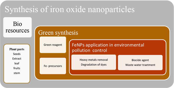
Green synthesis has emerged as a promising and eco-friendly approach for the synthesis of nanoparticles, including iron and iron oxide nanoparticles. This method utilizes plant extracts, microorganisms, or other natural resources as reducing and stabilizing agents instead of toxic chemicals to produce nanoparticles with desired properties. One advantage of green synthesis is the ability to control the size, shape, and crystalline structure of the nanoparticles, which can be analyzed using techniques such as powder X-ray diffraction (XRD). The two variable oxidation states (+2 and + 3) provide an opportunity for multiple products with different designing and crystallite structures. Iron and Iron oxide nanoparticles are both important for biological and photochemical activities. The method for green synthesis decides what kind of particles will one get and for what activity it is suitable. Iron nanoparticles are more suitable for biological activities like antibacterial, antimicrobial, anti-cancerous, and iron oxide for photoelectrical like band gap studies, conduction and photo-catalysis.
Department of Chemistry, Government College University Lahore, Lahore, Pakistan
Aliya Zahid
Department of Chemistry, Government College University Lahore, Lahore, Pakistan
Syeda Taskeen Shahid
Department of Chemistry, Government College University Lahore, Lahore, Pakistan
*Address all correspondence to: shaista.ali.19@gmail.com;, shaista.ali@gcu.edu.pk
1. Introduction
Nanochemistry is a branch of chemistry that focuses on the synthesis, characterization, and manipulation of nanoparticles and nanomaterials. Over the past few years, the field of nanoscience and technology has gained increasing importance in research and development areas. Nanoparticles and nanomaterials have diverse properties that can be tuned to meet specific requirements, making them useful in various applications, such as electronics, energy storage devices, and drug delivery systems. Nanoparticles can also be used for environmental remediation, water treatment, and catalysis, among others [1].
Iron is hard and brittle metal, as the fourth most abundant element in the earth’s crust; it makes up the majority of the planet’s core by weight. Researchers believe that iron has been used by humanity for about 5000 years. The most commonly found sources of iron are the minerals magnetite and hematite. However, smaller quantities of iron can also be derived from the minerals siderite, taconite, and limonite. It contains four fundamental structures, alpha, beta, gamma, and delta, where particles join under various circumstances [2].
Ferrites are categorized into four groups: alpha (attractive), beta, gamma, and omega. The symbol for iron in the periodic table is Fe, which has a nuclear weight of 55.845 and a nuclear number of 26 with a thickness of 7.874 g/cm3. Iron has high melting and boiling point of 1538 and 2862°C, respectively, making it a useful material for applications where high temperatures are involved, such as in the manufacturing of steel or in metallurgical processes. There are 33 different isotopes in existence, but only four of them are stable. The most well-known is iron-56, which has a usual overflow of 91.75% [2].
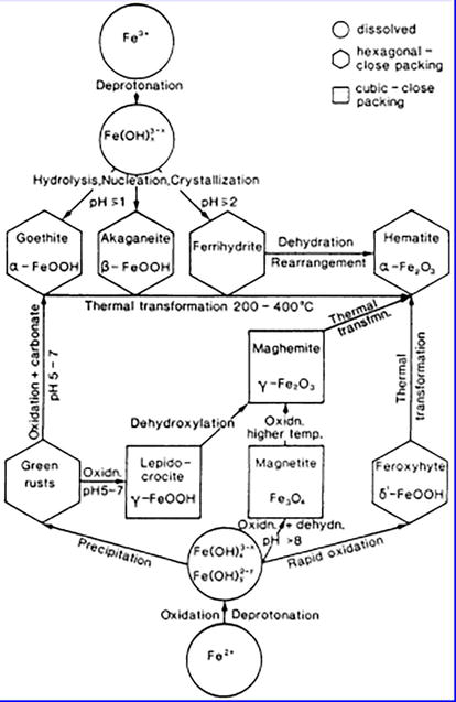
Iron has been the subject of several recent scientific efforts due to its lithophilicity and chalcophilicity, which result in the production of minerals such as pyrite (FeS2), magnetite (Fe3O4), hematite (Fe2O3), siderite (FeCO3), and others. Ferritin, a type of protein that stores iron in the body, also contains iron oxide (Fe3O4). Magnetite, in particular, has garnered significant attention in recent years. It is not uncommon to find magnetite in the Magnesian area of Asia Minor, where it occurs in two forms: reduced ferrous iron species and oxidized ferric iron. This intriguing chemical has the potential to be employed in a range of applications and exhibits a variety of unique properties. The features of magnetite vary according to the mix methods, and the size of the material (from bulk to nanoscale), the structure of magnetite was originally determined in 1995 using X-ray diffraction (Figure 1). When magnetite was initially identified in the region, its structure was first discovered using X-ray diffraction. Magnetite is naturally ferromagnetic and has an inverted spinal structure as a result of the alternating lattices of iron (II) and iron (III). The fact that it includes both divalent and trivalent iron sets it apart from other iron oxide [2].
2. Classification of iron and iron oxide nanoparticles
Fe2O3 is primarily used in the fields of sensors, coatings, UV radiation blocking, and coloring. Fe2O3 nanoparticles are also ideal for exploring nanoparticle polymorphism, magnetic transition, and structural phase transition.
Alpha Fe2O3 is an antiferromagnetic material with hexagonal structure corundum, its magnetic momentum is extremely low, and at 1 emu/cc, the transition is seen in alpha Fe2O3. The alpha structure exhibits mild ferromagnetism in the transition above 260 K, whereas it exhibits anti-ferromagnetism below 960 K. This is a low-temperature transition, and it is dependent on the size of the particles.
The beta Fe2O3 is a material that is paramagnetic and has a cubic structure.
The gamma polymer is a ferromagnetic molecule with a cubic spinal structure. At room temperature, it has a magnetic moment of 430 emu/cc.
The orthorhombic structure of the epsilon exhibits ferromagnetic molecules (Figure 2) [2].
Figure 2.
Formation and transformation of iron and iron oxides nanoparticles [3].
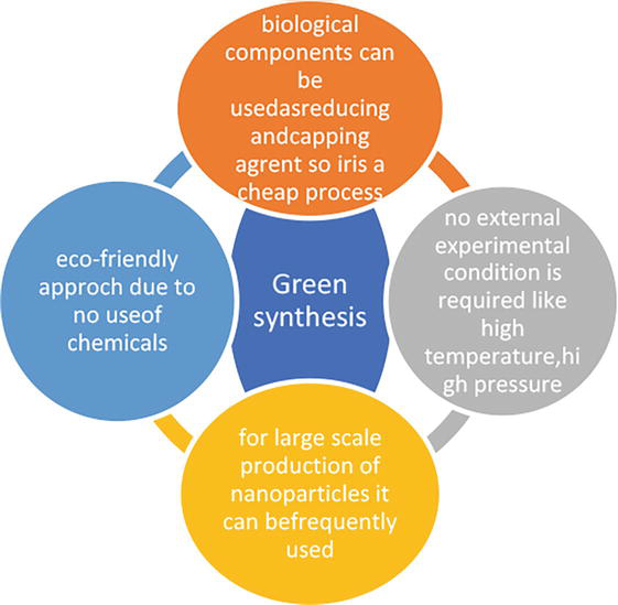
2.1 Benefits of green synthesis
The following advantages of green/biological synthesis (Figure 3) methods are comparable to those of frequently used physical and chemical synthesis protocols:
A method that is safe for the environment and uses no toxic substances.
By acting as a capping and reducing agent, the active biological component, which may include an enzyme, lowers the total cost of the manufacturing process.
Even during mass manufacturing and external experimental circumstances like high energy, small nanoparticles can be created [4].
Mostly two synthesis methods are used in the procedures, which are following
Wet chemistry method
Nonwet chemistry methods.
Precipitation, co-precipitation, hydrothermal, reduction, solvothermal, cryochemical synthesis, and spray/laser paralysis are an example of wet chemistry methods. The last three processes exhibit variation from the surrounding temperature. Cryochemical synthesis takes place below room temperature, and solvothermal synthesis and spray pyrolysis take place above it. Nonwet chemical synthesis includes inert gas condensation, laser ablation, laser-assisted synthesis, and plasma-assisted synthesis [5].
At present, there is no consensus on the mechanism underlying the synthesis of metallic nanoparticles mediated by plant extracts. However, it is widely recognized that secondary metabolites found in various plant components play a critical role in the process. These secondary metabolites include polyphenols, flavonoids, tannic acids, terpenoids, ascorbic acids, carboxylic acids, aldehydes, and amides. Many investigations have employed the IR spectroscopic technique to confirm the existence of numerous reducing sugars, which are often present in plants. Plant extracts containing phytochemicals with appropriate redox characteristics can effectively reduce metal precursors and convert them into matching metallic nanoparticles. According to one hypothesis, radical tannins “R” reduce metal under the influence of pH.
4.1 Method
The following methods can be used to initiate the bio-reduction process:
FeCl3+H2O→FeH2On+3+H2OE1
R+FeH2On+3+H2O→FeH2On+2+H++R−OHE2
FeH2On+22R−+H++OH−→Fe+2O−2+O−2+R−H+R−OHE3
The iron-polyphenol complex nanoparticles (Fe-P NPs) structure was produced by eucalyptus leaves, in a different research. The polyphenols found in eucalyptus extract possess the ability to convert Fe3+ into Fe2+ and at its reduction potential. However, the extract does not entirely convert the Fe2+ to zero-valent iron [6]. Due to the presence of polyphenol ligands, Fe2+ is firmly stabilized; yet, Fe3+polyphenol complexes are quickly formed when Fe2+oxidizes in the presence of oxygen, which is known as autoxidation. As a result, a black nano-iron colloid is produced when an iron metal solution reacts with a plant extract. An experiment using the X-ray absorption (XAS) spectroscopy technique indicated that plant polyphenols were located in a globular position and chelated with ferric ion (Fe3+). A similar response mechanism was hypothesized for Sage (Salvia). Plant polyphenols can be cross-linked via polyphenol condensation following a reaction with iron chloride (FeCl3) (Eq. (1)) [4, 5, 7].
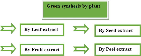
Compared to microorganisms, plants generate more stable metal nanoparticles and are considered the best candidates for large-scale and quick synthesis. Plants naturally contain a wide range of organic reducing chemicals that are well-suited to the synthesis of nanoparticles. Higher levels of antioxidants found in seeds, fruits, leaves, and stems are masked by the diversity of herbs and plant sources. Therefore, the use of plant-based phytochemicals in the entire synthesis and construction of nanoparticles creates an essential synergy between natural/plant sciences and nanotechnology. This affiliation offers nanotechnology a distinctly “green” perspective, known as green nanotechnology, which can be used without causing significant environmental contamination, setting new benchmarks for highly viable clean and green technology [4, 7]. In contrast to microbe-based synthesis, the creation of nanomaterials from plants is more uniform and faster. Various plant components, including the leaf, stem, seed, and root, are commonly used in the easiest, most affordable, and reproducible method of creating metallic nanoparticles (Table 1 and Figure 4) [27].
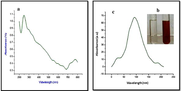
Peaks at 991,1070, 1631, 2800, and 3467 cm−1 in the Fourier transform infrared (FTIR) spectrum of green-synthesized iron nanoparticles (INPs) were caused by the bonds C▬O, C〓O, C▬H, and OH, respectively (Figure 6).
Figure 6.
FTIR spectrum of FeNPs [29].
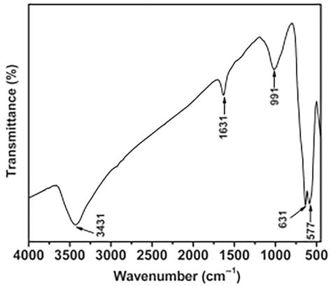
6.3 X-ray diffraction spectroscopy
The characteristics peak of iron nanoparticles were observed in the XRD pattern at 2θ-values 18.97, 29.81, 35.24, 39.53, and 48.30°. The iron nanoparticles were crystalline in nature. The reduction of metal ions by the extract is clearly showing the iron nanoparticles by X-ray diffraction. By using the Scherer formula we can find out the average particle size of iron oxide nanoparticles [30]. The average size of iron oxide nanoparticles is 45.09 nm (Figure 7).
Figure 7.
XRD analysis of FeNPs [31].
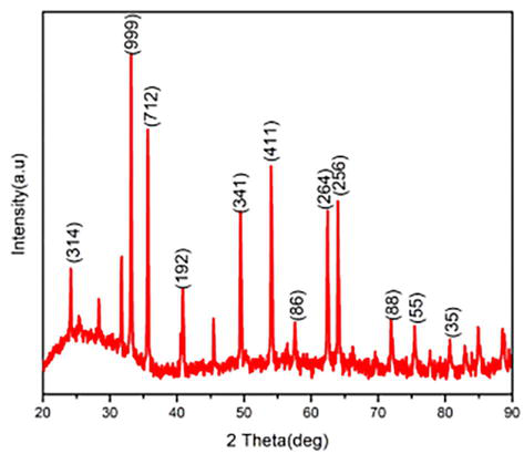
6.4 Scanning electron microscopes
This scanning electron microscope (SEM) is mostly used to identify the structure of the nanomaterial; it can tell us the morphology of the nanoparticles, and the image of iron nanoparticles obtained by the scanning electron microscope looks like a honeycomb. The image of iron nanoparticles in a scanning electron microscope is seen as nonuniform, and its size ranges from 10 to 40 nm with a smooth surface, but they are not uniform (Figure 8).
Figure 8.
SEM spectrum of FeNPs [32].
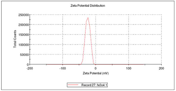
6.5 Field emission-scanning electron microscopy and zeta potential
Although there is much disagreement on the size threshold to separate NPs from bulk materials, size is a crucial factor in defining NPs [33, 34, 35]. The particles were suspended in water that had a viscosity of 0.887, a refractive index of 1.33, and a dielectric constant of 78.5. The zeta potential reflects the potential difference between the electrophoretically mobile particles EDL (electric double layer) and the layer of dispersant surrounding them at the sliding plane. The potential at the slipping/shear plane of a colloid particle moving through an electric field is also known as electrokinetic potential. The particle size distribution is a result (Tables 2–4 and Figure 9).
Plants
FTIR (cm−1)
Azadirachta indica (leaf extract)
O▬H at 3268.9 (stretching) and COOH at 1636.3 (stretching) [10]
P.granatum (seed extract)
C〓C at 1500 C〓N at 1640 C〓O at 1720 N▬H(amine salt) at 3000 N▬H at 3400 [11]
P.orientalis
C▬H at 3196(stretching) H▬C▬H at 1315(bending) C▬O at 1410 C▬C at 1000–1450 Fe▬O▬Fe at 663,462,426 [12, 13]
D.mezereum
Fe▬O at 516 O▬H of carboxylic acid at 3448 C〓C at 1635 (ring stretching) [14]
L.siceraria
O▬H bond at 3354 C〓O at 1701 Inorganic compound at 624 [15]
Spinancia oleracea (leaf extract)
OH at 3500–3200 C▬H at 2800 C〓O at 1658 C〓C at 1540,1402 Fe NPs at 670, 612.63 [6, 16, 17]
A. marina
M▬O bond at 400–450 Fe▬O at 618 and 467 Aromatic comp. at 2923,2853 C▬H bonds at 3422 O▬H group at 1630 [18]
G. mauritiana
OH at 3334.7 (stretching) C☰N at 2115.9 (stretching) (▬C〓O) at 1632 [21]
Camellia sinensis assamica
C▬O▬C at 1064 C〓C at 1616 (stretching) O▬H at 3355 C〓C at 1608 (ring stretching) [23]
Murraya koenigii
OH at 3454 (stretching) C▬H at 2427 (stretching) C〓O at 1767 ester group (stretching) The amino acid at 1637 Germinal CH3 group at 1384 Fe▬O at 831, 537, and 459 (stretching) [36, 37]
Urtica dioica
C▬O at 1070 C〓O at 1636 C▬H at 2800 OH at 3467 [23, 37]
Pinus eldarica
O▬H at 3420.97 C〓O at 1616.05 C▬O at 1069.05 C–H at 2918 Fe▬O at 640 and 450 [7, 36]
The sharp peaks at 18.38°, 30.18°, 35.55°, 43.25°, 53.60°, 57.11°, 62.76°, 71.30°, and 74.50° 141 corresponded to the crystal planes of 142 (111), (220), (311), (400), (422), (511), (440), (620), and (533). This demonstrated that the majority of the 143 IONPs synthesized were Fe3O4.
The XRD pattern of Fe▬O revealed the presence of three peak of αFe2O3,γFe2O3 whoes hkI value is (012),(410) and (221) respectively.
A. marina [PDF No.: 46–1312 JCPDS] Kingdom: Plantae Family: Acanthaceae Genus: Avicennia Species: A. marina Order: Lamiales
The XRD pattern of FeO-NPs revealed the presence of FeO at five peaks: 36.42°,42.24°,61.29°,73.24°, and 77.07°, which correspond to the (111), (200), (220), (311), and (222) planes of FeO, respectively.
It is discovered that the exits strong diffraction peaks with 2 values of 28.26° and 32.28°, which correlate to the hkl value of (220),(222), denoting the crystalline phase of Fe3O4-NPs.
The Fe3O4 XRD pattern revealed six typical diffraction peaks at planes 2θ = (220) at 30.3° (311) at 35.6°, (400) at 43.3°, (422) at 53.2°, (511) at 57.1°, and (440) at 62.8°.
The significant strong characteristic peaks of iron oxide particles are found at 2θ = 24.14°, 33.14°, 35.61°, 40.84°, 49.45°, 54.06°, 62.42°, and 64.00°, which correspond to iron oxide amorphous structures (220), (311), (400), (442), (511), and (440).
M.indica [(α-Fe2O3) JCPDS 87–1164] [(γ-Fe2O3) JCPDS 39–1346] Common name: mango Family name: Anacardiaceae
Hematite (-Fe2O3) JCPDS 87–1164, and maghemite (-Fe2O3) JCPDS 39–1346 in gamma phase. –Fe2O3 appears (in black) in diffracted planes (311) and (220), where -Fe2O3 appears (in blue) at (012), (104), (110), (113), (024), and (116).
S.aromaticum [JCPDF card #39–1346] Common name: clove Plant part is taken: buds Family name: Myrtaceae
The Fe2O3 characteristic XRD peaks suggest a hematite structure and are indexed as (012), (110), (222), (421), (422), and Fe3O4 is characterized as (111), (311), (222), (400), (422) and (440), indicating the co-existence of iron oxide phases.
Table 4.
XRD pattern result of the iron and iron oxides from different plants.
Figure 9.
Zeta potential spectrum of FeNPs [38].
6.6 Application of nanoparticles
Magnetic nanoparticles have a variety of uses, including in biomedicine, healthcare, agriculture, food production, environmental management systems, energy, textiles, electronics, building materials, machines, etc. (Figure 10).
Figure 10.
Application of iron and iron oxides nanoparticles.
This study focuses on the engineering of iron nanoparticles using several environmentally friendly techniques and their potential to remove contaminants from the environment (Table 5). To some extent, efforts are made to emphasize the many environmentally friendly agents for the production of iron nanoparticles, including polymers, amino acids, bacteria, fungi, plant extracts, etc. This review also covers the relationship between particle size, morphology, and other aspects and the characteristics of materials, processes, and protocols. According to the literature, several plants and components linked to plants have been used to easily synthesize iron nanoparticles, which have proven to be effective catalysts for a variety of environmental applications. As a result, plant materials appear increasingly viable as producers of iron nanoparticles because of their environmental benefits and high economic worth. To better understand the phytochemistry involved in the creation of iron nanoparticles, which has not yet been thoroughly defined. Additional study is required to examine more nearby and easily accessible resources for the production of iron nanoparticles in order to accomplish the sustainability of nanomaterial synthesis. Any novel methodology must first comprehend the biochemical processes involved in nanoparticle creation, and any solution must be cost-effective relative to traditional approaches. Utilizing local resources can help keep costs down in the long run as their development. A clearer explanation of biomolecules and their function in modulating the creation of nanoparticles will be provided in future studies with a more in-depth investigation. The objective is to alter the rate of synthesis and boost the stability of nanoparticles. Additionally, research should be done to boost the reactivity of iron nanoparticles during manufacture in order to accelerate the breakdown of environmental pollutants with the fewest possible eco-toxicological effects. Few investigations have confirmed that biosynthesized nanoparticles are less hazardous than manufactured nanoparticles. Additionally, a thorough risk evaluation of green-fabricated Fe NPs should be carried out, taking into account the nanoparticles kinetics, fate, transport, aggregation, and dissolution during processing. A variety of biochemical or functionalized nanoparticles can be produced using the green nanotechnology processes described in this paper as a solid foundation. These new products could be used in the environmental restoration sectors.
Field
Application of iron Nanoparticles
Biomedical
Cellular therapies (cell labeling/tissue repair/cell separation and handling/purifying cell populations/magneto-reception, diseases of the musculoskeletal system/severe inflammation, disability, and pain are all possible uses for magnetic nanoparticles (especially those coated with liposomes) for drug delivery [39].
Health care
Nanophotothermolysis with pulsed lasers for the treatment of cancer, hepatitis B virus, respiratory syncytial virus, influenza virus, antiviral agents against HIV-1, Monkeypox virus, herpes simplex virus type 1, and Tacaribe virus; delivering antigens for a vaccine; nanoscale biosensors and imaging; nanocoatings on surfaces; implants; nanocarrier for vaccination; antimicrobial activities; SLN in drug delivery and research; delivering antigens for a particular disease into the bloodstream; preventing aging of the skin [39]
Agriculture and food
Crop yields are increased by nano-based products (nano-fertilizers, nano-fungicides, and nano-pesticides), designed NPs, and CNTs; pyrite NPs are employed as a seed treatment for different plants before the seeds are sown, and more leaf quantity, greater leaf size, and higher biomass. Improved starch breakdown after storage. This suggests that iron pyrite nanoparticles might be created as a commercial seed treatment product (pro-fertilizer). As no NPs are introduced into the soil throughout the procedure, the method is secure. Fewer doses are needed than with chemical fertilizers. There are no negative impacts on plant development. Nanoporous membranes, gene transfer (crop enhancement), nano-composites, nanosensors, nanofood, encapsulation, food packaging, nanocoatings, and precision farming [39].
Environmental Remediation
Detection, monitoring, and treatment of pollution. Treatment of wastewater (adsorption, membrane filtration, and permeable reactive barriers). Palladium (Pd), climate change (carbon capture), synthetic leaves for CO2 sequestration, mineral carbonation, biomimetic carbonation, N2O breakdown, and methane combustion are some examples of catalyst coatings. Enhances manufacturing processes (efficiency, waste reduction), dematerialization (reduction in material quantity), sensing (pollutant sensors, nanoporous membranes, chemical and bionanosensors, nanowire sensor for explosives), and energy (heat distribution, for example, ceramic-like materials that sufficiently provide the structure’s dependability and durability) [39].
Energy
The conversion of waste heat from computers, cars, homes, power plants, etc. into usable electrical power using photovoltaic film coatings, more efficient fuel production and consumption, fuel cells, prototype batteries, aerogels, and thermoelectric materials [39].
Military and aerospace
Smart materials, nano-composites, nanocoatings, sensors, electronics, fuel additives, and energy devices [39].
Construction
Smart materials, concrete additives, nanoscale sensors, nano-composites, and nanocoatings. In order to color concrete, brick, tile, and other building materials, iron oxide pigments are utilized [39].
Electronics
Printed electronics, carbon nanotubes, nanowires, NEMS, spintronics, and quantum dots [39] are examples of emerging technologies.
1.Alagarasi A. Chapter 1. In: Introduction to Nanomaterials. 2011. p. 76. Available from: https://www.researchgate.net/publication/259118068
2.Rukhsar M, Ahmad Z, Rauf A, Zeb H, Ur-Rehman M, Hemeg HA. An overview of iron oxide (Fe3O4) nanoparticles: From synthetic strategies, characterization to antibacterial and anticancer applications. Crystals. 2022;12(12):1809. DOI: 10.3390/cryst12121809
3.Hock S. Precipitation of Hematite and Recovery of Hydrochloric Acid from Aqueous Iron (II, III) Chloride Solutions by Hydrothermal Processing. McGill University; 2009
4.Fahmy HM, Mohamed FM, Marzouq MH, Mustafa ABED, Alsoudi AM, Ali OA, et al. Review of green methods of iron nanoparticle synthesis and applications. BioNanoScience. 2018;8(2):491-503. DOI: 10.1016/j.jece.2020.104569
5.Monalisa P, Nayak PL. Eco-friendly green synthesis of iron nanoparticles from various plants and spices extract. International Journal of Plant, Animal and Environmental Sciences. 2013;3(1):68-78
6.Turakhia B, Turakhia P, Shah S. Green synthesis of zero-valent iron nano-particles from Spinaciaoleracea (spinach) and its application in wastewater treatment. Journal of Advance Research in Applied Sciences. 2018;5(1):46-51
7.Kheshtzar R, Berenjian A, Taghizadeh SM, Ghasemi Y, Asad AG, Ebrahiminezhad A. Optimization of reaction parameters for the green synthesis of zero-valent iron nanoparticles using pine tree needles. Green Processing and Synthesis. 2019;8(1):846-855. DOI: 10.1515/gps-2019-0055
8.Mahmoud R, Kotp AA, El-Ela FIA, Farghali AA, Moaty SA, Zahran HY, et al. Green synthesis of iron nanoparticles of clove and green coffee origin with an in vivo hepatoprotective investigation. Journal of Environmental Chemical Engineering. 2021;9(6):106320. DOI: 10.1016/j.jece.2021.106320
9.Huang L, Weng X, Chen Z, Megharaj M, Naidu R. Green synthesis of iron nanoparticles by various tea extracts: A comparative study of the reactivity. Spectrochimica Acta Part A: Molecular and Biomolecular Spectroscopy. 2014;130:295-301. DOI: 10.1016/j.saa.2014.04.037
10.Zambri NDS, Taib NI, Abdul Latif F, Mohamed Z. Utilization of neem leaf extract on the biosynthesis of iron oxide nanoparticles. Molecules. 2019;24(20):3803. DOI: 10.3390/molecules24203803
11.Bibi I, Nazar N, Ata S, Sultan M, Ali A, Abbas A, et al. Green synthesis of iron oxide nanoparticles using pomegranate seeds extract and photocatalytic activity evaluation for the degradation of textile dye. Journal of Materials Research and Technology. 2019;8(6):6115-6124. DOI: 10.1016/j.jmrt.2019.10.006
12.García DG, Garzón-Romero C, Salazar MA, Lagos KJ, Campaña KO, Debut A, et al. Bioinspired synthesis of magnetic nanoparticles based on iron oxides using Orange waste and their application as photo-activated antibacterial agents. International Journal of Molecular Sciences. 2023;24:4770. DOI: 10.3390/ijms24054770
13.Devi HS, Boda MA, Shah MA, Parveen S, Wani AH. Green synthesis of iron oxide nanoparticles using Platanusorientalis leaf extract for antifungal activity. Green Processing and Synthesis. 2019;8(1):38-45. DOI: 10.1515/gps-2017-0145
14.Beheshtkhoo N, Kouhbanani MAJ, Savardashtaki A, Amani AM, Taghizadeh S. Green synthesis of iron oxide nanoparticles by aqueous leaf extract of Daphne mezereum as a novel dye removing material. Applied Physics A. 2018;124:1-7. DOI: 10.1007/s00339-018-1782-3
15.Kanagasubbulakshmi S, Kadirvelu K. Green synthesis of iron oxide nanoparticles using Lagenariasiceraria and evaluation of its antimicrobial activity. Defence Life Science Journal. 2017;2(4):422-427. DOI: 10.14429/dlsj.2.12277
16.Bunea A, Andjelkovic M, Socaciu C, Bobis O, Neacsu M, Verhé R, et al. Total and individual carotenoids and phenolic acids content in fresh, refrigerated and processed spinach (Spinaciaoleracea L.). Food Chemistry. 2008;108(2):649-656. DOI: 10.1016/j.foodchem.2007.11.056
17.Fayyaz B, Zahra MB, Haider MS. Screening of phenolic compounds from spinach (Spinaciaoleracea), green synthesis of iron-nanoparticles and determination of its anti-microbial effect on Escherichia coli. Research Square. 2022. DOI: 10.1016/j.foodchem.2007.11.056
18.Karpagavinayagam P, Vedhi C. Green synthesis of iron oxide nanoparticles using Avicennia marina flower extract. Vacuum. 2019;160:286-292. DOI: 10.1016/j.vacuum.2018.11.043
19.Ebrahiminezhad A, Taghizadeh S, Ghasemi Y, Berenjian A. Green synthesized nanoclusters of ultra-small zero valent iron nano-particles as a novel dye removing material. Science of the Total Environment. 2018;621:1527-1532. DOI: 10.1016/j.scitotenv.2017.10.076
20.Manquián-Cerda K, Cruces E, Rubio MA, Reyes C, Arancibia-Miranda N. Preparation of nanoscale iron (oxide, oxyhydroxides and zero-valent) particles derived from blueberries: Reactivity, characterization and removal mechanism of arsenate. Ecotoxicology and Environmental Safety. 2017;145:69-77. DOI: 10.1016/j.ecoenv.2017.07.004
21.Amutha S, Sridhar S. Green synthesis of magnetic iron oxide nanoparticle using leaves of Glycosmismauritiana and their antibacterial activity against human pathogens. Journal of Innovations in Pharmaceutical and Biological Sciences. 2018;5(2):22-26. DOI: 10.1155/2021/8822645
22.Ben-Arfa BA, Salvado IMM, Ferreira JM, Pullar RC. Clove and cinnamon: Novel anti–oxidant fuels for preparing magnetic iron oxide particles by the sol–gel auto–ignition method. Journal of Alloys and Compounds. 2019;786:71-76. DOI: 10.1016/j.jallcom.2019.01.306
23.Saif S, Tahir A, Chen Y. Green synthesis of iron nanoparticles and their environmental applications and implications. Nanomaterials. 2016;6(11):209. DOI: 10.3390/nano6110209
24.Afsheen S, Tahir MB, Iqbal T, Liaqat A, Abrar M. Green synthesis and characterization of novel iron particles by using different extracts. Journal of Alloys and Compounds. 2018;732:935-944. DOI: 10.1016/j.jallcom.2017.10.137
25.Kumar KM, Mandal BK, Kumar KS, Reddy PS, Sreedhar B. Biobased green method to synthesis palladium and iron nanoparticles using Terminaliachebula aqueous extract. Spectrochimica Acta Part A: Molecular and Biomolecular Spectroscopy. 2013;102:128-133. DOI: 10.1016/j.saa.2012.10.015
26.Carrapiço A, Martins MR, Caldeira AT, Mirão J, Dias L. Biosynthesis of metal and metal oxide nanoparticles using microbial cultures: Mechanisms, antimicrobial activity and applications to cultural heritage. Microorganisms. 2023;11(2):378. DOI: 10.3390/microorganisms11020378
27.Herlekar M, Barve S, Kumar R. Plant-mediated green synthesis of iron nanoparticles. Journal of Nanoparticles. 2014:9. Article ID 140614. DOI: 10.1155/2014/140614
28.Murugan K, Dinesh D, Nataraj D, Subramaniam J, Amuthavalli P, Madhavan J, et al. Iron and iron oxide nanoparticles are highly toxic to Culexquinquefasciatus with little non-target effects on larvivorous fishes. Environmental Science and Pollution Research. 2018;25:10504-10514. DOI: 10.1007/s11356-017-0313-7
29.Hwang SW, Umar A, Dar GN, Kim SH, Badran RI. Synthesis and characterization of iron oxide nanoparticles for phenyl hydrazine sensor applications. Sensor Letters. 2014;12(1):97-101. DOI: 10.1166/sl.2014.3224
30.Kamath V, Chandra P, Jeppu GP. Comparative study of using five different leaf extracts in the green synthesis of iron oxide nanoparticles for removal of arsenic from water. International Journal of Phytoremediation. 2020;22(12):1278-1294. DOI: 10.1080/15226514.2020.1765139
31.Balu P, Asharani IV, Thirumalai D. Catalytic degradation of hazardous textile dyes by iron oxide nanoparticles prepared from Raphanussativus leaves’ extract: A greener approach. Journal of Materials Science: Materials in Electronics. 2020;31:10669-10676. DOI: 10.1007/s10854-020-03616-z
32.Rajendran S, Abuthahir S, Syedzahirullah S, Vignesh C, Vikrant K, Vignesh R. International journal of chemical concepts green synthesis of Nano iron oxide particles from mild steel. International Journal of Chemical Concepts. 2017;3(2):196-200
33.Desalegn B, Megharaj M, Chen Z, Naidu R. Green synthesis of zero valent iron nanoparticle using mango peel extract and surface characterization using XPS and GC-MS. Heliyon. 2019;5(5):e01750. DOI: 10.1016/j.heliyon.2019.e01750
34.Ruqeishi MS, Mohiuddin T, Al-Saadi LK. Green synthesis of iron oxide nanorods from deciduous Omani mango tree leaves for heavy oil viscosity treatment. Arabian Journal of Chemistry. 2019;12(8):4084-4090. DOI: 10.1016/j.arabjc.2016.04.003
35.Jamzad M, Kamari Bidkorpeh M. Green synthesis of iron oxide nanoparticles by the aqueous extract of Laurusnobilis L. leaves and evaluation of the antimicrobial activity. Journal of Nanostructure in Chemistry. 2020;10:193-201. DOI: 10.1007/s40097-020-00341-1
36.Ebrahiminezhad A, Zare-Hoseinabadi A, Berenjian A, Ghasemi Y. Green synthesis and characterization of zero-valent iron nanoparticles using stinging nettle (Urticadioica) leaf extract. Green Processing and Synthesis. 2017;6(5):469-475. DOI: 10.1515/gps-2016-0133
37.Samarawickrama KGR, Wijiayapala UGS, Fernando CAN. Green synthesis of iron nanoparticles using curry leaves (Murrayakoenigii) extract. Malaya Journal of Matematik. 2022;219:4309-4317
38.Chandrasekar N, Kumar K, Balasubramnian KS, Karunamurthy K, Varadharajan R. Facile synthesis of iron oxides, iron-cobalt and zero-valent iron nanoparticles and evaluation of their antimicrobial synthesis, free radical scavengining activity and antioxidant assay. Digest Journal of Nanomaterials and Biostructures (DJNB). 2013;8(2):765-775
39.Khan I, Saeed K, Khan I. Nanoparticles: Properties, applications and toxicities. Arabian Journal of Chemistry. 2019;12(7):908-931. DOI: 10.1016/j.arabjc.2017.05.011
Written By
Shaista Ali, Aliya Zahid and Syeda Taskeen Shahid
Submitted: 23 March 2023Reviewed: 24 April 2023Published: 24 June 2023
 Open access peer-reviewed chapter