 Open access peer-reviewed chapter
Abstract
Effective treatment of glioma; the most aggressive primary brain tumor has been a worrisome medical challenge across the globe. Owing to the architectural uniqueness of the brain coupled with the presence of the blood-brain barrier hijacks the success of conventional treatment strategies. In this context, magnetic nanocarriers (MNCs) have garnered significant attention over the past decade as efficient imaging and targeted drug delivery platforms in glioma. In many recent research, ferrite-based drug carriers have shown preferential anticancer activity against glioma cells both in vitro and in vivo. Under the influence of an externally applied magnetic field, anticancer drug-loaded MNCs could be directed at specific tumor locations and can release the cytotoxic drugs more precisely at the tumor area, thereby reducing off-target toxic effects. Among the ferrite-based MNCs, superparamagnetic nanocarriers are considered more biocompatible. Further, the outer surface of MNCs is coated with biodegradable hydrophilic polymers like PEG to make them suitable for in vivo applications. Additionally, MNCs can be functionalized with specific ligands like monoclonal antibodies, peptides, aptamers, etc., to improve tumor-specific targeting. The chapter highlights research advancements in MNCs-based drug targeting investigated over the past years for the treatment of glioma along with key challenges on the roads of technology transfer for industrial viability.
Keywords
- magnetic nanocarriers
- glioma
- drug targeting
- advancements
- challenges
1. Introduction
Glioma is a type of brain tumor that originates in the glial cells, which are supportive cells of the central nervous system. Glial cells provide structural support, nourishment, and insulation to neurons. Gliomas are the most common type of primary brain tumor, accounting for approximately 80% of all malignant brain tumors, they originate within the brain or spinal cord, as opposed to spreading from other parts of the body [1]. Gliomas can occur in individuals of any age but are more frequently diagnosed in adults. The exact causes of gliomas are not well understood, although certain genetic and environmental factors may play a role [2]. Exposure to ionizing radiation, genetic predisposition, and some rare genetic disorders have been associated with an increased risk of developing gliomas. Gliomas can occur in people of all ages, but they are most commonly diagnosed in adults between the ages of 45 and 65 [3]. These tumors can be classified into different types based on the specific glial cell they originate from. The three main types of gliomas are astrocytoma, oligodendroglioma, and ependymoma. Astrocytomas are the most common type of glioma and arise from astrocytes, which are star-shaped glial cells. They are further categorized into grades I–IV based on their level of aggressiveness, with grade IV being the most malignant form, known as glioblastoma multiforme (GBM) [4]. Oligodendrogliomas originate from oligodendrocytes, which are responsible for producing myelin, a substance that insulates nerve fibers. These tumors are usually slow-growing and occur more commonly in adults. Ependymomas develop from ependymal cells that line the ventricles of the brain and the center of the spinal cord. They are more commonly found in children and young adults [5]. Treatment options for glioma depend on several factors, including the tumor grade, location, and overall health of the patient. The main treatments for glioma include surgery, radiation therapy, and chemotherapy. In some cases, a combination of these treatments may be used. The goal of treatment is to remove as much of the tumor as possible, slow down its growth, and manage symptoms to improve the patient’s quality of life [6]. Symptoms of gliomas can vary depending on the size, location, and rate of growth of the tumor. Common symptoms may include headaches, seizures, cognitive and memory problems, changes in behavior or personality, difficulty with balance and coordination, and neurological deficits. The diagnosis of gliomas typically involves a combination of medical history evaluation, neurological examination, imaging tests such as MRI or CT scans, and sometimes a biopsy to confirm the presence of tumor cells [7].
Treatment of glioma, the most common form of brain tumor has been a tough challenge to treat and often necessitates novel targeting strategies [8]. Gliomas are highly aggressive and invasive, and their location in the brain makes them difficult to access and treat. The current standard treatments, including surgery, chemotherapy, and radiation therapy, have several limitations, such as low therapeutic efficacy, high toxicity, and drug resistance. Hence, there is an urgent need for novel and effective therapeutic strategies for glioma [9].
Nanotechnology has recently become a potential method for treating gliomas. Nanoparticles have unique physical and chemical properties that make them suitable for drug delivery applications. Among the various types of nanoparticles, magnetic nanocarriers have gained significant attention due to their ability to selectively target and deliver drugs to the tumor site, enhance drug accumulation, and reduce systemic toxicity [10].
Magnetic nanocarriers are composed of a magnetic core, which allows for magnetic targeting, and a shell, which can be functionalized with various ligands to improve tumor-specific accumulation. The magnetic core is typically composed of iron oxide, which is biocompatible and biodegradable [11]. The shell can be made of various materials, such as lipids, polymers, and silica, depending on the desired properties. One of the main advantages of magnetic nanocarriers is their ability to selectively target the tumor site using an external magnetic field. As a result, the loaded drug can be more precisely accumulated at the tumor site, while reducing systemic toxicity [12]. Additionally, magnetic nanocarriers can be functionalized with various ligands, such as antibodies, peptides, and aptamers, to improve tumor-specific targeting and reduce off-target effects. Another advantage of magnetic nanocarriers is their ability to overcome the blood-brain barrier (BBB), which is a major obstacle in the treatment of glioma [13]. The BBB is a complex structure of endothelial cells, which tightly regulates the passage of substances between the bloodstream and the brain. The BBB limits the penetration of most chemotherapeutic drugs into the brain, which hinders the efficacy of glioma treatment. However, magnetic nanocarriers can cross the BBB using magnetic targeting and can deliver drugs directly to the tumor site [14].
2. Blood-brain barrier
The blood-brain barrier (BBB) serves as a diffusion barrier that prevents poisons and other substances from entering the brain while safeguarding the brain’s neuronal tissues. Two different types of cellular junctions, the paracellular tight junction and the intercellular adherens junction, are present in the BBB based on its molecular construction [15]. Adherens junction, which is made up of cadherin, vascular endothelium (VE), catenin, and actinin maintains the BBB’s functional quality. Tight junctions, however, retain the BBB’s primary functioning because they are principally in charge of permeability via the BBB. A complicated cellular network makes up the BBB in adults. Brain endothelial cells, a highly specialized basal membrane, many pericytes surrounding the basal membrane, and astrocytic end-feet were the primary elements of this system [16].
2.1 Brain endothelial cells
For effective barrier construction and interactions with neighboring cells, such cells are necessary. Also, they are referred as brain microvascular endothelial cells (BMECs). The following are some ways that BMECs are different from endothelial cells found in other organs: (i) Consistent tight connections among brain endothelial cells stop chemicals from moving across cells, (ii) BMECs contain more mitochondria and fewer cytoplasmic vesicles, & (iii) BMECs do not exhibit identifiable transendothelial pathways such as intracellular vesicular transport [17]. Complex intercellular tight junctions prevent chemicals from passively diffusing into the brain, which causes blood vessels to have exceptionally high transendothelial electrical resistance (TEER) in
2.2 Basal membrane
The whole surface of the capillary endothelial cell layers is covered by this structure, which is made up of type IV collagen, laminin, and fibronectin. This membrane has pericytes embedded inside it, and astrocytic end-feet surround them. This membrane’s possible use is to prevent the solutes from moving around freely [19].
2.3 Pericytes
Pericytes are the contractile cells that surround the endothelial cells. These cells regulate the expression of BBB-specific genes in endothelial cells, induce polarization of astrocytic end-feet encircling CNS blood vessels, and prevent CNS immune cells from impairing the proper formation of BBB. Additionally, these cells assist in decreasing the expression of substances that raise vascular permeability [20].
2.4 Astrocytic end-feet
Previously, it was believed that the astrocytic end-feet surrounding endothelial cells did not significantly contribute to BBB maintenance [21]. On the other hand, recent research by Nuriya et al. (2013) revealed the variability of diffusion patterns surrounding astrocytic end-feet. They established the presence of certain astrocytic end-feet that can organize into compact networks and prevent molecules from diffusing freely through them. The variability of diffusion patterns is influenced by the different blood vessels & physical features of the gliovascular interface, such as the distance between endothelial cells & astrocytic end-feet. As a result, these networks firmly surround blood vessels, which raises the possibility that astrocytic end-feet serve functional purposes [22].
3. Molecular composition of BBB
The molecular composition of the BBB is complex and dynamic, and it is controlled by a number of variables, including the surrounding environment and the specific needs of the brain. Some of the key components of the BBB include:
Tight junction proteins: The endothelial cells that make up the BBB are connected by tight junctions, which form a barrier that prevents the free passage of molecules. Numerous proteins, such as claudins, occluding, and junctional adhesion molecules, are found in tight junctions [23].Transport proteins: The BBB is highly selective in the molecules that allows to cross from the blood into the brain. This is achieved through the expression of specialized transport proteins that selectively move molecules across the endothelial cell layer. Examples of these transporters include glucose transporters, amino acid transporters, and efflux transporters that pump out potentially harmful substances [24].Enzymes: The BBB also contains a variety of enzymes that regulate the transport and metabolism of molecules. For example, the enzyme monoamine oxidase helps to break down neurotransmitters like dopamine and serotonin [25].Adhesion molecules: Adhesion molecules are proteins that help to maintain the structure and function of the BBB. These molecules include integrins and cadherins, which are important for cell-cell interactions and communication [26].Extracellular matrix components: The extracellular matrix is a network of proteins and other molecules that provides structural support to cells. The BBB contains specific extracellular matrix components, such as laminin and collagen, which help to maintain the integrity of the endothelial cell layer [27].Overall, the molecular composition of the BBB is highly specialized and tightly regulated, allowing for the selective transport of molecules between the blood and the brain while maintaining the overall health and function of the brain [28].
4. Targeting strategy of magnetic nanocarrier vs. conventional therapy
Nanocarriers are a promising approach for the treatment of glioma due to their ability to selectively target the tumor site, enhance drug accumulation, and reduce systemic toxicity [29]. In comparison, conventional therapies such as surgery, chemotherapy, and radiation therapy have several limitations, such as low therapeutic efficacy, high toxicity, and drug resistance. The targeting strategy of magnetic nanocarriers involves the use of an external magnetic field to guide the nanoparticles to the tumor site [30]. The magnetic field can be applied locally, allowing for precise targeting & increased drug accumulation at the tumor site, while reducing off-target effects. This targeted approach can also overcome the blood-brain barrier, which is a significant obstacle to the treatment of glioma, and deliver drugs directly to the tumor site [31].
In contrast, conventional therapies have limited tumor specificity, and a significant amount of the drug is distributed throughout the body, which results in systemic toxicity. Additionally, the blood-brain barrier limits the penetration of most chemotherapeutic drugs into the brain that blocks the efficacy of glioma treatment [32]. The strategy of drug targeting by MNCs in bypassing BBB in comparison to conventional drug therapy has been depicted in Figure 1. While surgery can remove a significant portion of the tumor, it is often not feasible for tumors located in critical brain regions. Moreover, conventional therapies often lead to drug resistance, limiting their long-term efficacy. Gliomas are highly heterogeneous, and the tumor cells can develop resistance to chemotherapy and radiation therapy, leading to treatment failure [33]. Magnetic nanocarriers can overcome drug resistance by delivering drugs directly to the tumor site, allowing for higher drug concentrations, and reducing the likelihood of drug resistance. In conclusion, magnetic nanocarriers offer several advantages over conventional therapies for the treatment of glioma, including targeted drug delivery, enhanced drug accumulation, reduced systemic toxicity, and overcoming drug resistance. While there are still several challenges to overcome, including nanoparticle design optimization and magnetic targeting parameters, for the treatment of gliomas in the future, magnetic nanocarriers show enormous potential [34]. The effectiveness of magnetic nanocarriers over conventional therapy in tumor targeting has been summarized in Figure 1.

Figure 1.
Magnetic carrier vs. conventional drug therapy.
5. Types of magnetic nanocarrier
Magnetic nanocarriers are a type of nanoparticle that can be guided to specific cells or tissues using an external magnetic field. They have potential applications in a variety of fields, including drug delivery, imaging, and therapy. There are several types of magnetic nanocarriers, including:
Iron oxide nanoparticles: Iron oxide nanoparticles are among the most commonly used magnetic nanocarriers due to their biocompatibility and magnetic properties. They can be coated with various materials to improve their stability, targeting, and drug loading capacity [35].Magnetic liposomes: Magnetic liposomes are liposomal structures that contain magnetic nanoparticles. They can be functionalized with various ligands to target specific cells or tissues and can be used for both diagnostic and therapeutic purposes [36].Magnetic polymeric nanoparticles: Magnetic polymeric nanoparticles are polymeric structures that contain magnetic nanoparticles. They can be designed to be biodegradable, biocompatible, and capable of controlled drug release [37].Magnetic dendrimers: Dendrimers are branched, synthetic molecules that can be functionalized with magnetic nanoparticles. They can be used for targeted drug delivery, imaging, and diagnostics [38].
6. Method of synthesis of MNCs
6.1 Physical methods
It consists of two processes like bottom-up & top-down. In the top-down process, larger particles were made into smaller ones. It is challenging to produce a uniform shape and size for an MNP via the mechanical method. In the bottom-up method, well dispersed ultra-micron size particles can be easily formed employing some physical processes like laser evaporation, inert gas condensation, etc. [39]. The processes are as follows,
6.1.1 Ball milling method/mechanical method
It helps to produce the MNCs in bulk amounts by a top-down approach. It is a process in which the grinding of coarse material into finer particles occurs. The basic operation involves placing raw materials in a jar with steel balls acting as a crushing component. Through the kinetic energy generated by balls, a collision occurs between the balls and the larger materials, which are crushed into smaller ones. The powder ball ratio, milling time, and vibration speed were chief controllers to form a nano-/micron-sized crystal. But due to the presence of iron balls and uncertain temperature inside the jar may causes some contamination to the materials [40].
6.1.2 Laser evaporation
It is processed exactly the opposite of the first method known as a bottom-up strategy where MNCs are prepared by condensation from a gaseous phase or liquid. It is also called laser ablation where the high-energy laser is applied. In this method, the coarse particles are evaporated under a laser beam, and the ingredients are positioned at the base of the liquid solution and passed through a laser beam. The irradiation occurs and when the vapor cools into a gaseous state, nucleation takes place which helps in the formation of MNCs. It is cheaper and does not require any high-cost chemicals, processes, and instruments [41].
6.1.3 Wire explosion method
It is a physiochemical technique that is a very effective and useful strategy to make the MNCs. Here no additional steps are required to separate out the NPs from a solution and again not involved in any processes to form by-products. It is a fully organized process and no contamination chances because it requires minimum energy [42].
6.2 Chemical methods
It processes through different bottom-up parts. Here are some comprehensive methods through which well-organized MNCs were formed by adding some newer technologies and some new ingredients.
6.2.1 Solution precipitation
In this process, a precipitating agent must be added to the meta precursor which creates a precipitate of insoluble materials and after precipitation it shows a high yield of products. The advantage is the uniformity of particles, nucleation, and growth of particles. Here the solution is prepared by taking FeCl3 and NaOH solution at 50–100C. The solution is made more efficient by increasing temperature and by stirring process. Here the disadvantage is it requires a prolonged time to produce a complete solution followed by precipitation [43].
6.2.2 Co-precipitation method
Here magnetic nanoparticles are prepared in an aqueous solution of iron in anaerobic conditions at ambient temperature. Here some nanocrystals like Manganese ferrite (MnFe2O4) are prepared by using manganese chloride (MnCl2) & ferric chloride (FeCl3) as metal ions and salt of NaOH as a precipitant. From here nanocrystals are precipitated by the addition of NaOH which creates some metal ions at a certain temperature. But the main problem is to make it reproducible by allowing proper synthetic conditions. However, some ferrimagnetic materials by the process of oxidation adjust the pH, ionic strength, etc. [44].
6.2.3 Thermal decomposition approach
Here some organometallic precursors are immersed followed by surfactants are used under anaerobic zones as a capping agent for rotting in organic solvents which produces as MNCs. Fatty acids are employed as stabilizing agents in this instance to create MNCs. Which helps the material to be in a proper shape and size. Iron oxide MNCs must undergo oxidation in order to be formed; iron valent precursors Fe(CO)5 are thermally decomposed to produce MNCs. To control the size, shape, and dispersion behavior they must possess it into some liquid organic material and vapor phases but in the presence of oxygen there must be some chemical reaction that creates challenges [45].
6.3 Polyol method
It is used in MNCs to synthesize uniform MNCs to show magnetic resonance imaging. These methods were water-compatible and chelation which processes to nuclei formation and helps to control the size, stability, and dispersibility of the product. Some polyols are PEG, TEG, Glycerol, etc. This part creates a layer on the MNCs to make it more shiner in texture having a proper size through which we can place it inside the brain and the magnetic field also accepts the material having a controlled release [46].
6.4 Microemulsion synthesis
It is prepared through thermodynamically stable isotropic preparation in the presence of interfacial layers of surfactant molecules, two identical micro emulsions are mixed then the appearance must be formed as a micelle by which the extraction is made through centrifugation. These are turbid emulsions having both hydrophilic and lipophilic phases and can be miscible with the help of surfactants and co-surfactants. This process is coordinated through two types i.e., O/W type, W/O type, supplied water & oil. It is also responsible for maintaining the shape and size of MNCs provided with the help of the effectiveness of the surfactants which is used. The majority of iron oxide MNCs are made using an emulsion of the W/O type and it’s also known as nanoreactors to produce MNCs having some transition materials [47].
6.5 Hydrothermal method
It is also known as the solvothermal route to prepare MNCs and ultrafine powders, at different temperatures and different pressures. Here the main part to produce the MNCs are hydrolysis and oxidation. Due to the incorporation of an aqueous medium or water, the chances of crystal formation are dependent. The shape and crystallinity of MNCs are further explored through solvent mixing, time, temperature, and pressure quantity. This process needs high temperature and pressure. So, comparatively, it is preferred over the Sol-gel method for preparing proper shape, size, and high crystalline products. The challenge is the slowness of kinetic at certain temperatures which increases the rate of crystallization. If we use some reaction mixtures at a certain temperature and pressure, then the formation will be uniform [48].
6.6 Sol-Gel method
In this part, gel formation is needed first by hydrolysis and polycondensation at room temperature. Some metal salts are also used to prepare a sol solution and other colloidal solutions by dissolving in water or other solvents. The Vander Waals force is created in between the particles of the heated mixtures till all solvents are eliminated and the solution is dried completely. This method is very effective for the control of the composition of MNCs and somehow size and shape of MNCs. The mixture of FeCl3 and NaOH is required at 50–100C. Sol-gel method does not require any instrument that’s why it is very cheap method to prepare MNCs. But in some cases, it may cause some contamination from by-product reactions. The disadvantage is it may involve some toxic organic solvents [49].
6.7 Biological synthesis
It is processed through green chemistry by chemical manipulation which focuses on eliminating the toxic material from the environment. It starts with some microorganisms & plants like algae, fungi, actinomycetes, & bacteria. The advantages of these methods are efficiency, eco-friendly, & an organized procedure; however, a drawback is the nanoparticles’ poor dispersion. Particles having a size range of 60 nm ferromagnetic magnetite were ideal for biological synthesis. The mechanism of preparation of MNCs is the activity of nitrate reductase, mixed mechanism, and shuttle electrons quinones. But till now it is not very clear to prepare. So, Fe3O4 magnetic materials are used through a catalyst named photo-catalyst. Some metallic ingredients are carried through tissue, extract, and enzymes of living organisms to process a strong, nontoxic product [50].
7. Mechanism of magnetic nanocarrier for the treatment of glioma
The mechanism of magnetic nanocarriers for the treatment of glioma involves several steps, which are discussed below. Further, the overall mechanism of magnetic-oriented drug targeting at tumor cells has been depicted in Figure 2.
Magnetic targeting : Magnetic nanocarriers are guided to glioma cells using an external magnetic field. The magnetic field can be applied externally or implanted locally in the brain, allowing for precise targeting of the nanocarriers to the tumor site [51].Penetration across the BBB : Once the magnetic nanocarriers are in proximity to the BBB, they can penetrate the endothelial cell layer and cross the BBB through several mechanisms. These may include passive diffusion, receptor-mediated transport, or transcytosis across the endothelial cells [52].Uptake by glioma cells : Once the magnetic nanocarriers have crossed the BBB and reached the glioma cells, they can be taken up by the cells through various mechanisms, such as receptor-mediated endocytosis or phagocytosis [53].Controlled release of loaded cargo : Magnetic nanocarriers can be designed to release their payload of drugs or other therapeutic agents in a controlled manner once they are inside the glioma cells. This can be achieved through various mechanisms, such as pH-dependent release, thermal triggering, or enzymatic degradation [54].Improved therapeutic effect : The drugs or therapeutic agents released from the magnetic nanocarriers can exert their therapeutic effect on the glioma cells, inhibiting their growth or inducing apoptosis (programmed cell death) [55].
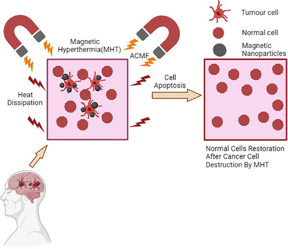
Figure 2.
Mechanism of magnetic nanocarrier for the treatment of glioma.
8. MNCs in brain tumor-targeted imaging and therapy
Magnetic nanoparticles (MNCs) have emerged as promising tools for brain tumor-targeted imaging and therapy. MNCs can be functionalized with specific ligands or antibodies that can target the nanoparticles to brain tumor cells, enabling precise localization and characterization of the tumor. MNCs can also be used to deliver therapeutic agents directly to the tumor site, increasing treatment efficacy while minimizing systemic toxicity [56].
Here are some of the ways that MNCs are being used for brain tumor-targeted imaging and therapy:
Magnetic resonance imaging (MRI): MNCs can be used as contrast agents for MRI. The unique magnetic properties of MNCs can generate strong signal changes in MRI, allowing for high-resolution imaging of the brain tumor. Targeted MNCs can be functionalized with specific ligands or antibodies that bind to receptors on the surface of brain tumor cells, enabling precise localization of the tumor [10].Magnetic hyperthermia: MNCs can be used to generate heat through the application of an external magnetic field. This approach, known as magnetic hyperthermia, can induce tumor cell death and increase the efficacy of chemotherapy or radiation therapy. Targeted MNCs can be used to selectively heat tumor cells while sparing healthy tissue [57].Drug delivery: MNCs can be used to deliver therapeutic agents directly to the brain tumor site. The MNCs can be functionalized with specific ligands or antibodies that target the tumor cells, allowing for precise delivery of the therapeutic agent to the tumor. This approach can increase the efficacy of the treatment while reducing systemic toxicity [10].Photodynamic therapy: MNCs can also be used in combination with photodynamic therapy (PDT) to treat brain tumors. In this approach, targeted MNCs are functionalized with photosensitizing agents that generate reactive oxygen species (ROS) upon exposure to light. This approach can selectively induce cell death in tumor cells while sparing healthy tissue [58].
8.1 MNCs for hyperthermia
The basic principle of magnetic hyperthermia involves the application of an alternating magnetic field (AMF) to MNCs that have been localized to the tumor site. The AMF causes the MNCs to oscillate, generating heat through hysteresis losses in the surrounding tissue. The amount of heat generated by the MNCs can be controlled by adjusting the frequency and intensity of the applied AMF [59].
Here are some key factors that need to be considered when using MNCs for hyperthermia:
Particle size and shape : The size and shape of MNCs can affect their heating efficiency. Smaller particles tend to heat up more efficiently, but may also be more prone to aggregation. The shape of the particles can also affect their heating efficiency, with elongated particles often exhibiting higher heating efficiencies compared to spherical particles [60].Magnetic properties : The magnetic properties of MNCs can also affect their heating efficiency. MNCs with high magnetic anisotropy exhibit larger hysteresis losses and higher heating efficiencies. However, the magnetic properties of MNCs can also affect their stability and biocompatibility [61].Targeting moieties : Targeting moieties can be used to direct MNCs to the tumor site, allowing for selective heating of tumor cells while sparing normal tissue. Various targeting moieties, such as antibodies, peptides, and aptamers, have been investigated for use with MNCs [62].AMF parameters : The frequency and intensity of the applied AMF can be adjusted to control the amount of heat generated by the MNCs. The optimal AMF parameters may vary depending on the size and magnetic properties of the MNCs, as well as the tumor microenvironment [63].
9. Recent advancements in MNCs based drug targeting
In a study published in the journal Nanoscale, researchers used magnetic nanoparticles loaded with doxorubicin (a chemotherapy drug) to treat glioma in mice. They found that the nanoparticles were able to cross the blood-brain barrier and accumulate in the tumor tissue, resulting in significant tumor growth inhibition [64].
Another study, published in the Journal of Controlled Release, investigated the use of magnetic nanoparticles to deliver small interfering RNA (siRNA) to glioma cells. The researchers found that the nanoparticles were able to effectively deliver siRNA, resulting in a significant knockdown of the targeted genes and inhibition of tumor growth [65].
In a study published in the journal Theranostics, researchers used magnetic nanoparticles in combination with alternating magnetic field hyperthermia to treat glioma in mice. They found that the combination therapy significantly inhibited tumor growth and prolonged the survival of the mice [66].
A recent review article published in the journal Frontiers in Pharmacology summarized the current state of research on magnetic nanoparticles for the treatment of glioma. The authors highlighted several promising approaches, including the use of magnetic nanoparticles for targeted drug delivery, hyperthermia, and multimodal therapy [67].
The creation of engineered MNCs that are able to be used as theranostic tools is progressing continuously. Although MNP platforms have made crucial progress, there are still technical knowledge gaps that prevent bench-to-bedside translation [68]. Before receiving regulatory clearance for human use, all MNP formulations should undergo thorough toxicological testing, taking into account the impact on various biological functions at the cellular & molecular level. Furthermore, there are restrictions with regard to magnetic targeting, in which the external magnetic field that is applied must be sufficiently powerful to capture the MNP-drug complexes at the location of choice [69]. Inducing and maintaining temperatures above the systemic temperature (37.5°C) in a specified target volume as well as achieving homogeneous heat distributions inside the intended organ are additional issues connected with hyperthermia-based treatment. Strong magnetic cores for MRI imaging, external magnets to assist with site-specific delivery of the MNCs to the targets, initiation of hyperthermia for temperature-triggered release of drugs, and a recognition layer for active targeting are all likely to be part of the MNCs of the future [70]. Finally, the therapeutic will likely be loaded to the coating material. Although the clinical uses of magnetic delivery of drugs are developing more slowly, the theranostic application, which will lead to personalized medicines, holds enormous potential. A ray of optimism exists for the clinic implementation of MNP-based theranostics thanks to the success of MRI and the development achieved so far in clinical trials [71].
In a different investigation, a formulation of zinc ferrite nanoparticles (ZFNP) with docetaxel (DTX) loaded on it was created and tested against C6 glioma cells. The findings suggest that ZFNP might be employed as a docetaxel nanodrug carrier for glioma cell delivery [72]. According to a recent review titled “Targeting to Brain Tumour: Nanocarrier-Based Drug Delivery Platforms, Opportunities, and Challenges,” developments in the field of molecular neuroscience have actually revolutionized the use of nanotechnology-based approaches for the treatment of brain tumors that have spread to other parts of the body. In the upcoming years, it is anticipated that drug delivery systems based on nanocarriers will advance to new levels and significantly alter cancer research [73].
10. Challenges
While magnetic nanoparticles hold promise for the treatment of glioma, there are several challenges that need to be addressed before they can be widely used in clinical practice. Here are some of the main challenges:
Blood-brain barrier (BBB) penetration: The BBB is a major obstacle to the delivery of therapeutics to the brain. While magnetic nanoparticles can be functionalized to target glioma cells, they may still have difficulty crossing the BBB to reach the tumor site. Strategies to overcome this challenge include the use of focused ultrasound to temporarily disrupt the BBB and improve nanoparticle delivery.Specificity : While magnetic nanoparticles can be targeted to glioma cells, it is important to ensure that they do not accumulate in healthy brain tissue. This requires the development of highly specific targeting strategies that can distinguish between glioma cells and normal brain cells.Toxicity : Magnetic nanoparticles can potentially cause toxicity if they accumulate in non-target tissues or if they induce an immune response. It is important to carefully evaluate the safety of magnetic nanoparticles in preclinical studies before they can be used in humans.Clinical translation : While there have been promising results in preclinical studies, it can be challenging to translate these findings into effective clinical therapies. The development of magnetic nanoparticle-based therapies for glioma will require extensive testing in clinical trials to evaluate their safety and efficacy.Cost : Magnetic nanoparticle-based therapies can be expensive to develop and manufacture, which may limit their accessibility for patients.One of the most difficult challenges is that the in
vivo effects of diverse nanocarriers are sometimes considerably different from their invitro activities. A significant gap in thein vitro in vivo correlation always exists for nanodrug carriers, making regulatory clearance difficult.
11. Conclusion
In conclusion, MNC-based drug targeting represents an emerging avenue for the treatment of glioma, providing a means to overcome the limitations associated with conventional glioma therapy. With continued advancements in nanotechnology, MNC-based strategies have the potential to revolutionize the field of glioma treatment, improving patient outcomes and quality of life. Moreover, the combination of MNCs with other therapeutic modalities, such as hyperthermia or photodynamic therapy, has shown synergistic effects in glioma treatment, further highlighting the potential of MNC-based approaches. However, in spite of MNCs holding tremendous promise, several challenges remain in their way of practical utilization, industrial scalability, or during technology transfer. Optimizing the design and synthesis of MNCs to achieve desirable properties, improving the understanding of MNC interactions with the BBB, checking industrial viability, and ensuring long-term in
Acknowledgments
The authors are very much grateful to Prof. Manoj Ranjan Nayak, President, Siksha ‘O’ Anusandhan (Deemed to be University) for providing the necessary facilities and encouragement.
Disclosure statement
The authors of the article have no conflict of interest to declare.
References
- 1.
Davis ME. Epidemiology and overview of gliomas. Seminars in Oncology Nursing. 2018; 34 (5):420-429. WB Saunders. DOI: 10.1016/j.soncn.2018.10.001 - 2.
Wiemels JL, Wiencke JK, Sison JD, Miike R, McMillan A, Wrensch M. History of allergies among adults with glioma and controls. International Journal of Cancer. 2002; 98 (4):609-615. DOI: 10.1002/ijc.10239 - 3.
Molinaro AM, Taylor JW, Wiencke JK, Wrensch MR. Genetic and molecular epidemiology of adult diffuse glioma. Nature Reviews Neurology. 2019; 15 (7):405-417. DOI: 10.1038/s41582-019-0220-2 - 4.
Gladson CL, Prayson RA, Liu WM. The pathobiology of glioma tumors. Annual Review of Pathology: Mechanisms of Disease. 2010; 5 :33-50. DOI: 10.1146/annurev-pathol-121808-102109 - 5.
Nuriya M, Shinotsuka T, Yasui M. Diffusion properties of molecules at the blood-brain interface: Potential contributions of astrocyte endfeet to diffusion barrier functions. Cerebral Cortex. 2013; 23 (9):2118-2126 - 6.
Fernandes C, Costa A, Osório L, Lago RC, Linhares P, Carvalho B, et al. Current Standards of Care in Glioblastoma Therapy. Brisbane, Australia: Exon Publications; 2017. pp. 197-241. DOI: 10.15586/codon.glioblastoma.2017.ch11 - 7.
Armstrong TS, Bishof AM, Brown PD, Klein M, Taphoorn MJ, Theodore-Oklota C. Determining priority signs and symptoms for use as clinical outcomes assessments in trials including patients with malignant gliomas: Panel 1 report. Neuro-Oncology. 2016; 18 (suppl_2):ii1-ii2. DOI: 10.1093/neuonc/nov267 - 8.
Bush NA, Chang SM, Berger MS. Current and future strategies for treatment of glioma. Neurosurgical Review. 2017; 40 :1-4. DOI: 10.1007/s10143-016-0709-8 - 9.
Carlsson SK, Brothers SP, Wahlestedt C. Emerging treatment strategies for glioblastoma multiforme. EMBO Molecular Medicine. 2014; 6 (11):1359-1370. DOI: 10.15252/emmm.201302627 - 10.
Sun C, Lee JS, Zhang M. Magnetic nanoparticles in MR imaging and drug delivery. Advanced Drug Delivery Reviews. 2008; 60 (11):1252-1265. DOI: 10.1016/j.addr.2008.03.018 - 11.
Kim DK, Dobson J. Nanomedicine for targeted drug delivery. Journal of Materials Chemistry. 2009; 19 (35):6294-6307. DOI: 10.1039/b902711b - 12.
Mohseni M, Connell JJ, Payne C, Patrick PS, Baker R, Yu Y, et al. Scalable magnet geometries enhance tumour targeting of magnetic nano-carriers. Materials & Design. 2020; 191 :108610. DOI: 10.1016/j.matdes.2020.108610 - 13.
Li X, Tsibouklis J, Weng T, Zhang B, Yin G, Feng G, et al. Nano carriers for drug transport across the blood-brain barrier. Journal of Drug Targeting. 2017; 25 (1):17-28. DOI: 10.1080/1061186X.2016.1184272 - 14.
De Vries NA, Beijnen JH, Boogerd W, Van Tellingen O. Blood-brain barrier and chemotherapeutic treatment of brain tumors. Expert Review of Neurotherapeutics. 2006; 6 (8):1199-1209. DOI: 10.1586/14737175.6.8.1199 - 15.
Kadry H, Noorani B, Cucullo L. A blood-brain barrier overview on structure, function, impairment, and biomarkers of integrity. Fluids and Barriers of the CNS. 2020; 17 (1):1-24. DOI: 10.1186/s12987-020-00230-3 - 16.
Engelhardt B, Sorokin L. The blood-brain and the blood–cerebrospinal fluid barriers: Function and dysfunction. Seminars in Immunopathology. 2009; 31 :497-511. Springer-Verlag. DOI: 10.1007/s00281-009-0177-0 - 17.
Pong S, Karmacharya R, Sofman M, Bishop JR, Lizano P. The role of brain microvascular endothelial cell and blood-brain barrier dysfunction in schizophrenia. Complex Psychiatry. 2020; 6 (1-2):30-46. DOI: 10.1159/000511552 - 18.
Furtado D, Björnmalm M, Ayton S, Bush AI, Kempe K, Caruso F. Overcoming the blood-brain barrier: The role of nanomaterials in treating neurological diseases. Advanced Materials. 2018; 30 (46):1801362. DOI: 10.1002/adma.201801362 - 19.
Thomsen MS, Routhe LJ, Moos T. The vascular basement membrane in the healthy and pathological brain. Journal of Cerebral Blood Flow & Metabolism. 2017; 37 (10):3300-3317. DOI: 10.1177/0271678X1772243 - 20.
Bhowmik A, Khan R, Ghosh MK. Blood brain barrier: A challenge for effectual therapy of brain tumors. BioMed Research International. 2015:1-20. DOI: 10.1155/2015/320941 - 21.
Abbott NJ. Astrocyte–endothelial interactions and blood–brain barrier permeability. Journal of Anatomy. 2002; 200 (5):523-534. DOI: 10.1046/j.1469-7580.2002.00047_13.x - 22.
Galea I. The blood–brain barrier in systemic infection and inflammation. Cellular & Molecular Immunology. 2021; 18 (11):2489-2501. DOI: 10.1038/s41423-021-00757-x - 23.
Greene C, Campbell M. Tight junction modulation of the blood brain barrier: CNS delivery of small molecules. Tissue Barriers. 2016; 4 (1):e1138017. DOI: 10.1080/21688370.2015.1138017 - 24.
Redzic Z. Molecular biology of the blood-brain and the blood-cerebrospinal fluid barriers: Similarities and differences. Fluids and Barriers of the CNS. 2011; 8 (1):1-25. Available from:http://www.fluidsbarrierscns.com/content/8/1/3 - 25.
Youdim MB, Edmondson D, Tipton KF. The therapeutic potential of monoamine oxidase inhibitors. Nature Reviews Neuroscience. 2006; 7 (4):295-309. DOI: 10.1038/nrn1883 - 26.
Motz CT, Kabat V, Saxena T, Bellamkonda RV, Zhu C. Neuromechanobiology: An expanding field driven by the force of greater focus. Advanced Healthcare Materials. 2021; 10 (19):2100102. DOI: 10.1002/adhm.202100102 - 27.
Baeten KM, Akassoglou K. Extracellular matrix and matrix receptors in blood–brain barrier formation and stroke. Developmental Neurobiology. 2011; 71 (11):1018-1039. DOI: 10.1002/dneu.20954 - 28.
Serlin Y, Shelef I, Knyazer B, Friedman A. Anatomy and physiology of the blood–brain barrier. Seminars in Cell & Developmental Biology. 2015; 38 :2-6. Academic Press. DOI: 10.1016/j.semcdb.2015.01.002 - 29.
Kumar R, Shin WS, Sunwoo K, Kim WY, Koo S, Bhuniya S, et al. Small conjugate-based theranostic agents: An encouraging approach for cancer therapy. Chemical Society Reviews. 2015; 44 (19):6670-6683. DOI: 10.1039/c5cs00224a - 30.
Cheng Z, Li M, Dey R, Chen Y. Nanomaterials for cancer therapy: Current progress and perspectives. Journal of Hematology & Oncology. 2021; 14 (1):1-27. DOI: 10.1186/s13045-021-01096-0 - 31.
Dilnawaz F, Sahoo SK. Therapeutic approaches of magnetic nanoparticles for the central nervous system. Drug Discovery Today. 2015; 20 (10):1256-1264. DOI: 10.1016/j.drudis.2015.06.008 - 32.
Laquintana V, Trapani A, Denora N, Wang F, Gallo JM, Trapani G. New strategies to deliver anticancer drugs to brain tumors. Expert Opinion on Drug Delivery. 2009; 6 (10):1017-1032. DOI: 10.1517/17425240903167942 - 33.
Sathornsumetee S, Reardon DA, Desjardins A, Quinn JA, Vredenburgh JJ, Rich JN. Molecularly targeted therapy for malignant glioma. Cancer. 2007; 110 (1):13-24. DOI: 10.1002/cncr.22741 - 34.
Spandana KA, Bhaskaran M, Karri VR, Natarajan J. A comprehensive review of nano drug delivery system in the treatment of CNS disorders. Journal of Drug Delivery Science and Technology. 2020; 57 :101628. DOI: 10.1016/j.jddst.2020.101628 - 35.
Vangijzegem T, Stanicki D, Laurent S. Magnetic iron oxide nanoparticles for drug delivery: Applications and characteristics. Expert Opinion on Drug Delivery. 2019; 16 (1):69-78. DOI: 10.1080/17425247.2019.1554647 - 36.
Al-Jamal WT, Kostarelos K. Liposomes: From a clinically established drug delivery system to a nanoparticle platform for theranostic nanomedicine. Accounts of Chemical Research. 2011; 44 (10):1094-1104. DOI: 10.1021/ar200105p - 37.
Ulbrich K, Hola K, Subr V, Bakandritsos A, Tucek J, Zboril R. Targeted drug delivery with polymers and magnetic nanoparticles: Covalent and noncovalent approaches, release control, and clinical studies. Chemical Reviews. 2016; 116 (9):5338-5431. DOI: 10.1021/acs.chemrev.5b00589 - 38.
Sun W, Mignani S, Shen M, Shi X. Dendrimer-based magnetic iron oxide nanoparticles: Their synthesis and biomedical applications. Drug Discovery Today. 2016; 21 (12):1873-1885. DOI: 10.1016/j.drudis.2016.06.028 - 39.
Thiruvengadathan R, Korampally V, Ghosh A, Chanda N, Gangopadhyay K, Gangopadhyay S. Nanomaterial processing using self-assembly-bottom-up chemical and biological approaches. Reports on Progress in Physics. 2013; 76 (6):066501. DOI: 10.1088/0034-4885/76/6/066501 - 40.
Ali A, Shah T, Ullah R, Zhou P, Guo M, Ovais M, et al. Review on recent progress in magnetic nanoparticles: Synthesis, characterization, and diverse applications. Frontiers in Chemistry. 2021; 9 :629054. DOI: 10.3389/fchem.2021.629054 - 41.
Semaltianos NG, Karczewski G. Laser synthesis of magnetic nanoparticles in liquids and application in the fabrication of polymer–nanoparticle composites. ACS Applied Nano Materials. 2021; 4 (7):6407-6440. DOI: 10.1021/acsanm.1c00715 - 42.
Ali A, Ovais M, Cui X, Rui Y, Chen C. Safety assessment of nanomaterials for antimicrobial applications. Chemical Research in Toxicology [Internet]. 2020; 33 (5):1082-1109. DOI: 10.1021/acs.chemrestox.9b00519 - 43.
Tartaj P, del Puerto MM, Veintemillas-Verdaguer S, Gonzalez-Carreno T, Serna CJ. The preparation of magnetic nanoparticles for applications in biomedicine. Journal of Physics D: Applied Physics. 2003; 36 (13):R182. DOI: 10.1088/0022-3727/36/13/202 - 44.
Paswan SK, Kumar P, Singh RK, Shukla SK, Kumar L. Spinel ferrite magnetic nanoparticles: An alternative for wastewater treatment. In: Pollutants and Water Management: Resources, Strategies and Scarcity. New Jersey, USA: Wiley; 2021. pp. 273-305. DOI: 10.1002/9781119693635.ch11 - 45.
Mokhosi SR, Mdlalose W, Nhlapo A, Singh M. Advances in the synthesis and application of magnetic ferrite nanoparticles for cancer therapy. Pharmaceutics. 2022; 14 (5):937. DOI: 10.3390/pharmaceutics14050937 - 46.
Mollarasouli F, Zor E, Ozcelikay G, Ozkan SA. Magnetic nanoparticles in developing electrochemical sensors for pharmaceutical and biomedical applications. Talanta. 2021; 226 :122108. DOI: 10.1016/j.talanta.2021.122108 - 47.
Kale SN, Deore SL. Emulsion micro emulsion and nano emulsion: A review. Systematic Reviews in Pharmacy. 2017; 8 (1):39. DOI: 10.5530/srp.2017.1.8 - 48.
Dudchenko N, Pawar S, Perelshtein I, Fixler D. Magnetite nanoparticles: Synthesis and applications in optics and nanophotonics. Materials. 2022; 15 (7):2601. DOI: 10.3390/ma15072601 - 49.
Zha J, Roggendorf H. Advance material. In: Brinker CJ, Scherer GW, editors. Sol–Gel Science, The Physics and Chemistry of Sol–Gel Processing. Vol. 3. No. 10, xiv Boston: Academic Press; 1990. p. 908. bound—ISBN 0-12-134970-5. DOI: 10.1002/adma.19910031025 - 50.
Nair GM, Sajini T, Mathew B. Advanced green approaches for metal and metal oxide nanoparticles synthesis and their environmental applications. Talanta Open. 2022; 5 :100080. DOI: 10.1016/j.talo.2021.100080 - 51.
Liu HL, Hua MY, Yang HW, Huang CY, Chu PC, Wu JS, et al. Magnetic resonance monitoring of focused ultrasound/magnetic nanoparticle targeting delivery of therapeutic agents to the brain. Proceedings of the National Academy of Sciences. 2010; 107 (34):15205-15210. DOI: 10.1073/pnas.1003388107 - 52.
Barbu E, Molnàr É, Tsibouklis J, Górecki DC. The potential for nanoparticle-based drug delivery to the brain: Overcoming the blood–brain barrier. Expert Opinion on Drug Delivery. 2009; 6 (6):553-565. DOI: 10.1517/17425240902939143 - 53.
Hartl N, Adams F, Merkel OM. From adsorption to covalent bonding: Apolipoprotein E functionalization of polymeric nanoparticles for drug delivery across the blood–brain barrier. Advanced Therapeutics. 2021; 4 (1):2000092. DOI: 10.1002/adtp.202000092 - 54.
Navya PN, Kaphle A, Srinivas SP, Bhargava SK, Rotello VM, Daima HK. Current trends and challenges in cancer management and therapy using designer nanomaterials. Nano Convergence. 2019; 6 :1-30. DOI: 10.1186/s40580-019-0193-2 - 55.
Zhang Y, Xi K, Fu X, Sun H, Wang H, Yu D, et al. Versatile metal-phenolic network nanoparticles for multitargeted combination therapy and magnetic resonance tracing in glioblastoma. Biomaterials. 2021; 278 :121163. DOI: 10.1016/j.biomaterials.2021.121163 - 56.
Parveen S, Misra R, Sahoo SK. Nanoparticles: A boon to drug delivery, therapeutics, diagnostics and imaging. Nanomedicine: Nanotechnology, Biology and Medicine. 2012; 8 (2):147-166. DOI: 10.1016/j.nano.2011.05.016 - 57.
Murase K, Takata H, Takeuchi Y, Saito S. Control of the temperature rise in magnetic hyperthermia with use of an external static magnetic field. Physica Medica. 2013; 29 (6):624-630. DOI: 10.1016/j.ejmp.2012.08.005 - 58.
Sarbadhikary P, George BP, Abrahamse H. Recent advances in photosensitizers as multifunctional theranostic agents for imaging-guided photodynamic therapy of cancer. Theranostics. 2021; 11 (18):9054. DOI: 10.7150/thno.62479 - 59.
Liu X, Zhang Y, Wang Y, Zhu W, Li G, Ma X, et al. Comprehensive understanding of magnetic hyperthermia for improving antitumor therapeutic efficacy. Theranostics. 2020; 10 (8):3793. DOI: 10.7150/thno.40805 - 60.
Obaidat IM, Issa B, Haik Y. Magnetic properties of magnetic nanoparticles for efficient hyperthermia. Nanomaterials. 2015; 5 (1):63-89. DOI: 10.3390/nano5010063 - 61.
Lanier OL, Korotych OI, Monsalve AG, Wable D, Savliwala S, Grooms NW, et al. Evaluation of magnetic nanoparticles for magnetic fluid hyperthermia. International Journal of Hyperthermia. 2019; 36 (1):686-700. DOI: 10.1080/02656736.2019.1628313 - 62.
Wadajkar AS, Menon JU, Kadapure T, Tran RT, Yang J, Nguyen KT. Design and application of magnetic-based theranostic nanoparticle systems. Recent Patents on Biomedical Engineering (Discontinued). 2013; 6 (1):47-57. DOI: 10.2174/1874764711306010007 - 63.
Gavilán H, Avugadda SK, Fernández-Cabada T, Soni N, Cassani M, Mai BT, et al. Magnetic nanoparticles and clusters for magnetic hyperthermia: Optimizing their heat performance and developing combinatorial therapies to tackle cancer. Chemical Society Reviews. 2021; 50 (20):11614-11667. DOI: 10.1039/d1cs00427a - 64.
Norouzi M, Yathindranath V, Thliveris JA, Kopec BM, Siahaan TJ, Miller DW. Doxorubicin-loaded iron oxide nanoparticles for glioblastoma therapy: A combinational approach for enhanced delivery of nanoparticles. Scientific Reports. 2020; 10 (1):11292. DOI: 10.1038/s41598-020-68017-y - 65.
Gandhi NS, Tekade RK, Chougule MB. Nanocarrier mediated delivery of siRNA/miRNA in combination with chemotherapeutic agents for cancer therapy: Current progress and advances. Journal of Controlled Release. 2014; 194 :238-256. DOI: 10.1016/j.jconrel.2014.09.001 - 66.
Rodrigues HF, Capistrano G, Bakuzis AF. In vivo magnetic nanoparticle hyperthermia: A review on preclinical studies, low-field nano-heaters, noninvasive thermometry and computer simulations for treatment planning. International Journal of Hyperthermia. 2020; 37 (3):76-99. DOI: 10.1080/02656736.2020.1800831 - 67.
Jain KK. A critical overview of targeted therapies for glioblastoma. Frontiers in Oncology. 2018; 8 :419. DOI: 10.3389/fonc.2018.00419 - 68.
Cole AJ, Yang VC, David AE. Cancer theranostics: The rise of targeted magnetic nanoparticles. Trends in Biotechnology. 2011; 29 (7):323-332. DOI: 10.1016/j.tibtech.2011.03.001 - 69.
Gupta R, Xie H. Nanoparticles in daily life: Applications, toxicity and regulations. Journal of Environmental Pathology, Toxicology and Oncology. 2018; 37 (3):209-230. DOI: 10.1615/JEnvironPatholToxicolOncol.2018026009 - 70.
Kumar CS, Mohammad F. Magnetic nanomaterials for hyperthermia-based therapy and controlled drug delivery. Advanced Drug Delivery Reviews. 2011; 63 (9):789-808. DOI: 10.1016/j.addr.2011.03.008 - 71.
Siafaka PI, Okur NÜ, Karantas ID, Okur ME, Gündoğdu EA. Current update on nanoplatforms as therapeutic and diagnostic tools: A review for the materials used as nanotheranostics and imaging modalities. Asian Journal of Pharmaceutical Sciences. 2021; 16 (1):24-46. DOI: 10.1016/j.ajps.2020.03.003 - 72.
Panda J, Satapathy BS, Sarkar R, Tudu B. A zinc ferrite nanodrug carrier for delivery of docetaxel: Synthesis, characterization, and in vitro tests on C6 glioma cells. Journal of Microencapsulation. 2022; 39 (2):136-144. DOI: 10.1080/02652048.2022.2053757 - 73.
Kumar LA, Pattnaik G, Satapathy BS, Swapna S, Mohanty D. Targeting to brain tumor: Nanocarrier-based drug delivery platforms, opportunities, and challenges. Journal of Pharmacy & Bioallied Sciences. 2021; 13 (2):172. DOI: 10.4103/jpbs.JPBS_239_20