 Open access peer-reviewed chapter
Abstract
Arsenic has become a major toxicological concern due to its rising concentrations in aquatic bodies. It is added to the water either by natural sources including weathering of rocks, sediments, volcanic eruptions and aquifers, or by anthropogenic sources including herbicides, wood preservatives, metal smelting, drugs, pesticides, burning of coal, agriculture runoff and petroleum refining processes among others. The untreated and uncontrolled discharge of arsenic by industries into the natural water bodies poses serious threat to aquatic fauna by deteriorating water quality and making it unsuitable for fishes. Fish is an important bioindicator of aquatic bodies and excessive arsenic concentration causes its bioaccumulation in fish organs and muscles. This deposited arsenic in the fish imposes serious damage to physiology, biochemical disorders such as poisoning of gills, livers, decrease fertility, tissue damage, lesions, and cell death. It also enters in the cell and produces reactive oxygen species which increases the level of stress which further concentrates the oxidative enzymes and cortisol levels in fish. The uncontrolled discharge of arsenic and its devastating impact on fish diversity is a major concern for aquaculture progress and economic stability. This, along with its other implications is the scope of this chapter.
Keywords
- arsenic toxicity
- fish
- arsenic sources
- aquatic arsenic
- arsenic impact
1. Introduction
Heavy metals are gaining attention as significant pollutants due to their toxicity problems in ecosystem at different levels [1]. Some of these metals are commonly known as pollutants that include arsenic, cadmium, copper, nickel and lead which pose threats of serious nature to aquatic environment and to the living organisms in the aquatic ecosystem [2]. Arsenic is a metalloid element that is abundant in the aquatic environment as a result of both natural and anthropogenic processes. It is a significant and ubiquitous environmental contaminant that causes health issues to all living organisms [3]. Arsenic in herbicides, fungicides, pesticides and rodenticides is the significant source of environmental contamination [4]. Arsenic mainly enters into the environment through two channels: (a) natural activities, and (b) man-made activities. Natural activities such as volcanic eruption, forest fires and weathering of rocks add a significant amount of arsenic in aquatic environment. While, man-made activities, such as different industries including paint, pharmaceutical, pesticide, detergent and electronic industries are the main source of arsenic discharge in water bodies [5]. Besides all these sources, the smelting and mining operations along with the domestic and agriculture run-off continuously add arsenic in natural waters [6]. In water, arsenic trivalent, arsenite is oxidized in water in the presence of dissolved oxygen and converted into arsenate that remain intact in sediments for long period of time and pose serious threats to aquatic fauna. Arsenic toxicity has been reported in many countries including China, Pakistan, Bangladesh, India and other South Asian countries along with many parts of the United States [7]. According to IARC, three chemical forms of arsenic are present: organic arsenic, inorganic arsenic and arsenic gas [8]. Fish is most sensitive bioindicator of pollution and cannot be safe from harmful impacts of these pollutants [9]. These have potential to induce biochemical and physiological changes which ultimately effect overall behavior, growth pattern and ultimately leads to death [10, 11]. Arsenic can enter in to fish body through oral cavity with contaminated food and absorption through skin and gills. Arsenic hasa tendency to accumulate in fish tissues and organs and cause serious damages to gills, gastrointestinal tract, kidneys, heart, brain and other organ. Such damages alter fish behavior, homoeostasis, hematology and biochemical mechanisms [12].
2. Sources of arsenic
In natural environment, arsenic is a common crystalline metalloid having characteristics of both metal and non-metal. It is the 14th and 20th abundant element in saltwater and earth crust respectively [13] Arsenic contamination occurs due to both natural (such as volcanic eruptions, rock weathering) and anthropogenic activities (such as the production of alloys, pesticides, glass, and medicinal items,
2.1 Natural sources of arsenic
The presence of arsenic in natural water is influenced by the aquifer’s local geology, hydrogeology and geochemical properties. Climate change and human activities also play a part and influence its presence. Natural sources of arsenic in water have been attributed to a variety of natural geochemical processes including oxidation of arsenic-bearing sulphides, desorption of arsenic from (hydro)oxides (e.g., iron, aluminum, and manganese oxides), reductive dissolution of arsenic-bearing iron (hydro)oxides, release of arsenic from geothermal water, and as well as leeching from sulphides.
2.1.1 Anthropogenic sources
Nonferrous metal mining and smelting, fossil fuel processing, combustion, wood preservation, pesticide production and its application in agricultural fields, municipal and industrial waste disposal and incineration are the main anthropogenic activities that may release arsenic into the environment [15, 16]. The majority of anthropogenic arsenic is released into the soil, primarily through pesticides or solid wastes. A significant amount, however, is also released into the air and water [17]. Arsenic, in its soluble forms, enters into the ground water and water bodies through runoff and leeching [18].
Mining tailing contain a significant amount of arsenic in the form of arsenopyrite, arsenian pyrite, arsenates and in association with iron oxyhydroxides. Arsenic can be produced by roasting arsenopyrite which is the most abundant ore mineral of arsenic, and by smelter dust of some metals such as gold, copper and lead [19]. Arsenic is found in approximately 11 million tonnes of copper and lead resources worldwide. In 2007, the total global production of arsenic trioxide was 59 thousand tonnes [20]. Arsenic is a highly toxic mineral found in the earth’s crust that can enter the food chain through soil, water and plants. The main anthropogenic sources of arsenic in Canada are smelter and base-metal refinery facilities as well as thermal and power-generation stations. It was estimated that the Canadian base-metal smelters and refineries released approximately 15 tonnes of arsenic per year in liquid effluent, 310 tonnes in the atmosphere and 770 tonnes in solid waste [21]. The majority of this emissions (almost 90%) came from coal-fired power or thermal power-generation stations, as well as smelter and metallurgical facilities. Arsenic concentrations have been found to be elevated in the vicinity of these sources.
Another significant anthropogenic source of arsenic is the widespread use of arsenical wood preservatives. Chromated copper arsenate (CAA) is one of the most common wood preservatives used worldwide on large scale containing 34 percent arsenic content [22]. It has been estimated that a considerable amount of CAA (7800 to 78,000 mg/kg) remained in the treated wood [23]. Leaching of preservative components from in-service treated wood has thus been a source of arsenic in the environment. Furthermore, widespread arsenic contamination around wood preservation occurs as a result of raw material handling, spills, sludge deposition and dripping from freshly impregnated wood or rain water leaching from impregnated wood piles, particularly under low pH conditions, at these sites [24]. Exposure to sunlight and weathering both increase the rate of leaching from the treated wood. As a result, elevated arsenic levels have been found in soils surrounding treated woods [25, 26]. Arsenic is a widely used component in pesticides and most commonly available as lead arsenate, calcium aresenate, magnesium arsenate, zinc arsenate, arsenite and Paris green. These are commonly used in apple orchard of Canada [6].
2.1.2 Arsenic species in air
Methylated form of arsenic is mainly common in atmosphere. Arsenate is predominated form of arsenic and is most likely present near smelters, volcanic eruptions and coal burning sites [3]. Peat and landfills are major sources of volatile arsine in air. Some volatile species of arsenic are emitted into the air due to microbial activities in soil and water bodies which further oxidize and reactive with atmospheric sulfur oxide and ozone [6, 27]. This atmospheric arsenic reaches soil and water bodies by snow, dry deposition and through rain fall. Some of the atmospheric arsenic particles also combine with dust and may then be inhaled or ingested by living organisms [28]. In rural areas, the arsenic concentration in rain and snowfall are comparatively low (0.00003 mg/L), while its concentration around coal burning industries is relatively high (0.0005 mg/L) (Figure 1) [29].
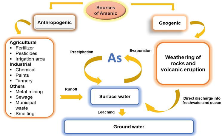
Figure 1.
Sources and routes of arsenic to approach water bodies. Arsenic has two main sources to enter water bodies: Anthropogenic and geogenic. Addition of arsenic in surface water bodies is by direct discharge. Gaseous arsenic from different sources also backs its way in to the water bodies through precipitation. Arsenic in soil and water enters the ground water through leeching.
3. Impacts of arsenic on fish
3.1 Prevalence of arsenic in the environment
Arsenic-exposed organisms may absorb arsenic by ingestion, inhalation and penetration through skin or mucous membranes which allows it to enter the cells mainly by active transport [30]. Inhaled arsenic induces severe biochemical and physiological changes such as poisoning, impaired growth and reproduction, immune system abnormalities, cell and tissue damage, oxidative stress and apoptosis in aquatic organisms.
3.2 Factors affecting toxicity of arsenic
3.2.1 Arsenic speciation
The toxic effect of Arsenic in the aquatic ecosystem depends on its form such as inorganic or organic and its level of oxidation [5]. Arsenite (As-III), arsine (As-III), Arsenate (AsV) and arsenic are the four oxidation states of arsenic [6]. Inorganic arsenic (iAs); As-III and As-V are prevalent forms in aquatic habitats. As-III is limited in extremely reduced conditions with a low redox potential while As-V is stable in oxygen-rich environments [31]. Inorganic metallic compounds are typically more hazardous than organometallic compounds. Likewise, inorganic Arsenic is more soluble in water than organic Arsenic it, thus, can accumulate in tissues more quickly [32]. Arsenic is converted into less- or non-toxic metabolites like arsenocholine (AsC) and arsenobetaine (AsB) for excretion when it is ingested by living organisms [33].
3.2.2 Biotic and abiotic factors
Many biotic and abiotic factors like exposure time, arsenic speciation, water temperature, pH, organic content, phosphate concentration, suspended particles, and presence of other chemicals and toxicants significantly alter the toxic and other effects of arsenic on aquatic life [34]. Median survival time of fishes usually reduces as the temperature and arsenic concentration increases. It is helpful to identify different arsenic species when investigating for arsenic exposure because they vary in their origin and toxicity [35].
By the biomethylation process, harmful inorganic arsenic is transformed into less toxic pentavalent (Met-A) forms such as monomethylarsonate (MMAv), dimethylarsenate (DMAv) and trimethylarsine (DMAIII). Nevertheless, monomethylarsenite (MMAIII), dimethylarsenite (DMAIII) and trimethylarsonic oxide (TMAOv) are more hazardous than inorganic arsenic and are produced through biomethylation. In general, AsB (arsenobetaine), arsenocholic (AsC), and DMAA make up around 85 to 90% of the arsenic found in edible parts of marine fish, whereas iAs (inorganic arsenic) species make up about 10%. Little is known about the types of arsenic found in freshwater fishes, but what is available indicates that AsB and DMAA are the dominant arsenic species in freshwater fishes [36].
3.2.3 Bioaccumulation
The process by which some toxic chemicals (heavy metals including arsenic and other toxicants) present in the environment accumulate in living organisms is known as bioaccumulation. Their absorption is considered passive, with diffusion gradients formed by metal adsorption or binding to tissue and cell surfaces [37]. Arsenic accumulation in tissue will depend on the rates at which various organs take in and eliminate arsenic [38]. Every organism metal concentration is determined by a variety of mechanisms, including its intake, excretion, storage, and transformation. Bioaccumulation differs among metal species and in fish species because of variations in their permeability, metabolic rates and the amount and types of metal binding ligands present at the organismic surface. Although accurate quantification of all these activities may not be necessary, understanding their proportional role in the overall pattern of metal turnover is sometimes the only way to evaluate tissue residue data.
The inhabitants of aquatic environments, such as fish, are unable to escape the negative impacts of arsenic [39]. The amount of toxic metal found in various fish organs is used as an indicator for the ecosystem metal contamination. This is thought as an important approach for highlighting the significance of higher metal levels in aquatic organisms [40]. Arsenic has a multidimensional impact on fish as they bioaccumulate in various tissues and can harm their immunological, respiratory, digestive, excretory, reproductive, neurological, and endocrine systems.
Mostly, arsenic accumulates in all of the fish critical organs. The most common site for the highest concentrations of arsenic in fish are the liver, kidney, and gills [41] as well as rarely the gut [42]. To determine the impact of arsenic contaminated water, several studies have been carried out. The gills and liver of tilapia have shown to accumulate significantly more arsenic thus the gill was noted to be most significantly impacted organ [43]. Great Slave Lake, Northwest Territories, Canada inhabitant fish arsenic concentration were determined in the gill, liver, muscle and skin of fishes
4. Effect of arsenic on behavioral changes
Fish exposed to chemicals exhibit quantifiable behavioral changes that provide unique information that cannot be discovered using traditional toxicological techniques [45]. The relationship between behavior and an organism physiology, ecology, and environment offers a special point of view [46]. Even a tiny quantity of some toxicants can make fish behave abnormally due to impaired sensory sensitivity. Numerous abnormal behaviors, including erratic movement, fast opercula movement, jumping out of the test medium, lateral swimming and loss of balance were observed by exposure to sodium arsenate [47]. Within a few minutes of exposure, especially at higher concentrations, 2.250 mgL−1 of sodium arsenate, the treated fish began to exhibit their first obvious responses. However, depending on the concentrations in the exposure medium, fish exposed to low concentrations, i.e., below 0.08 mgL−1 of sodium arsenate, had no or little behavioral alterations. Neurotoxic effects and sensory system irritability were the root causes of the aberrant behaviors. The fish avoidance responses to arsenic are indicated by their jumping and back-and-forth movements. The excessive mucus secretion was likely caused by sodium arsenate directly irritating the skin. The dysfunction of the nervous system may be the cause of lateral swimming and loss of balance [48].
5. Effect of arsenic on fish major organs and organ system
5.1 Effects on organs
Arsenic is hazardous to fish organs, including the skin, liver, kidney, lung, gastrointestinal tract and muscles [49]. Among them liver and kidneys, are essential organs in vertebrates that carry out detoxification processes, protein synthesis, homeostasis and excretion of nitrogenous waste respectively. Acute and sub-acute effects of arsenic may involve many organ systems particularly respiratory, cardiovascular, gastrointestinal, neurological and hematological systems. At 9.64 mgL−1 waterborne arsenic and 43.1–60 μgg−1 dietary arsenic concentration exposure anemia, liver degeneration and gallbladder inflammation were noted [50].
5.1.1 Skin
Fish skin serves as its outermost protective barrier. Because of the presence of club and mucous cells in the tissue, the skin and gill tissue of fish are keratinized and have a mucous covering [51]. It is susceptible to several water-dissolved toxins because of constant contact. Skin of
5.1.2 Gills
Gas exchange, ion control, and excretion of metabolic waste products are the three primary functions performed by gills. Consequently, by serving as a first barrier, gills can significantly contribute to the body defense against hazardous chemicals by reducing the number of poisonous compounds that are taken up by other organs [54]. Gills serve as the initial sites where waterborne contaminants are concentrated because of their constant exposure to the outside environment. One of the first signs of toxicant poisoning is respiratory discomfort. Fish are particularly susceptible to the toxicity of arsenic due to the high rate at which it is absorbed through the gills [55]. The presence of arsenic in gill tissue indicates that the gills were in direct touch with arsenic-contaminated water. The majority of research on arsenic toxicity in freshwater fish has focused on the effects of arsenic intake through the gills and nutritional absorption by fish that feed on benthic organisms [56]. Fish, upon arsenic exposure, show difficult breathing because coagulated mucus blocks the gills, and fish suffer direct damage from arsenic ions to their blood vessels which led to vascular collapse in the gills and anoxia [57]. After being exposed to sodium arsenite,
5.1.3 Liver
By synthesizing proteins, detoxifying metabolites and aiding in digestion, the liver plays a significant role in metabolic regulation. Fish liver plays a crucial role in the absorption, bioaccumulation, biotransformation and elimination of arsenic [32]. Liver is the main target organ of arsenic poisoning. Arsenic is efficiently metabolized in fish tissue, particularly in the liver and gut and it tends to accumulate in fish such as the green sun fish and the
5.1.4 Kidney
Kidney along gills is primary pathways for waste excretion in fish body. When fish are exposed to arsenic contaminants, histopathological alterations occur. Upon arsenic exposure, kidney enzyme, glutathione decreases [40]. Upon non-lethal doses (3.8 mgL−1 and 7.6 mgL−1 arsenic) exposure
5.1.5 Gastrointestinal tract
The gastrointestinal system is the main pathway for dietary arsenic intake and absorption. Arsenic is delivered to the body organs via the circulatory system after being absorbed by the digestive tract [44]. Dietary exposure to arsenic has been demonstrated to damage the mucosal lining of the lake whitefish gastrointestinal system, causing mucosal sloughing and increased mucosal production [32]. Gastrointestinal disorders might result from acute arsenicexposure. Although the gastrointestinal effects are most noticeable immediately after ingesting arsenic, they can also develop with chronic exposure through other means. The primary gastrointestinal lesion appears to be increased small blood vessel permeability, which results in fluid loss and hypotension [45]. Exposure to a high concentration of arsenic, 20 mgL−1, displayed disorganized, and consequent fusion of mucosa, lamina propria and edema, damaged serosa and degeneration [46]. Different Fish species exposed to various arsenic species included lake whitefish, walleye, northern pike,
5.1.6 Brain
Brain is extremely sensitive to arsenic because of its high rate of polyunsaturated fatty acids, oxygen consumption and extremely high rate of oxygen free radical formation without correspondingly large levels of arsenic [49]. It is thought that the arsenic poisoning altered the important biochemical components of the
5.1.7 Muscles
The muscles, which make up to 80% of the fish itself, are what give the fish its swimming propulsion. The fish may move in any direction due to the muscle numerous orientations of arrangement (myomeres). Fish muscle, which makes up the majority of its bulk, is the part that people often eat [52]. The least amount of arsenic accumulated in the muscles across all experimental groups compared to other soft tissues. Muscle tissue does not directly come into touch with toxicants, so it is active detoxification site. As a result, arsenic is not transferred from other tissues to muscles. The least amount of arsenic has been found in the muscle of the
5.1.8 Gonads
Fish reproduction was thought to be a reliable predictor of endocrine disruption caused by chemical substances, especially arsenic, in aquatic environments [55]. An earlier monitoring study in the Mekong Delta of Vietnam found a link between arsenic accumulation and gonad development in the catfish,
6. Biochemical and physiological changes
In aquatic medium, toxicants often exhibit their effects at the cellular or molecular level, which causes significant alterations in biochemical markers. Heavy metal pollution also has an impact on the body primary building blocks, such as lipids, proteins, and carbohydrates, which are crucial for building the body and generating energy [58]. Among these blood glucose level utilized as an indicator of environmental stress and showed how carbohydrate metabolism changed in the presence of hypoxia and stress. When Indian catfish,
6.1 Carbohydrate
When fish are under stress, carbohydrates are main and immediate energy sources whereas, protein is spared. Changes in the levels of glucose, lactic acid and glycogen are among the effects of arsenic stress on fish carbohydrate metabolism. Among this blood glucose level was utilized as an indicator of environmental stress and showed how carbohydrate metabolism changed in the presence of hypoxia and stress [58]. Three important Indian carps,
6.1.1 Protein
Due to anoxic or hypoxic conditions, which increase carbohydrate consumption, heavy metal stress that affects glucose levels indicates a change in energy requirements and expenditure. As glycogen stores run short, tissue proteins use the process of deamination of amino acids to supply keto acids. Thus, a study of serum protein composition is needed to understand how energy requirements and expenditure change under metal stress. In order to determine fish overall nutritional status, estimation of total protein, albumin, and globulin in serum is of great diagnostic significance [61]. The ratio of albumin to globulin is a helpful measure for monitoring changes in the relative proportion of serum protein. Furthermore, hepatic tissue necrosis may cause a reduction in protein synthesis [62]. In
6.1.2 Lipid
Lipid bilayers make up biomembranes, which also have different kinds of protein embedded in or attached to them. All biomembranes mostly consist of phospholipids. For the biomembrane to function properly, the lipid component composition must be maintained. Biomolecules including stored lipids, proteins, and carbs assist fish in dealing with stress. When a fish is under acute stress or toxicity, stored glycogen heals it; however, when a fish is exposed to arsenic continuously, the degree of the stress increases and lipids and proteins begin to play a role. Either the oxidation process or gradual saturation can mobilize lipids to supply the energy demand [64]. The integrity of the cell membrane is maintained by phospholipids and cholesterol. High-density lipoproteins (HDL), which are good cholesterol, and low-density lipoproteins (LDL), which are bad cholesterol, both exist in the body and aid in the removal of harmful cholesterol from the blood. The HDL cholesterol level should be as high as possible. As compared to LDL cholesterol, very-low-density lipoprotein (VLDL) is similar in that it is mostly made up of lipids with little protein. HDL is the primary serum lipoprotein in rainbow trout, followed by LDL and VLDL [65]. The primary form of reserve lipids, triacylglycerols, are mobilized prior to phospholipids during starvation. It is well-recognized that arsenic alter lipid levels. Hence, lipid profile analysis also functions as a biomarker for fish health. During arsenic intoxication serum total lipid levels in
7. Effect on oxidative status and other enzymes
Enzymes are biological macromolecules that regulate an organism metabolism. Much work has been conducted on how arsenic exposure affects the enzyme activity of certain fish [68]. Arsenic is absorbed through the gills, it has the potential to disturb the antioxidant system and impact the body reactions to oxidants by boosting glutamate cysteine ligase activity and glutathione levels Glucose-6-phosphate dehydrogenase (G6PDH) was significantly increased in fish gills upon exposure of arsenic, which modified antioxidant responses to an arsenic pro-oxidant challenge. As the produced nicotinamide adenine dinucleotide phosphate (NADPH) is an important element for the H2O2-scavenging pathway of cells [69] and for glutathione metabolism, there is indication that glucose-6-phosphate dehydrogenase, a major enzyme of the pentose phosphate pathway, has an important function in antioxidant systems [70]. Effect of sodium arsenate on
8. Hematological changes
The hematopoietic system is influenced by short- and long-term arsenic exposure. Hematological profiles of fish are commonly used to detect the environmental contamination in aquatic ecosystem [76]. Different fish blood parameters are used to determine the effects of sub-lethal arsenic. Hematological and biochemical examinations of blood parameters in fish exposed to pollutants are crucial for determining the animal structural and functional status [77]. Red and white blood cell formation may be reduced as a result of arsenic exposure [78]. Leukocyte numbers were reduced as a result of chronic arsenic exposure which had an impact on the structure of the head kidney [50]. Arsenic induces changes to hematological markers and oxidative stress in the fish liver [79]. Numerous studies [80, 81, 82] have noted an anemic state of the fish during acute and sub-lethal treatment, which led to a low level of hemoglobin (Hb) in the arsenate-treated fish. Another potential explanation is that the toxicity of arsenic may inhibit erythropoiesis due to its effect on membranes. It was also that the fish exposed to toxicants had a lower amount of red blood cells [83]. In
9. Immunotoxic effects
Arsenic as an immunotoxic substance has an impact on a variety of immunological responses, including altering co-receptor expression, lowering delayed hypersensitivity reactions, reducing mitogen-activated T-cell proliferation, production of freer intracellular Ca2+and releasing lymphokines [87]. The primary immune-competent fish organs are the head kidney, spleen and thymus [88]. The head kidney macrophages (HKM) are essential for the activation of fish innate immunity, and arsenic-induced macrophage mortality is going to impair the immunological system of the exposed fish. Arsenic accumulates in fish liver and kidney and it can impair the fish immune system by decreasing the synthesis of antibodies and cytokines [89]. Sub-lethal fish become immunocompromised and vulnerable to infections as a result of arsenic exposure, which alters the functional arms of their innate and acquired immune systems [90]. In fish, adaptive immunity develops later, therefore the impact of ecotoxins on innate immunity may be more substantial [91]. In zebra fish, the system expressed crucial antiviral genes and generated enough tumor necrosis factor (TNF-α) to fall within the range of arsenic [92]. Arsenic has a significant impact on the immune system in fish, with the two immunologically significant organs, the head kidney and spleen, responding to its toxic effects in various ways. Arsenic caused a drop in both T- and B-lymphocytes cell responses in the head kidney and spleen, although its effects seem to be more prominent on the B-cells. The phagocytic capability of macrophages was similarly impacted by fish exposure to different arsenic concentrations, which helped in the spread and duration of bacterial and viral infections [42].
10. Cytogenotoxic effects
Arsenic is recognized as a possible sulfhydryl-reactive substance that may bind to and aggregate many cell surface proteins [93]. Arsenic increases nitric oxide generation at the level of transcriptional activation along with inducing ribosylation, polyadenosine diphosphate, DNA strand breaks, depletion of nicotinamide adenine dinucleotide and the development of micronuclei, like other oxygen radical-producing stressors [94]. Cell death may be caused by the accumulation of cellular proteins, the generation of reactive oxygen species, or the stimulation of protein tyrosine kinases by arsenic [36]. Furthermore, denaturing of biological enzymes and changing gene regulation are toxic consequences of inorganic arsenic. For the purpose of researching the cytotoxicity of various arsenic compounds, fish cell lines may be used as sensitive substitutes for entire fish. In JF cells, arsenite may cause apoptosis by the induction of oxidative stress, however in TO-2 fish cell lines, it disrupts the cell cycle without the induction. Arsenic may impair cell division by disrupting the spindle apparatus [95]. In addition, it causes sister chromatid exchange, the formation of micronuclei, DNA-protein crosslinking, and different types of mutations [96]. In reaction to arsenic, a duration- and dose-dependent increase in the formation of micronuclei in the gill cells of
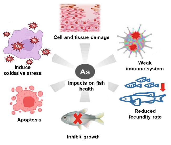
Figure 2.
Impact of arsenic on different organs of fish. Induction of oxidative stress, inhibition of fish growth, weakness of the immune system, reduced fecundity rate, damaging of the cell and tissues and enhancement of the cell death through apoptosis are the effects of arsenic toxicity in fish.
References
- 1.
Jaishankar M, Tseten T, Anbalagan N, Mathew BB, Beeregowda KN. Toxicity, mechanism and health effects of some heavy metals. Interdisciplinary Toxicology. 2014; 7 (2):60 - 2.
Lambert TW, Lane S. Lead, arsenic, and polycyclic aromatic hydrocarbons in soil and house dust in the communities surrounding the Sydney, Nova Scotia, tar ponds. Environmental Health Perspectives. 2004; 112 (1):35-41 - 3.
Rossman TG. Mechanism of arsenic carcinogenesis: An integrated approach. Mutation Research/Fundamental and Molecular Mechanisms of Mutagenesis. 2003; 533 (1-2):37-65 - 4.
Nickson R, McArthur J, Burgess W, Ahmed KM, Ravenscroft P, Rahmanñ M. Arsenic poisoning of Bangladesh groundwater. Nature. 1998; 395 (6700):338 - 5.
Canivet V, Chambon P, Gibert J. Toxicity and bioaccumulation of arsenic and chromium in epigean and hypogean freshwater macroinvertebrate. Archives of Environmental Contamination and Toxicology. 2001; 40 :345-354 - 6.
Brammer H, Ravenscroft P. Arsenic in groundwater: A threat to sustainable agriculture in South and South-East Asia. Environment International. 2009; 35 (3):647-654 - 7.
Yoshida T, Yamauchi H, Sun GF. Chronic health effects in people exposed to arsenic via the drinking water: Dose–response relationships in review. Toxicology and Applied Pharmacology. 2004; 198 (3):243-252 - 8.
IARC Working Group on the Evaluation of Carcinogenic Risks to Humans. Arsenic, Metals, Fibres and Dusts. Lyon (FR): International Agency for Research on Cancer. (IARC Monographs on the Evaluation of Carcinogenic Risks to Humans, No. 100C.) ARSENIC AND ARSENIC COMPOUNDS. 2012. Available from: https://www.ncbi.nlm.nih.gov/books/NBK304380/ - 9.
Suganya A, Murugan K, Kovendan K, Mahesh Kumar P, Hwang JS. Green synthesis of silver nanoparticles using Murraya koenigii leaf extract against Anopheles stephensi and Aedes aegypt. Parasitology Research. 2013; 112 :1385-1397 - 10.
Beyers DW, Rice JA, Clements WH, Henry CJ. Estimating physiological cost of chemical exposure: Integrating energetics and stress to quantify toxic effects in fish. Canadian Journal of Fisheries and Aquatic Sciences. 1999; 56 (5):814-822 - 11.
Guba M, von Breitenbuch P, Steinbauer M, Koehl G, Flegel S, Hornung M, et al. Rapamycin inhibits primary and metastatic tumor growth by antiangiogenesis: Involvement of vascular endothelial growth factor. Nature Medicine. 2002; 8 (2):128-135 - 12.
Lavanya D, Rani DK. Analysis of feature selection with classification: Breast cancer datasets. Indian Journal of Computer Science and Engineering (IJCSE). 2011; 2 (5):756-763 - 13.
Popovic A, Djordjevic D, Polic P. Trace and major element pollution originating from coal ash suspension and transport processes. Environment International. 2001; 26 (4):251-255 - 14.
Bhattacharya P, Mukherjee AB, Jacks G, Nordqvist S. Metal contamination at a wood preservation site: Characterisation and experimental studies on remediation. Science of the Total Environment. 2002; 290 (1-3):165-180 - 15.
Nasser NA, Patterson RT, Roe HM, Galloway JM, Falck H, Sanei H. Use of Arcellinida (testate lobose amoebae) arsenic tolerance limits as a novel tool for biomonitoring arsenic contamination in lakes. Ecological Indicators. 2020; 113 :106177 - 16.
Slimak M, Delos C. Environmental pathways of exposure to 129 priority pollutants. Journal of Toxicology: Clinical Toxicology. 1983; 21 (1-2):39-63 - 17.
Duker AA, Carranza E, Hale M. Arsenic geochemistry and health. Environment International. 2005; 31 (5):631-641 - 18.
Pongratz R. Arsenic speciation in environmental samples of contaminated soil. Science of the Total Environment. 1998; 224 :133-141 - 19.
Smedley PL, Kinniburgh DG. A review of the source, behaviour and distribution of arsenic in natural waters. Applied Geochemistry. 2002; 17 (5):517-568 - 20.
Fields S. Caution–children at play: How dangerous is CCA? Environmental Health Perspectives. 2001; 109 (6):A262-A269 - 21.
Lund U, Fobian A. Pollution of two soils by arsenic, chromium and copper, Denmark. Geoderma. 1991; 49 (1-2):83-103 - 22.
Stephens RW, Brudermann GE, Morris PI, Hollick MS, Chalmers JD. Value assessment of the Canadian pressure treated wood industry Report to Canadian Forest Service; 1994 - 23.
Zagury GJ, Samson R, Deschênes L. Occurrence of metals in soil and ground water near chromated copper arsenate–treated utility poles. Journal of Environmental Quality. 2003; 32 (2):507-514 - 24.
Frank R, Braun HE, Ishida K, Suda P. Persistent organic and inorganic pesticide residues in orchard soils and vineyards of Southern Ontario. Canadian Journal of Soil Science. 1976; 56 (4):463-484 - 25.
Cullen WR, Reimer KJ. Arsenic speciation in the environment. Chemical Reviews. 1989; 89 :713-764 - 26.
Challenger F. Biological methylation. Chemical Reviews. 1945; 36 (3):315-361 - 27.
Rasmussen PE, Subramanian KS, Jessiman BJ. A multi-element profile of house dust in relation to exterior dust and soils in the city of Ottawa, Canada. Science of the Total Environment. 2001; 267 (1-3):125-140 - 28.
Andreae MO. Arsenic in rain and the atmospheric mass balance of arsenic. Journal of Geophysical Research Oceans. 1980; 85 (C8):4512-4518 - 29.
Genchi G, Lauria G, Catalano A, Carocci A, Sinicropi MS. Arsenic: A review on a great health issue worldwide. Applied Sciences. 2022; 12 (12):6184. DOI: 10.3390/app12126184 - 30.
Masuda H. Arsenic cycling in the Earth’s crust and hydrosphere: Interaction between naturally occurring arsenic and human activities. Progress in Earth and Planetary Science. 2018; 5 (1):1-1. DOI: 10.1186/s40645-018-0224-3 - 31.
Mandal BK, Suzuki KT. Arsenic round the world: A review. Talanta. 2002; 58 (1):201-235 - 32.
Camacho J, de Conti A, Pogribny IP, Sprando RL, Hunt PR. Assessment of the effects of organic vs. inorganic arsenic and mercury in Caenorhabditis elegans. Current Research in Toxicology. 2022; 1 (3):100071 - 33.
Byeon E, Kang HM, Yoon C, Lee JS. Toxicity mechanisms of arsenic compounds in aquatic organisms. Aquatic Toxicology. 2021; 237 :105901 - 34.
Ardini F, Dan G, Grotti M. Arsenic speciation analysis of environmental samples. Journal of Analytical Atomic Spectrometry. 2020; 35 (2):215-237 - 35.
Chen SJ, Yan XJ, Chen Z. Arsenic in Nature. In: Kretsinger RH, Uversky VN, Permyakov EA, editors. Encyclopedia of Metalloproteins. New York, NY: Springer; 2013.doi: 10.1007/978-1-4614-1533-6_489 - 36.
Neff JM. Ecotoxicology of arsenic in the marine environment. Environmental Toxicology and Chemistry: An International Journal. 1997; 16 (5):917-927 - 37.
Kumaresan M, Riyazuddin P. Overview of speciation chemistry of arsenic. Current Science. 2001; 10 :837-846 - 38.
Barral-Fraga L, Barral MT, MacNeill KL, Martiñá-Prieto D, Morin S, Rodríguez-Castro MC, et al. Biotic and abiotic factors influencing arsenic biogeochemistry and toxicity in fluvial ecosystems: A review. International Journal of Environmental Research and Public Health. 2020; 17 (7):2331. DOI: 10.3390/ijerph17072331 - 39.
Orloff K, Mistry K, Metcalf S. Biomonitoring for environmental exposures to arsenic. Journal of Toxicology and Environmental Health, Part B. 2009; 12 (7):509-524 - 40.
Šlejkovec Z, Bajc Z, Doganoc DZ. Arsenic speciation patterns in freshwater fish. Talanta. 2004; 62 (5):931-936 - 41.
Bryan GW, Darracott A. Bioaccumulation of marine pollutants. Philosophical Transactions of the Royal Society of London. B, Biological Sciences. 1979; 286 (1015):483-505 - 42.
Amiard-Triquet C, Amiard JC. Influence of ecological factors on accumulation of metal mixtures. In: Langston WJ, Bebianno MJ, editors. Metal Metabolism in Aquatic Environments. Boston, MA: Springer; 1998. doi: 10.1007/978-1-4757-2761-6_11 - 43.
Mendil D, Uluözlü ÖD, Hasdemir E, Tüzen M, Sari H, Suicmez M. Determination of trace metal levels in seven fish species in lakes in Tokat, Turkey. Food Chemistry. 2005; 90 (1-2):175-179 - 44.
Squadrone S, Prearo M, Brizio P, Gavinelli S, Pellegrino M, Scanzio T, et al. Heavy metals distribution in muscle, liver, kidney and gill of European catfish (Silurus glanis) from Italian Rivers. Chemosphere. 2013; 90 (2):358-365 - 45.
Farag AM, Stansbury MA, Bergman HL, Hogstrand C, MacConnell E. The physiological impairment of free-ranging brown trout exposed to metals in the Clark Fork River, Montana. Canadian Journal of Fisheries and Aquatic Sciences. 1995; 52 (9):2038-2050 - 46.
Wepener V, Van Vuren JH, Du Preez HH. Uptake and distribution of a copper, iron and zinc mixture in gill, liver and plasma of a freshwater teleost, Tilapia sparrmanii. Water SA. 2001; 27 (1):99-108 - 47.
Chetelat J, Cott PA, Rosabal M, Houben A, McClelland C, Belle Rose E, et al. Arsenic bioaccumulation in subarctic fishes of a mine-impacted bay on Great Slave Lake, Northwest Territories, Canada. PLoS One. 2019; 14 (8):e0221361 - 48.
Saglio P, Trijasse S. Behavioral responses to atrazine and diuron in goldfish. Archives of Environmental Contamination and Toxicology. 1998; 35 :484-491 - 49.
Kumari B, Kumar V, Sinha AK, Ahsan J, Ghosh AK, Wang H, DeBoeck G. Toxicology of arsenic in fish and aquatic systems. Environmental chemistry letters. 2017; 15 :43-64 - 50.
Baldissarelli LA, Capiotti KM, Bogo MR, Ghisleni G, Bonan CD. Arsenic alters behavioral parameters and brain ectonucleotidases activities in zebrafish (Danio rerio). Comparative Biochemistry and Physiology Part C: Toxicology & Pharmacology. 2012; 155 (4):566-572 - 51.
Tolins M, Ruchirawat M, Landrigan P. The developmental neurotoxicity of arsenic: Cognitive and behavioral consequences of early life exposure. Annals of Global Health. 2014; 80 (4):303-314 - 52.
Khan MI, Ahmad MF, Ahmad I, Ashfaq F, Wahab S, Alsayegh AA, et al. Arsenic exposure through dietary intake and associated health hazards in the Middle East. Nutrients. 2022; 14 (10):2136. DOI: 10.3390/nu14102136 - 53.
Singh AK, Banerjee TK. Toxic effects of sodium arsenate (Na2HAsO4x7H2O) on the skin epidermis of air-breathing catfish Clarias batrachus (L.). Veterinarski Arhiv. 2008;78 (1):73-88 - 54.
Cockell KA, Hilton JW, Bettger WJ. Hepatobiliary and hematological effects of dietary disodium arsenate heptahydrate in juvenile rainbow trout ( Oncorhynchus mykiss ). Comparative Biochemistry and Physiology Part C: Comparative Pharmacology. 1992;103 (3):453-458 - 55.
Rakers S, Gebert M, Uppalapati S, Meyer W, Maderson P, Sell AF, et al. ‘Fish matters’: The relevance of fish skin biology to investigative dermatology. Experimental Dermatology. 2010; 19 (4):313-324 - 56.
Kumar R, Banerjee TK. Analysis of arsenic bioaccumulation in different organs of the nutritionally important catfish, Clarias batrachus (L.) exposed to the trivalent arsenic salt, sodium arsenite. Bulletin of Environmental Contamination and Toxicology. 2012:445-449 - 57.
Kumar R, Banerjee TK. Analysis of arsenic bioaccumulation in different organs of the nutritionally important catfish, Clarias batrachus (L.) exposed to the trivalent arsenic salt, sodium arsenite. Bulletin of environmental contamination and toxicology. 2012; 89 :445-9 - 58.
Mdegela R, Myburgh J, Correia D, Braathen M, Ejobi F, Botha C, et al. Evaluation of the gill filament-based EROD assay in African sharptooth catfish ( Clarias gariepinus ) as a monitoring tool for waterborne PAH-type contaminants. Ecotoxicology. 2006;15 :51-59 - 59.
Lima AA, Furtado C, Valduriez P, Mattoso M. Parallel OLAP query processing in database clusters with data replication. Distributed and Parallel Databases. 2009; 25 :97-123 - 60.
Pedlar RM, Ptashynski MD, Wautier KG, Evans RE, Baron CL, Klaverkamp JF. The accumulation, distribution, and toxicological effects of dietary arsenic exposure in lake whitefish (Coregonus clupeaformis) and lake trout (Salvelinus namaycush). Comparative Biochemistry and Physiology Part C Toxicology & Pharmacology. 2002; 131 (1):73-91 - 61.
Mondal K, Samanta S. A review on arsenic contamination in fresh water fishes of West Bengal. Journal of Global Biosciences. 2015; 4 (5):2369-2374 - 62.
Ahmed MK, Habibullah-Al-Mamun M, Parvin E, Akter MS, Khan MS. Arsenic induced toxicity and histopathological changes in gill and liver tissue of freshwater fish, tilapia ( Oreochromis mossambicus ). Experimental and Toxicologic Pathology. 2013;65 (6):903-909 - 63.
Caumette G, Koch I, Estrada E, Reimer KJ. Arsenic speciation in plankton organisms from contaminated lakes: Transformations at the base of the freshwater food chain. Environmental Science & Technology. 2011; 45 (23):9917-9923 - 64.
Hossain K, Akhand AA, Kato M, Du J, Takeda K, Wu J, et al. Arsenite induces apoptosis of murine T lymphocytes through membrane raft-linked signaling for activation of c-Jun amino-terminal kinase. The Journal of Immunology. 2000; 165 (8):4290-4297 - 65.
Das S, Unni B, Bhattacharjee M, Wann SB, Rao PG. Toxicological effects of arsenic exposure in a freshwater teleost fish, Channa Punctatus . African Journal of Biotechnology. 2012;11 (19):4447-4454 - 66.
Carlson P, Smalley DM, Van Beneden RJ. Proteomic analysis of arsenic-exposed zebrafish (Danio rerio) identifies altered expression in proteins involved in fibrosis and lipid uptake in a gender-specific manner. Toxicological Sciences. 2013; 134 (1):83-91 - 67.
Das T, Goswami M. Histopathological and ultrastructural changes in the gill and liver of fresh water fish Channa punctatus exposed to sodium arsenite. Bioscience Biotechnology Research Communications. 2018;11 (3):434-441 - 68.
Allen T, Singhal R, Rana SV. Resistance to oxidative stress in a freshwater fish Channa punctatus after exposure to inorganic arsenic. Biological Trace Element Research. 2004;98 :63-72 - 69.
Roy S, Bhattacharya S. Arsenic-induced histopathology and synthesis of stress proteins in liver and kidney of Channa punctatus . Ecotoxicology and Environmental Safety. 2006;65 (2):218-229 - 70.
Ghosh D, Datta S, Bhattacharya S, Mazumder S. Long-term exposure to arsenic affects head kidney and impairs humoral immune responses of Clarias batrachus . Aquatic Toxicology. 2007;81 (1):79-89 - 71.
Datta S, Ghosh D, Saha DR, Bhattacharaya S, Mazumder S. Chronic exposure to low concentration of arsenic is immunotoxic to fish: Role of head kidney macrophages as biomarkers of arsenic toxicity to Clarias batrachus . Aquatic Toxicology. 2009;92 (2):86-94 - 72.
Gelberg H. Pathophysiological mechanisms of gastrointestinal toxicity. Comprehensive Toxicology. 2018:139-178. DOI: 10.1016/ B978-0-12-801238-3.10923-7 - 73.
Haque M, Roy SK. Acute effects of arsenic on the regulation of metabolic activities in liver of fresh water fishes (Taki) during cold acclimation. Jordan Journal of Biological Sciences. 2012; 5 (2):91-97 - 74.
Begum A, Mustafa AI, Amin MN, Banu N, Chowdhury TR. Accumulation and histopathological effects of arsenic in tissues of shingi fish (stinging catfish) Heteropneustes fossilis (Bloch, 1794). Journal of the Asiatic Society of Bangladesh, Science. 2013; 39 (2):221-230 - 75.
de Rosemond S, Xie Q , Liber K. Arsenic concentration and speciation in five freshwater fish species from Back Bay near Yellowknife, NT, Canada. Environmental Monitoring and Assessment. 2008; 147 :199-210 - 76.
Chauncey B, Schmid EC, Goldstein L. Arsenical and mercurial inhibition of tyrosine transport by the flounder intestine. Journal of Toxicology and Environmental Health, Part A Current Issues. 1988; 23 (2):257-265 - 77.
Ng SC, Furman R, Axelsen PH, Shchepinov MS. Free radical chain reactions and polyunsaturated fatty acids in brain lipids. ACS Omega. 2022; 7 (29):25337-25345. DOI: 10.1021/acsomega.2c02285 - 78.
Palaniappan PR, Vijayasundaram V. The effect of arsenic exposure and the efficacy of DMSA on the proteins and lipids of the gill tissues of Labeo rohita . Food and Chemical Toxicology. 2009;47 (8):1752-1759 - 79.
Palaniappan PL, Vijayasundaram V. The bioaccumulation of arsenic and the efficacy of Meso-2, 3-dimercaptosuccinic acid in the selected organ tissues of Labeo rohita fingerlings using inductively coupled plasma-optical emission spectrometry. WASJ. 2009; 6 :1247-1254 - 80.
Maher W, Goessler W, Kirby J, Raber G. Arsenic concentrations and speciation in the tissues and blood of sea mullet (Mugil cephalus) from Lake Macquarie NSW, Australia. Marine Chemistry. 1999; 68 (1-2):169-182 - 81.
D’Amico AR, Gibson AW, Bain LJ. Embryonic arsenic exposure reduces the number of muscle fibers in killifish (Fundulus heteroclitus). Aquatic Toxicology. 2014; 146 :196-204 - 82.
Dubińska-Magiera M, Daczewska M, Lewicka A, Migocka-Patrzałek M, Niedbalska-Tarnowska J, Jagla K. Zebrafish: A model for the study of toxicants affecting muscle development and function. International Journal of Molecular Sciences. 2016; 17 (11):1941. DOI: 10.3390/ijms1711194 - 83.
Arcand-Hoy LD, Benson WH. Fish reproduction: An ecologically relevant indicator of endocrine disruption. Environmental Toxicology and Chemistry An International Journal. 1998; 17 (1):49-57 - 84.
Celino FT, Yamaguchi S, Miura C, Miura T. Arsenic inhibits in vitro spermatogenesis and induces germ cell apoptosis in Japanese eel (Anguilla japonica). Reproduction. 2009; 138 (2):279 - 85.
Javed M, Usmani N. Stress response of biomolecules (carbohydrate, protein and lipid profiles) in fish Channa punctatus inhabiting river polluted by thermal power plant effluent. Saudi Journal of Biological Sciences. 2015;22 (2):237-242 - 86.
Datta S, Saha DR, Ghosh D, Majumdar T, Bhattacharya S, Mazumder S. Sub-lethal concentration of arsenic interferes with the proliferation of hepatocytes and induces in vivo apoptosis in Clarias batrachus L. Comparative Biochemistry and Physiology Part C, Toxicology & Pharmacology. 2007; 145 (3):339-349 - 87.
Garg S, Gupta RK, Jain KL. Sublethal effects of heavy metals on biochemical composition and their recovery in Indian major carps. Journal of Hazardous Materials. 2009; 163 (2-3):1369-1384 - 88.
Abdel-Tawwab M, Abdel-Rahman AM, Ismael NE. Evaluation of commercial live bakers’ yeast, Saccharomyces cerevisiae as a growth and immunity promoter for fry Nile tilapia, Oreochromis niloticus (L.) challenged in situ with Aeromonas hydrophila. Aquaculture. 2008;280 (1-4):185-189 - 89.
Jacobs MN, Covaci A, Schepens P. Investigation of selected persistent organic pollutants in farmed Atlantic salmon (Salmo salar), salmon aquaculture feed, and fish oil components of the feed. Environmental Science & Technology. 2002; 36 (13):2797-2805 - 90.
Ghaffar A, Hussain R, Aslam M, Abbas G, Khan A. Arsenic and urea in combination alters the hematology, biochemistry and protoplasm in exposed rahu fish ( Labeo rohita )(Hamilton, 1822). Turkish Journal of Fisheries and Aquatic Sciences. 2016;16 (2):289-296. DOI: 10.4194/1303-2712-v16_2_09 - 91.
Abhay DS. Comparative study of cholesterol alterations in a freshwater teleost fish, Amblypharyngodon mola exposure to heavy metals. The Bioscan Journal of Life Sciences. 2013; 8 (3):1001-1003 - 92.
Perrier H, Perrier C, Peres G, Gras J. The lipoproteins of the plasma of the rainbow trout (Salmo gairdnerii Richardson): Immunoelectrophoresis, selective precipitation and lipid composition. Comparative Biochemistry and Physiology Part B, Comparative Biochemistry. 1979; 62 (3):245-248 - 93.
Abdel-Tawwab M, Mousa MA, Abbass FE. Growth performance and physiological response of African catfish, Clarias gariepinus (B.) fed organic selenium prior to the exposure to environmental copper toxicity. Aquaculture. 2007;272 (1-4):335-345 - 94.
Vinodhini R, Narayanan M. The impact of toxic heavy metals on the hematological parameters in common carp ( Cyprinus carpio L.). Journal of Environmental Health Science & Engineering. 2009;6 (1):23-28 - 95.
Morales AE, Pérez-Jiménez A, Hidalgo MC, Abellán E, Cardenete G. Oxidative stress and antioxidant defenses after prolonged starvation in Dentex dentex liver. Comparative Biochemistry and Physiology Part C, Toxicology & Pharmacology. 2004; 139 (1-3):153-161 - 96.
Bagnyukova TV, Luzhna LI, Pogribny IP, Lushchak VI. Oxidative stress and antioxidant defenses in goldfish liver in response to short-term exposure to arsenite. Environmental and Molecular Mutagenesis. 2007; 48 (8):658-665 - 97.
Humtsoe N, Davoodi R, Kulkarni BG, Chavan B. Effect of arsenic on the enzymes of the Rohu carp, Labeo rohita (Hamilton 1822). The Raffles Bulletin of Zoology. 2007;14 :17-19 - 98.
Patlolla AK, Tchounwou PB. Serum acetyl cholinesterase as a biomarker of arsenic induced neurotoxicity in Sprague-Dawley rats. International Journal of Environmental Research and Public Health. 2005; 2 (1):80-83 - 99.
Gül Ş, Belge-Kurutaş E, Yıldız E, Şahan A, Doran F. Pollution correlated modifications of liver antioxidant systems and histopathology of fish (Cyprinidae) living in Seyhan Dam Lake, Turkey. Environment International. 2004; 30 (5):605-609 - 100.
Pal S, Chatterjee AK. Protective effect of methionine supplementation on arsenic-induced alteration of glucose homeostasis. Food and Chemical Toxicology. 2004; 42 (5):737-742