Open Access is an initiative that aims to make scientific research freely available to all. To date our community has made over 100 million downloads. It’s based on principles of collaboration, unobstructed discovery, and, most importantly, scientific progression. As PhD students, we found it difficult to access the research we needed, so we decided to create a new Open Access publisher that levels the playing field for scientists across the world. How? By making research easy to access, and puts the academic needs of the researchers before the business interests of publishers.
We are a community of more than 103,000 authors and editors from 3,291 institutions spanning 160 countries, including Nobel Prize winners and some of the world’s most-cited researchers. Publishing on IntechOpen allows authors to earn citations and find new collaborators, meaning more people see your work not only from your own field of study, but from other related fields too.
To purchase hard copies of this book, please contact the representative in India:
CBS Publishers & Distributors Pvt. Ltd.
www.cbspd.com
|
customercare@cbspd.com
Many diseases and signs of organophosphate poisoning share common symptoms with other illnesses. Misusing pesticides can lead to severe damage to both the environment and people’s health. Insects, birds and other animals are affected by the pesticide due to its effect on cholinesterase (ChE). This enzyme breaks down acetylcholine into smaller parts that accumulate in neuromuscular junctions and nerve terminals. In cholinergic toxicity, signs appear such as nicotine-like effects and muscarinic side effects in the nervous system. In accordance with this study, an apparatus for measuring ChE-activity in birds’ blood and tissues is presented. Literature regarding proper ChE-activity in both wild and hybrid birds was found by consulting the Mosul city library. Additionally, the electrometric method proved accurate and effective. This makes it a good method for exposing potentially exposed birds in the natural world while also creating a biological control to reduce environmental pollution by carbamates or organophosphates.
Environmental Science Department, Environmental Science and Technologies Mosul University, United States
*Address all correspondence to: ashraf.saddik.@uomosul.edu.iq
1. Introduction
Pesticides are released into the environment through a complex chain of events and can be transported through air, water, land or even within organisms affecting birds’ environment. The extent of movement and spread can be local or far-reaching and varies according to the types of pesticides and their impact on the issue of food security and the provision of food for the ever-increasing world population while ignoring its effects on the birds that mainly protect crops from agricultural pests such as insects, caused their death and a significant decrease in their numbers with insect resistance to agricultural pesticides. Different types of pesticides belonging to other groups have been produced. This poses substantial risks to human health, birds and the environment [1].
Exposure of wild birds to pesticides leads to reduced egg production, which leads to a significantly decreased in the birds [2, 3]. Organophosphorus and pyrethroid compounds are used in agriculture and veterinary medicine [4, 5]. To control pest-borne diseases [6, 7, 8]. Excessive use of pyrethroids and organophosphates can cause human and animal contamination [9, 10, 11, 12, 13]. Organophosphate poisoning occurs due to exposure or abuse [14, 15] and intoxication occurs due to inhibition of ChE activity, leading to accumulation of acetylcholine in nerve endings and the appearance of muscarinic, nicotine and CNS symptoms-related intoxication, eventually leading to death [16, 17] but the main target of pyrethroids is the sodium channel Causes delayed blockage of the channel, resulting in a continuous flow of sodium, and its effect on voltage-sensitive chloride channels. Resulting in decreased chlorine flow through depleted airway channels [18, 19].
Measuring ChE activity in tissue and blood helps to detect organophosphate and pyrethroid toxicity in the early stages of intoxication. A 25–30% decrease in ChE activity in plasma or erythrocytes of birds is evidence of exposure such compounds [5, 9]. There appear to be several methods for measuring the activity of ChE, namely Ellman’s [20, 21], radiometric [22] and electrometric [23].
The last method is simple, cheap and does not require complicated equipment (just a pH meter and a water bath). Several changes have been made to the method, such as: e.g., electrometry (Michael method), by using different buffer solutions (setup and structure), and the interaction of sample volume with different time and temperatures [24, 25]. Michael’s original method was only used in humans, so some modifications were made due to various natural variations in ChE activity in the blood and tissues of different animals [24]. The main changes were to increase the incubation temperature from 20 to 37°C to increase the blood volume of the sample at the time of interaction and to minimize the incubation time depending on the animal type to which this method was applied to sheep [6, 24, 26]. Electroassay for Measuring ChE Activity in Dairy Cows [27]. The method as electrometric was not utilized previously on Budgie for measuring the activity of (ChE). Also, there is no information on the normal values of the ChE activity in birds.
Use the electrometric method to measure the activity of (ChE) in different birds. It is important for detecting contamination with organophosphorus and carbamate pesticides that cause the death of birds by ingesting pesticides through contaminated water or food, such as fruits or insects, causing them to die. It also affects their young, causing them to die. Adequate information on normal levels of cholesterol activity in birds.
The present work uses blood and tissue samples from different birds, such as brain, liver, and muscle, ranging in sample size from 30 to 35 g. Blood samples were collected from the jugular vein (guillotine decapitation) and placed into tubes containing EDTA. Plasma was separated from blood by centrifugation at 3000 rpm for 15 minutes (Model 80-1, China). Place the brain, liver and thorax in a clean, dry plastic bag with crushed ice until homogenized at 3 mL per 100 mg of weight in barbiturate-phosphate saline (pH = 8.1) with a hand mixer. This is a sample preparation method for the purpose of screening for cholinesterase (ChE)-activity in samples that It was taken from natural birds.
3. Measure as stipulated in the method as electrometric
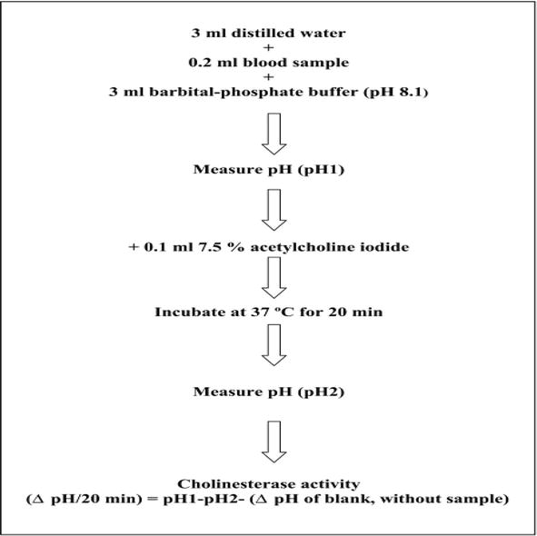
Blood samples and homogenized tissue were used to measure normal ChE activity. Measurements were done on the same day using electrometry Figure 1 [27, 28], using a pH meter type (pH 025 M, US) to measure the pH 1 of the samples, and then using a water bath type (HH-2 Digital Lab Thermostatic Water Bath, China) for use. After incubation at 37°C for 30 min, samples were taken directly from the incubator and pH 2 was measured. Measure the change in acid function using the following equation:
Figure 1.
Steps for the determination of blood cholinesterase activity by a modified electrometric method.
pH2−pH1−ΔpHof blank=ΔpH/30min.ChEactivityE1
ChE activity in budgie plasma and homogenate tissues was accurately measured electrometrically based on the mean and standard deviation [29], and the following coefficient of variation:
Coefficient=Standard deviationMean×100E2
The true and false percentages of ChE activity in the brain and plasma tissues of Budgie birds were calculated. Two sections of the plasma and brain tissue samples were separated. The first section of the samples was used to test the (ChE) activity (as previously described plus 40 μl of DW), and the second portion of the samples contained 40 μl of quinidine sulfate (0.1%) for each sample. Samples were incubated for 10 minutes at 37°C to inhibit pseudo-(ChE) [21, 30]. Through certain inhibitors, quinidine sulfate can prevent pseudo-(ChE) in brain and plasma tissues [20, 31]. Based on pseudo-(ChE) activity, the total residual (ChE) activity in brain and plasma tissues was identified as follows:
ChEactivity=totalChE−activity of realChEWithnoquinidinefollowing quinidine additionE3
Dichlorvos 50% (produced in China by Shenzhen Horizon Industry Co. Ltd.) and deltamethrin 2.5% were used to assess the inhibition of activity (ChE) in the brain tissues of Budgie birds in vitro (Indogulf company, India).
After obtaining the brains of seven Budgie birds for each experiment and incubating the samples for 10 minutes at 37°C to inhibit activity, inhibitor (ChE) incubation was carried out with concentrations of 0 (control), 0.5, and 1 μM/L for the treated group of Dichlorvos and concentrations of 0, 5 and 10 μM/L for the group of Deltamethrin [32]. Before inhibition, the percentage was calculated as follows:
Inhibition%ChEactivity=Activity ofChEfor control samplewithnodichlorovos or deltamethrin−Activity ofChEwith Dichlorovos or deltamethrinActivity ofChEfor control sample(with nodichlorovos or deltamethrin)×100E4
Table 1 shows the normal reference range values, 95% confidence interval and related statistics for plasma, brain, liver and pectoralis muscle ChE activities of the bird species studied. Using the technique of in vitro ChE inhibition for 10 min by 0.1% quinidine sulfate, the estimated percentages of true ChE activity in the plasma of the Quail, Large pin-tailed sand grouse, Starling, Rock dove (Columba livia), Local dove (Streptopelia Senegalensis) and Budgie birds were 77%, 69%, 71% and 73%, 75%, 75%, respectively (Table 2).
The cholinesterase activity values listed in this table were found out by an electrometric method in the bird species Quail, sand grouse, Starling, Rock dove (Columba livia), Local dove (Streptopelia Senegalensis) and Budgie birds. N = 10.
Cholinesterase values are the mean ± SE of 10 plasma samples for each of bird species.
Estimation of true cholinesterase activity as determined by the described electrometric method in the plasma of Quail, Sand grouse, Starling, Rock dove (Columba livia), Local dove (Streptopelia Senegalensis) and Budgie birds.
Quinidine sulfate was used to inhibit pseudo cholinesterase activity in the plasma.
Cholinesterase values are the mean ± SE of 10 plasma samples for each of bird species.
We conclude from the foregoing that measuring cholinesterase activity in the blood (plasma) and tissues (brain, liver, and muscles) of wild birds and domestic birds that live with humans is very important through which we discover the values of cholinesterase activity in wild birds, which reveal to us the extent of environmental pollution with these pollutants. (Pesticides) and their effect on wildlife, especially endemic and migratory birds, which cause the sudden death of large numbers of birds when these birds migrate through countries that use pesticides randomly and unstudied. Protecting our environment from pollution with organic phosphorus pesticides and carbamate pesticides that cause the death of birds directly or indirectly by poisoning and killing the organisms that birds depend on for their food, which causes the death of birds and the loss of this great environmental wealth. Because it maintains the ecological balance and is also a natural agricultural pesticide.
For their assistance and completion of this work, the author is grateful to the Environmental Science Department of the College of Environmental Science and Technologies at Mosul University in Iraq.
References
1.Karen DJ, Joab BM, Wallin JM, Johnson KA. Partitioning of chlorpyrifos between water and an aquatic macrophyte (Elodea densa). Chemosphere. 1998;37(8):1579-1586. DOI: 10.1016/S0045-6535(98)00141-6
2.Weir SM, Youssif MR, Anderson T, Salice CJ. Current progress in developing standardized methods for reptilian toxicity testing to inform ecological risk assessment. Bird and Reptile Species in Environmental Risk Assessment Strategies. 2023:130-150
3.Singh PP, Kumar A, Chauhan RS, Pankaj PK. How safe is the use of chlorpyrifos: Revelations through its effect on layer birds. Veterinary World. 2016;9(7):753-758. DOI: 10.14202/vetworld.2016
4.Bradberry SM, Cage SA, Proudfoot AT, Veale JA. Poisioning due to pyrethroid. Toxicological Review. 2005;24(2):93-106. DOI: 10.2165/00139709-200524020-00003
5.Thompson CM, Prins JM, George KM. Mass spectrometric analyses of organophosphate insecticide oxon proteinadducts. Environmental Health Perspectives. 2009;118(1):11-19. DOI: 10.1289/ehp.0900824
6.Alias A, Megdad MH, Ali MH, Esmaial YA. Study of (ChE)activity in adult ewes treated by the anthelmintic drug levamisole. Al-Rafidain Science Magazine. 2010;21(3):93-102. Available from: https://scholar.google.com/citations?
7.Kazal EH, Alias AS. Survey of some therapeutic formulation for anti-ectoparasites used in clinic veterinary in Tilkaif and clinic veterinary teaching Hospital in Nineveh province.Al-Rafidain. Science Magazine. 2014;25(1):57-66. Available from: https://www.iasj.net/iasj?func=article&aId=86108
8.Tomlin CD. The Pesticide Manual: A World Compendium. 14th ed. Alton, Hampshire, UK: British Crop Protection Council; 2006. pp. 186-187. Available from: https://www.scirp.org/(S(lz5mqp453edsnp55rrgjct55))/reference/ReferencesPapers.aspx?ReferenceID=1880037
9.Kumar S, Kaushik G, Villarreal-Chiu JF. Scenario of organophosphate pollution and toxicity in India: A review. Environmental Science and Pollution Research. 2016;23:9480-9491. DOI: 10.1007/s11356-016-6294-0
10.Vermeire T, Mcphail R and Waters M. Organophosphorous pesticides in the environment. In World Health Organization. IV. Meeting Report of the International Workshop on Approaches to Integrated Risk Assessment 2001; pp. 1-18. Available from: https://www.who.int/ipcs/publications /en/ch_3d.pdf
11.Lotti M. (ChE) inhibition : Complexities in interpretation. Clinical Chemistry. 1995;41(12 Pt 2):1814-1818. Available from: https://www.ncbi.nlm.nih.gov/pubmed/7497638
12.Ahmed A, Kai I, Wang JR, Olson MR, Bonner OH, Gaafar AR, et al. The impact of repeated organophosphorus pesticide exposure on biomarkers and neurobehavioral outcomes among adolescent pesticide applicators. Journal of Toxicology and Environmental Health, Part A. 2017;80(10-12):542-555. DOI: 10.1080/15287394. 2017. 1362612
13.Haug G, Hoffiman H. Chemistry of plant protection. In: Synthetic Pyrethroid Insecticides: Structures and Properties. New York: Springer-Verlag; 1990
14.Alias AS. The use of largactil as an anti-poisoning with organopgosphorus pesticides. Veterinary Practitioner. 2021;22(2):137-139
15.Ahmed AS. Change in acetylcholine activity and some blood parameters in adult sheep dipped in deltamethrin. Iraqi Journal of Veterinary Sciences. 2020;35(2):301-304
16.Moretto A. Experimental and clinical toxicology of anti (ChE). Toxicology Letters. 1988;28(102–103):509-513. Available fom: https://www.ncbi.nlm.nih.gov/pubmed/10022304
17.Tilson HA. New horizons: Future directions in neurotoxicology. Environ. Hlth Perspect. 2000;108:439-441. DOI: 10.1289/ehp.00108s3439
18.Sderlund DM, Clark JM, Sheets LP, Mullin LS, Piccirillo VJ, Sargent D, et al. Mechanisms of pyrthroid neurotoxicity: Implications for cumulative risk assessment. Toxicology. 2002;171(1):3-59. DOI: 10.1016/s0300-483x(01) 00569-8
19.Dong K. Insect sodium chaaels and insecticides resistance. Invertebrate Neuroscience. 2007;7(1):17-30. DOI: 10.1007/s10158-006-0036-9
20.Ellman GL, Courtney KD, Andres V, Featherstone RM. A new and rapid colorimetric determination of acetyl (ChE) activity. Biochemical Pharmacology. 1961;7:88-95. DOI: 10.1016/0006-2952(61)90145-9
21.Hestrin S. The reaction of acetylcholine and other carboxylic acid derivatives with hydroxylamine, and its analytical application. Journal of Biological Chemistry. 1949;180(1):249-261. Available from: https://www.ncbi.nlm.nih.gov/pubmed/18133390
22.Johnson CD, Russel RL. A rapid simple radiometric assay for (ChE)suitable for multiple determinations. Analytical Biochemistry. 1975;64:229-238. DOI: 10.1016/j.taap.2009.02.022
23.Michel HO. An electrometric method for the determination of red blood cell and plasma cholinesterase activity. Journal of Laboratory and Clinical Medicine. 1949;34:1564-1568. DOI: 10.1007/BF03161035
24.Mohammad FK, St Omer VE. Modifications of Michel’s electrometric method for rapid measurement of blood cholinesterase activity in animals: A minireview. Veterinary and Human Toxicology. 1982;24(2):119-121
25.Silvestri R. New techniques to measure blood cholinesterase activity in domesticated animals. American Journal of Veterinary Research. 1977;38(5):659-662. Available from: https://www.ncbi.nlm.nih.gov/pubmed/18075
26.Mohammad FK, Faris GA, Al-Kassim NA. A modified electrometric method for measurement of erythrocyte acetylcholinesterase activity in sheep. Veterinary and Human Toxicology. 1997;39(6):337-339. Available from: http://europepmc.org/abstract/med/9397501
27.Mohammad FK, Faris GAM, Alias AS, Al-Baggou B. Blood cholinesterase activities in cattle, sheep and goats measured by a modified electrometric method. Journal of Animal and Veterinary Advances. 2005;4:923-926. Available from: http://docsdrive.com/pdfs/medwelljournals/javaa/2005/923-926.pdf; https://www.Researchgate.net/publication/26591158_Blood_(ChE)_Activities_n_Cattle_Sheep_andGoats_Measured_by_a_Modified_Electrometric_Method
28.Mohammad FK, Alias AS, Ahmed OAH. Electrometric measurement of plasma, erythrocyte, and whole blood cholinesterase activities in healthy human volunteers. Journal of Medical Toxicology. 2007;3:25-30
29.Scarsella G, Toschi G, Bareggi SR, Giacobini E. Molecular forms of cholinesterases in cerebrospinal fluid, blood plasma, and brain tissue of the beagle dog. Journal of Neuroscience Research. 1979;4(1):19-24
30.Jabouri A, Hassan MM. Determination of (ChE) Activity in Goat Blood by Method as Electrometrics: Inhibition of Organophosphorus and Carbamate Pesticides. Mosul, Iraq: University of Mosul; 2004. DOI: 10.3390/ s18124281
31.Abass K. Validation of an Eiectrometeric method for cholinesterase measurement in the plasma of ducks. Tikrit Journal of Pure Science. 2006;11(1):1-4. Available from: https://www.iasj.net/iasj?func=article&aId=39819
32.Faris GA-M, Al-Dewachi OS, Said MO, Mohammad FK. Determination of plasma cholinesterase activity in cockerels by an electrometric method. Iraqi Journal of Veterinary Sciences. 1999;12:255-260
33.Alias AS, Mohammad FK. Electrometric measurement of plasma and tissue cholinesterase activities of four wild birds in Iraq. Journal of Medicl Toxicology. 2005;4:197-202. DOI: 10.2478/v10102-011-0022-x
34.Alias S. Effect of dichlorvos on cholinesterase activity in pigeons (rock dov). Iraqi Journal of Veterinary Sciences. 2006;20(2):191-202
35.Alias AS. The use of an electrometric method for measurement of cholinesterase activity in plasma and tissues of local doves. Iraqi Journal of Veterinary Sciences. 2009;23(1):9-14
36.Alias AS. Measuring of cholinesterase activity via electrometric method in blood and tissues of budgie bird’s. Veterinary Practitioner. 2020;21(2):P450-P454
Written By
Ashraf S. Alias
Submitted: 08 February 2023Reviewed: 12 February 2023Published: 13 April 2023
 Open access peer-reviewed chapter