Open Access is an initiative that aims to make scientific research freely available to all. To date our community has made over 100 million downloads. It’s based on principles of collaboration, unobstructed discovery, and, most importantly, scientific progression. As PhD students, we found it difficult to access the research we needed, so we decided to create a new Open Access publisher that levels the playing field for scientists across the world. How? By making research easy to access, and puts the academic needs of the researchers before the business interests of publishers.
We are a community of more than 103,000 authors and editors from 3,291 institutions spanning 160 countries, including Nobel Prize winners and some of the world’s most-cited researchers. Publishing on IntechOpen allows authors to earn citations and find new collaborators, meaning more people see your work not only from your own field of study, but from other related fields too.
To purchase hard copies of this book, please contact the representative in India:
CBS Publishers & Distributors Pvt. Ltd.
www.cbspd.com
|
customercare@cbspd.com
Goat diseases are economically significant and potential to achieve many national and international assurances on food security, poverty alleviation and improved nutritional standard. These diseases pose several constraints to the development of livestock sector to a country where is endemic. This sector constitutes a quantum of significant livestock production which serves as a source of meat, milk, wool and source of income to a farmer. Although, most of these diseases are quite responding to various treatment regimens with the exception of those few microbes which largely be control through timely recognition, movement restriction, vector control and moreover the use of effective high quality vaccines.
Desert Research Monitoring and Control Center, Yobe State University, Damaturu, Nigeria
*Address all correspondence to: babaganaalhaibukar@gmail.com
1. Introduction
Goat diseases can cause huge economic loss to the farmers due to high intensity to goat farming with poor management practices. Factors affecting livestock production in most countries includes diseases, poor management and lack of proper breeding policies [1]. Disease is an abnormal condition that negatively affects the structure or function of a body system of an animal. Various organisms like bacteria, fungal, parasite, protozoa, rickettsia and viruses are said to caused goat diseases, low quality feeds and poor management practice can predispose to metabolic disorders, which can caused losses due to reduced productivity and death [2]. Diseases are very important to farmers and affect the production of small ruminants in several ways [3]. It incurs increase in the cost of production, reduces production rate, which directly or indirectly affects the quantity and quality of animal products and causes a great loss to the farmer. Goats are usually exposed to vulnerable diseases and harsh conditions due to nonchalant attitudes of the farmers, where they allowed their animals scavenge freely on the streets without proper monitoring and sometimes subjected to extreme starvation with little or no concern to their well-being [1]. Several factors like overpopulated herd size, less ventilation and poor management system can predispose to disease. Fomites such as water and feed troughs, as well as bedding can also transmit disease for a short time, but do not remain infectious for long periods. Goats form an integral part of animal production in most rural and urban communities, their economic advantage is primarily associated with the case of handling as it favors small scale investment minimum risk of loss and high reproductive efficiency. Livestock production is a tremendous enterprise in East African countries where about 56% of livestock wealth in Africa is maintained [4]. Goats are mainly kept for meat, milk, manure, wool and immediate source of income. In most developing countries, the owner-ship of small ruminants varied from house-holds, farmers with mixed farming activities to some landless agriculture migrant workers [5]. A sound management practice is a basic tool to maintain animal health in the production of goats. There are some human health risks directly associate with dealing with diseased animals, while some diseases affecting goats do not have any zoonotic effect to human health. Small ruminants are the main farm animals owned by the poor in most developing countries which are considered as ‘mobile banks’ and are reared as source of not only milk and meat for family consumption but also as source of income that can easily be utilized for paying household expenditures [6]. Efforts to improve the productivity of goats have been hindered by a variety of factors including infectious diseases that results in a countless number of animal deaths [7]. The basic knowledge about diseases and management practice at the practitioner level on goats production deems necessary. Productivity of goats is affected due to increased incidence of diseases and poor management practices. Viral diseases like PPR, goat pox, contagious ecthyma and viral pneumonia and bacterial diseases like enterotoxaemia, tetanus, brucellosis, mastitis and metritis, mycotic diseases such as dermatophytosis and rickettsial infections like conjunctivitis are common causes of goat’s mortality in rural areas while, Gastrointestinal nematodiasis, fasciolopses and tape worm infestation causes less mortality but can cause severe depression in the growth and reproductive performance of goats [8]. This study therefore seeks to make an attempt to identify some common diseases of Goats and provide treatment and preventive measures to control goat diseases. In view of that, the socioeconomic aspect of the farmers, tendency to recognized common diseases (Table 1) and the professional methods of preventing goat diseases are established. It is also believed to be useful for scientists, extension service providers, veterinarians and para-veterinarians in designing appropriate preventive measures to minimize the risk for diseases in goat production.
This chapter is a detailed summary of the most important common diseases of goats and this can be a guide to veterinary students, field veterinarians, animal health workers, animal scientist and goat farmers regarding the impact of these diseases. This chapter also gives out a hint on the treatment and preventive measures associated with diseases of goats. Important diseases that are zoonotic and economically important like anthrax, brucellosis, caseous lymphadenitis, contagious caprine pleuropneumonia (CCPP), dermatophilosis, foot rot, candidiasis, cryptococcosis, babesiosis, cowdriosis, anaplasmosis, Peste des petits ruminants (PPR), goat pox, ecthyma and hypocalcaemia (Table 1) are vividly discussed. Each disease is dealt with various subsections like definition of the diseases, etiology, transmission, clinical signs, diagnosis, treatment and preventive measures.
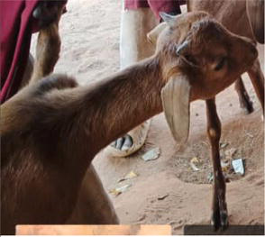
Anthrax is an infectious zoonotic disease of wild and domestic herbivores caused by a spore- forming bacterium, which is characterized by onset of high fever, oozing of unclotted blood from natural orifice and sudden death.
Transmission: Susceptible animals get infected by ingesting spores while grazing in highly contaminated soil or through the bite of certain flies. There is report of human infection through contact with the infected animals or contaminated animal tissue or products. The bacterium also penetrates body through lesion and sometime be acquired through ingestion of poorly cooked meat from infected animals.
Etiology: the disease is caused by Bacillus anthracis which is an aerobic or facultative anaerobic capsulated gram-positive, rod shaped spore forming bacterium. The spores can remain viable on soil for many years.
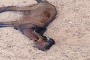
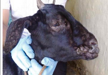
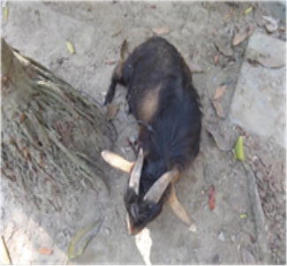
Clinical signs: Incubation period is typically 1–20 days. Most infections are noticeable after 3–7 days. Anthrax disease may be per acute, acute, sub-acute or chronic. The per acute form most often affects cattle, sheep and goats at the start of an outbreak and is characterized by staggering, trembling, difficulty in breathing, convulsions and death. Progression of the disease is rapid and premonitory signs may go unnoticed often animals are found dead with no rigormortis (Figure 1, Table 1). Blood may fail to clot due to the toxin released by B. anthracis. The acute or subacute form is common in cattle, sheep and horses and manifest high fever, increased heart rate, excitement, depression, incoordination, cessation of rumination, low milk production, bloody discharges, respiratory distress, convulsion, and death within 48–72 hours. But the chronic form is usually seen in less susceptible species like swine.
Figure 1.
Absence of rigor mortis in goat with anthrax by author.
Diagnosis: Careful microscopic examination of stained smears of blood, vesicular fluid, or edema may reveal the presence of B. anthracis. Biochemical and microbiologic tests provide a definitive diagnosis. It can also be isolated from skin lesions or respiratory secretions.
Treatment: Treatment of per acute case is usually untimely because of sudden death. Good recovery can be achieved through the use of anthrax antiserum in the early stage of the disease. Oxytetracycline at the dose rate of 5miligram/kilogram body weight parenterally can be effective if used from anthrax is most often seen in less susceptible species such as swine, but it has also been reported as developing in cattle, horses, dogs and cats. Route of infection in animals is most often ingestion, rather than inhalation or inoculation via skin lesions, initial suspicions of anthrax may be raised when livestock are found dead, bloated the onset of the disease. Penicillin streptomycin (Penstrep) also found effective if given concurrently with antiserum for 5 consecutive days.
Prevention: Anthrax can be prevented through annual vaccination programs. Rapid detection and reporting, quarantine, treatment of sub clinically affected animals (post exposure prophylaxis) and burning or burial of suspect and confirmed cases may prevent spore formation.
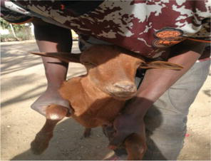
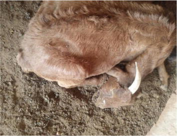
3.2 Brucellosis
Brucellosis is a bacterial infection that can affect goats and other livestock such as sheep and cows and wild ruminants such as deer, elk and bison. Brucellosis causes abortion or stillbirth in animals. Brucellosis is one of the widest spread zoonosis transmitted by animals and in endemic areas, human brucellosis has serious public health consequences.
Etiology: Brucellosis in goats is normally caused by a Gram-negative coccobacillary rod, Brucella melitensis although Brucella abortus may also cause clinical brucellosis.
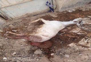
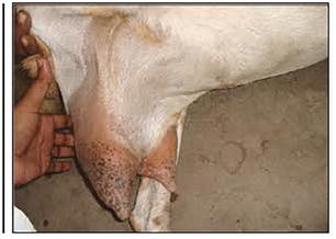
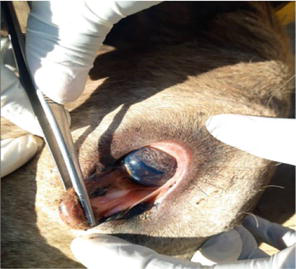
Clinical sign:Brucella melitensis is the most common cause of brucellosis in sheep and goats. It can cause abortion, retained placenta and swelling of the testicles. Abortion usually occur in late pregnancy in sheep and during the fourth month of pregnancy in goats (Figure 2, Table 1). Communicably, brucellosis is contagious to humans. Bacteria are present in milk, placenta, fetal fluids, fetus, vaginal discharges, semen and urine. Ruminants and other animals can shed bacteria long-term or lifelong.
Figure 2.
Abortion complicated with uterine prolapse in brucellosis. By author.
Diagnosis: History and clinical signs may be suggestive of the brucellosis. Demonstration of the bacteria in smears made from the samples of blood, bone marrow and other bod fluids can help confirming the diagnosis of the disease. Brucella can be isolated from the abomasal contents and lungs of the fetus, mammary glands, supramammary, retropharyngeal, parotid and mandibular lymph nodes and seminal vesicles by culturing on 5–10% blood or selective serum agar. Other serological methods like Serum Agglutination Test, Rose Bengal Plate Test, Enzyme Linked Immuno-Sorbent Assay (ELISA), Agar Gel Immuno-Diffusion (AGID) and Complement Fixation Test can be diagnostically used in the confirmation of brucellosis.
Treatment: There is no specific treatment of brucellosis that is successful, but long term antibiotics treatment can eliminate B. melitensis infections in valuable goats but the reproductive performance may be poor.
Prevention: Prompt vaccination of cattle, sheep and goats is recommended especially in endemic areas. Good hygiene practice such as milk pasteurization, proper meat processing, correct handling of stillbirths and animal carcasses are important strategy for the prevention of brucellosis in goats.
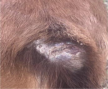
3.3 Caseous lymphadenitis (CLA)
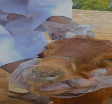
Caseous lymphadenitis is an infectious disease caused by the bacterium Corynebacterium pseudotuberculosis, that affects the lymphatic system, resulting in abscesses in the lymph nodes (Figure 3, Table 1) and internal organ of Goats and Sheep.
Figure 3.
Lymphnode abscess seen in clinical CLA in goat.
Transmission: The disease is highly contagious that affects sheep and goats [9]. When abscess ruptures, it releases a huge number of bacteria on to the skin and wool and it results to the consequent contamination of the surrounding environment. Animals may get infected when come in contact with the affected animals or indirectly via already contaminated fomites [10]. Infected animals may contaminate feed and water, which may become source of infection. The disease is also easily spread through the materials that are used during the operation of the animals such as castration, identification with ear tags or by tattooing. It is thought to also be spread by coughing or even by flies [11].
Etiology: CLA is caused by a gram-positive, nonmotile pleomorphic rods bacterium known as Corynebacterium pseudotuberculosis that has a characteristic Chinese letter arrangement in the smear. When this organism successfully established in the host, it surrounds and subdue the immune system, as a result it causes chronic infection that may remain in the animal for life but not pestilent [10].
Clinical signs: In CLA, there is abscessation in the region of peripheral lymph nodes especially the submandibular, parotid, prescapular and prefemoral nodes, which is termed superficial or cutaneous form. The internal form of CLA more commonly presents as dyspnea, loss of weight and failure to thrive. Other clinical signs include large pus filled cyst on the neck, sides and udders, cough, purulent nasal discharge, fever and tachypnea with abnormal lung sounds may be observed.
Diagnosis: A provisional diagnosis of the disease can be based on clinical features and physical examination of lesions associated with lymphnodes. Confirmation of the disease is achieved by bacterial culture of suspected lesions and purulent materials from an intact abscess. Serologic tests are available but their reliability is unrealistic.
Treatment: Treatment of CLA is often unsuccessful, but supportive care can be helpful. However, CLA abscesses must be treated to prevent ruptures and further contamination of other animals and environment. Parenteral antibiotics may be used in severe cases. Surgical drainage of the affected lymph nodes is recommended.
Prevention: The prevention of CLA can be achieved through strict biosecurity measures, immediate isolation of the affected animals from the flock. Surgical procedures such as castration, shearing or mass vaccination should be carried out through aseptic means and affected premises should be disinfected thoroughly.
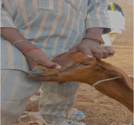
3.4 Contagious caprine pleuro-pneumonia (CCPP)
Contagious caprine pleuro-pneumonia (CCPP) is a highly contagious and rapidly spreading mycoplasmal disease that affects a vast majority of goat’s population, which is characterized by severe respiratory distress associated with sero-mucoid nasal discharge, dyspnea, coughing, pyrexia and general malaise.
Transmission: Main route of transmission occurs through inhalation of infected aerosol. Airborne transmission can result in distant spread [12]. Transmission by direct contact is also reported [13]. Infected objects, vectors, fomites and animal products are yet to be known in transmission role [14].
Etiology: Contagious caprine pleuro-pneumonia is a highly fatal disease that is caused by Mycoplasma capricolum capripneumoniae or Mccp (previously Mycoplama biotype F38), in Africa, Asia and the Middle East. Morbidity is often 100% and mortality may reach 80%.
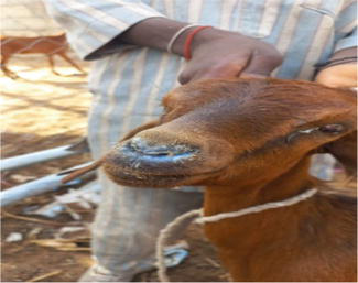
Clinical signs: The disease is characterized by anorexia, dullness, depression, weakness and lethargy, pyrexia, weight loss and decreased production. Also have respiratory signs of bilateral nasal discharge (Figure 4, Table 1), dyspnea, tachypnea and coughing. Occasionally, the only sign seen is sudden death.
Figure 4.
Sign of bilateral nasal discharge in CCPP. By author.
Diagnosis: The simplest and quickest procedure in field diagnosis is the detection of antibodies by Latex agglutination (LAT) as is easy to run and has a long shelf life. Other diagnosis include growth inhibition disc tests (GI), direct and indirect fluorescent antibody tests, complement fixation test (CFT), indirect hemaglutination test, ELISA and PCR. Isolation of M. capricolum capripneumonia from clinical samples is the only way to definitively diagnose the infection but is not normally performed as it is difficult and time consuming.
Treatment: Macrolides, tetracycline and quinolones are very active against M. capricolum capripneumonia. Antibiotics like tylosin at 1 ml/10 kg and enrofluxacin at 1 ml/20 kg body weight can be helpful in the treatment of CCPP.
Prevention: Vaccination has been an important aspect of CCPP prevention in a country where it is prevalent. Quarantine of affected animals and strict biosecurity measures for the introduction of new animals is necessary to reduce transmission and losses due to CCPP.
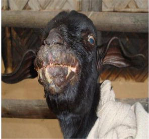
3.5 Dermatophilosis
Detmatophilosis is contagious bacterial disease of skin that affects sheep and goats. It is an infection affecting multiple species of animals world-wide, most common in young or immunosuppressed animals or in animals that are chronically exposed to wet conditions.
Transmission: Dermatophilosis is believed to be spread by direct contact between animals, through contaminated environments or possibly through biting insects.
Etiology: The disease is caused by a dimorphic bacterium, Dermatophilus congolensis that has two characteristic morphologic forms: filamentous hyphae and motile zoospores. Is a gram positive, non-acid fast, facultative anaerobic actinomycete, which is the only currently accepted species in the genus but, a variety of strains can be present within a group of animals during an outbreak [15].
Clinical signs: In severe generalized dermatophilosis, there is often loss of condition and motion, scab formation on the lips, muzzle, nose, ears (Figure 5, Table 1), feet and scrotom which if severely affected make prehension difficult. The scabs can become detached and reveal a yellow, creamy or hemorrhagic exudate. Allopecia can occur if the scabs are rubbed off. There is tufted papules and crusts that resembles paintbrushes. Concurrent infection with orf virus and other stress factors like malnutrition, pregnancy and lactation exacerbate the disease [16]. Most infections are mild thus render susceptible animal with normal functioning immune system spontaneously recover in time.
Figure 5.
Scab formation on the lips, muzzle and nose. By author.
Diagnosis: Clinical and cytological examinations of fresh lesions are suggestive of the disease. A definitive diagnosis is made by demonstration of the organism in cytological preparations, isolation on culture and/or via skin biopsy. Indirect fluorescent antibody technique and a single dilution ELISA test have been developed for large serologic and epidemiologic surveys.
Treatment: The causal organism is susceptible to a wide range of antimicrobials. High doses of penicillin-streptomycin are effective in severely affected animals, if administered in early stage of the disease. Heavy doses of long acting tetra cline (20 mg/kg) may be used and topical application of lime sulfur is a cost-effective adjuvant to antibacterial therapy. Insect repellent can be use externally to control biting insects.
Prevention: Isolation and culling of clinically affected animals can be helpful in preventing the disease. Ectoparasites control is a method used in breaking the infective cycle of the parasite. Keeping susceptible animals dry and frequent checking of the zinc sulphate and copper sulphate level in feeds have been found useful in reducing the spread and incidence of the disease.
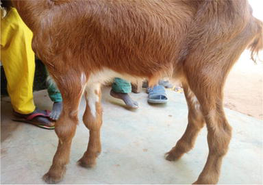
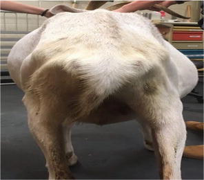
3.6 Mastitis
Mastitis is an inflammation of mammary gland due to physical injury, stress, bacterial or viral infections. It can either be clinical or subclinical. It is characterized clinically by clots or serum formation in the milk, swollen udder (Figure 6, Table 1), hot and tender to touch. Sub clinically, can be detected using California Mastitis Test (CMT), milk culture or Somatic Cell Count (SCC).
Figure 6.
Swollen udder in mastitic goat. By author.
Transmission: In goats, both vertical and horizontal transmissions are likely to occur. But vertical transmission presents very low occurrence. The introduction of mastitis is favored mainly by the factors that intervene in the horizontal transmission of pathogen. The pathogens can be eliminated by milk, feces, urine and oronasal secretions. The hands of the milker, milking equipment, vectors and fomites as a general way. The most often entry is via the galatogenic route [17]. All animals are susceptible, increasing the predisposition mainly according to age and number of lactations [18].
Etiology: The disease has a multiple etiolo gy but Staphylococcus aureus and Streptococcus agalactiae are the commonest bacteria isolated from cases of mastitis in small ruminants. Other bacteria identified include Corynebacterium pyogenes, Klebsiella spp, Mycobacterium spp and Brucella spp [16].
Clinical signs: The clinical manifestations of acute mastitis include edema, elevated fever above 1050 F, increased pulse rate, loss of appetite, depression, apathy, dyspnea, swelling and redness of the mammary gland, enlargement of the retro-mammary lymph nodes and lethargic movement are observed. Agalactia or lack of milk and hard lumps are common features of chronic mastitis. Claudication is a common sign in which small ruminants limps in order not to tamper with the inflamed mammary gland [19]. However, in subclinical mastitis, there is no evidence of clinical signs, but alterations in milk composition can occur and positive respond to CMT or other suggestive tests [20].
Diagnosis: History and clinical features are suggestive for tentative diagnosis. Microbiological culture can be reliable to determine the presence of organism in milk sample; California Mastitis Test (CMT) and somatic cell count (SCC) are commonest tests for mastitis. Other tests like multiplex-PCR and Enzyme-Linked Immuno Sorbent Assay (ELISA) are important techniques used in the diagnosis of mastitis.
Treatment: Glucocorticoids is recommended in the early course of disease, antibiotics like penicillin streptomycin (penstrep) at 200 mg/ml for 3–5 consecutive days is effective, oxytetracycline, benzylpenicillin, cloxacillin, amoxicillin,, ampicillin, or erythromycin have been recommended to treat mastitis. Some strains of S. aureus have found to be resistant to penicillin, hence drug sensitivity test is recommended before the use of such drugs on the treatment of mastitis. Topical application of antibiotic cream can be helpful.
Prevention: Proper milking practices, good hygiene for milking utensils and culling persistent infectors can help in reducing the incidence of the disease. Kidding pens and Bedding should be disinfected daily to avoid growth of pathogenic bacteria. Abscess draining and proper wound dressing can be carried out regularly. The hygienic-sanitary management aimed at preventing mastitis involves a number of factors including the choice of antimicrobial, microorganism susceptibility, duration of treatment, dosage employed, and the animal’s immune status [21].
3.7 Foot rot
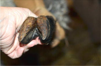
Foot rot is a contagious disease of the hooves in goats and sheep, characterized by ulceration and necrosis of the sensitive laminae of the foot (Figure 7, Table 1) and lameness. This disease is prevalent in the Southern region of the United States due to wetness and humidity of the environment.
Figure 7.
Ulceration and necrosis of laminae of foot in goat.
Transmission: Transmission is mainly enhanced by genetics, stocking rate and environmental factors. The disease can be spread from infected animal to non-infected susceptible animals (direct transmission). Incidence of overgrown hooves can predispose animals to foot rot. During the rainy season, infected animals can contaminate the soil and muddy pens which can enhance disease transmission to other animals If not treated; sick animals can become permanently infected [22]. The organism can also be transported to the soil by visitor’s shoes.
Etiology: Foot rot is disease caused by a large Gram-negative rod-shaped bacterium, Fusobacterium necrophorum and Dichelobacter nodosus which are mostly common in contaminated soil.
Clinical signs: Foot rot results in lameness, inappetence, loss of weight, and necrotic lesions in the interdigital space with foul smelling of the foot, there is elevated temperature and reduced production performance, lethargy, grazing on knees and abnormal gait. This condition may result increase in production losses, cost of treatment and prevention. Affected animals will lose value due to the infection [23]. The disease is very difficult to eradicate when it affects a herd/flock.
Diagnosis: The first signs of hoof rot are limping, holding limbs above the ground, grazing on knees, and abnormal gait, should be sufficient for diagnosis. If laboratory confirmation is required, submit smears and swabs of interdigital exudate and necrotic tissue from multiple animals for bacteriology.
Treatment: Systemic administration of antibiotics (penicillin streptomycin or oxytetracycline) and dry underfoot conditions usually resolve even severe lameness after a few days without the need to pare away dead horn. To curate in-between hooves with potassium per manganite and topical application of aerosol sprays of cetrimide or oxytetracycline may be helpful. Foot bathing in formalin or zinc sulphate (with surfactant) is another success achievable option.
Prevention: Quarantine all newly purchased animals before introducing in to the flock, isolate affected animals and give a deserving treatment, keep barn dry and clean to avoid contamination, Provide good drainage to areas of pastures and paddock, trim hooves regularly, supplement trace minerals and vitamins and give adequate nutrition.
Candidiasis is a mucocutaneous fungal disease caused by a yeast-like fungus. It is a normal inhabitant of gastro intestinal tract, nasopharynx and outer genitalia of different species of animals.
Etiology: Candidiasis is caused by a yeast-like candida specie most commonly Candida albicans that usually affects immunocompromized animals and opportunistic in causing diseases. The disease is distributed worldwide in a different spp. of animals [24].
Clinical signs: Signs noticed are defined patch of red itchy skin, pustules and scabs (Figure 8, Table 1). There is a local overgrowth of candida spp. on the tongue and mucosa of the mouth that appears as white plaques. Anorexia, dehydration, watery diarrhea, loss of weight and sometime death. Affected kids may develop listlessness, inappettance and stunted growth.
Figure 8.
Area of pustules and scabs in goats with candidiasis. By author.
Diagnosis: Diagnosis can be made by microscopic examination of scrapings or biopsy specimens from mucocutaneous lesions. The fungus can be seen visibly on staining with Wrights, methylene or Gram stain techniques. It can also be confirmed on culture of a sample in blood or tissue agar.
Treatment: Antifungal like amphotericin B or nystatin ointment can be effective when use topically, 1% iodine solution may also be used in the treatment of cutaneous candidiasis.
4.2 Cryptococcosis
Cryptococcosis is a dimorphic potential fungal disease of mainly the lung and brain that is distributed worldwide and affects immunocompromised animals especially goats and sheep causing pneumonia and or meningitis [25].
Etiology: At least 322 species of the genus Cryptococcus (Tremellales, Agaricomycotina) have been described [26]. However, only Cryptococcus neoformans (var. neoformans and var.grubii) and C. gattii have been described as causing disease in humans and domestic animals [27]. The pathogenic species of Cryptococcus are the only species of the order Tremellales able to grow well at temperatures >30°C, and their capacity to grow at 37°C is one of their main virulence factors [28].
Clinical signs: In cryptococcosis signs observed are; pyrexia, paraplegia, depression, severe dyspnea due to the obstruction of the nostrils, swelling of the nasal region, purulent nasal discharge, with abundant granulation tissue and hemorrhagic exudate in the nostrils [29]. Goats may have severe respiratory disease, including cough, anorexia, fever and severe weight loss [27]. Neurologic signs may also be observed in goats [30] and or meningitis (Figure 9, Table 1). The infection is sub-acute to chronic, with a clinical course of 2–6 week in goats and sheep [29].
Figure 9.
A sign of cryptococcal meningitis in goat. By author.
Diagnosis: Diagnosis. A definitive diagnosis can be achieved by cytologic evaluation of cerebrospinal fluid, skin and nasal exudates or isolation of C. neoformans from blood or body fluids such as CSF. Cryptococcal antigen latex agglutination serology (CALAS) can be performed on serum or body fluids but only provides presumptive evidence [31]. Gram stain is also useful.
Treatment: Prompt anti-fungal treatment such as Amphotericin B plus flucytosine, fluconazole, itraconazole or ketoconazole was found effective in the treatment of Cryptococcus in goats especially when treatment with antibiotics did not give any result. Success with oral fluconazole (5 mg/Kg/day orally for 6 months) was established in a goat with abdominal wall infection with C. gatti [32].
Prevention: It is difficult to prevent exposure to Cryptococcus in goats, since it is commonly found in the environment. Avoidance and environmental control of bird droppings (especially pigeons) are important [33]. Good hygiene and environmental sanitation is paramount in the prevention of the Cryptococcus.
4.3 Ringworm
Ringworm is a skin lesion usually circular and hairless, caused by a fungal infection of the hair follicle and outer layer of skin. Ringworm is a zoonotic disease. Sheep and goats develop crusty, scaly, circular patches that may or may not be pruritic (itchy).
Transmission: It is transmitted by close contact between animals or via animals contracting infective spores in the environment or by direct or indirect contact with contaminated equipment or environment.
Etiology: Ringworm is sometime called wool fungus, which is typically from the Trichophyton or Mycosporum genera. Ringworm is highly contagious and zoonotic in nature.
Clinical Signs: The primary signs observed are alopecia, scaling, crusting and poor growth. Sheep and goats develop moist and reddened skin, but later gray, scaly and dry, circular patches (Figure 10, Table 1) which is due to coalesced lesions that may or may not be pruritic (itchy). In more severely affected animals, lesions become confluent to produce an extensive areas of infection. Sheep and goats used for exhibition are at a much higher risk of contracting ringworm due to shearing practices, which cause exposure of the skin and the spread of fungal spores [34].
Figure 10.
Gray scaly dried circular area consumed by ringworm. By author.
Diagnosis: Diagnosis is typically made by visual examination and/or microscopic examination of biopsy of lesions, hair or skin scrapings. A definitive diagnosis and identification of the organism is made by a fungal culture.
Treatment: Treatments may not shorten the time to complete healing of lesions. Treatment with ketoconazole is found effective. Topical application of charmil gel can be helpful. The use of imidazole spray may stop progression of lesions and lower the spread to other animals.
Prevention: It is important to isolate infected animals so as not to spread ringworm to the rest of the flock or herd. Minimize mixing of animals in pre-confinement periods. Not only is it important to treat the animal, it is important to disinfect pens and anything with which the infected sheep or goat may have been in contact [34]. Thorough hand washing is also recommended after treating the animals.
4.4 Aspergillosis
Aspergillus spp. may cause infections in a variety of domestic animals. They are saprobes that are widely distributed in nature. Spread occurs via aerosols of spores present in soil, decaying vegetation, and occasionally animal tissues [35], a specialized hyphal structure of some fungi that produce conidia (asexually produced spores borne externally to the cells), can be observed in highly oxygenated tissues, such as those of the respiratory system [36].
Etiology: Aspergillosis is caused by several species of aspergillus, and there are more than 300 species of Aspergillus [37]. A. fumigatus is known to be directly associated with infection. Other spps like A. niger, A. flavus, A. terreus, and A. nidulans are opportunistic pathogens that are being recognized commonly with the use of molecular techniques.
Clinical signs: In ruminants, signs include moist cough, nasal discharge, pyrexia and shallow respiration. Pulmonary aspergillosis in sheep and goats is characterized clinically by anorexia, dyspnea, apathy, cough and nasal discharge [38]. The lungs are mottled, firm and heavy. There may be loss of condition associated with necrosis of the nasal mucosa in goats with nasal form of aspergillosis which may result in severe dyspnea (Figure 11, Table 1).
Figure 11.
Nasal and cutaneous aspergillosis in goat. By author.
Diagnosis: Aspergillosis can be confirmed by immunohistochemical, molecular and culture-based diagnostic methods. Macroscopic and histologic lesions can be used as presumptive diagnosis. There is another alternative diagnostic approach which involves the use of pan-fungal PCR on animal tissues [39]. Immunodiffusion, complement fixation, and ELISA can be used to detect antibodies against Aspergillus spp [40].
Treatment and prevention: Antifungal agents are currently unlicensed, but management of the disease usually relies on preventative measures such as ensuring clean bedding, good hygiene and good husbandry.
Parasites commonly found in goats can be divided into two general categories: Internal (Endo) and External (Ecto) parasites:
5.1 Endo parasites of goats
Endo parasites are worms that live in the body or inside an organ and there are multiple types. The most common internal parasites in goats are: Nematodes (roundworms) e.g. lung worms (Dictyocaulus spp. or Muellerius capillaris), Tapeworms, for example, moniezia, Liver flukes, for example, Fasciola hepatica, and intestinal parasites like Coccidia, for example, Eimeria or Isospora and Cryptosporidia. Parasites grow and reproduce in certain environments. Goats that live in those environments are at high risk of becoming infested [41].
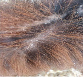
Clinical signs: The clinical signs of endoparasitism in goats include: reduced weight gain, decreased milk yield, Diarrhea, Rough hair coat (Figure 12, Table 1), loss of condition, Weakness, Anemia, Fever, hyperpnea, Coughing and Bottle jaw.
Figure 12.
Diarrhea and rough haircoat in goat affected by internal parasite. By author.
Diagnosis: Endo parasite of goats can be diagnosed mainly based on laboratory tests. The commonly used laboratory tests for the diagnosis of endo parasitic diseases include blood packed cell volume, etiological examination, which involves the detecting of the parasitic larvae or eggs from stool, blood, nasal secretions as well as tissue of the animals, serological assay such as Enzyme linked immunosorbent assay, indirect hemagglutination test or fluorescence immunoassay and molecular diagnostic techniques such as polymerase chain reaction (PCR) or DNA sequencing.
Treatment: Benzimidazoles (oxfendazole, febantel, fenbendazole and albendazole) Macrocyclic lactones: Avermectins (ivermectin, doramectin and eprinomectin) Milbemycins Cholinergic agonists: Imidazothiazoles (levamisole) Tetrahydropyrimidines (pyrantel and morantel) are found effective for internal parasite. However, only morantel, thiabendazole, fenbendazole, albendazole and phenothiazine are approved for goats [42]. Dewormer resistance occurs when there is less than 95% reduction in fecal egg count 14 days after administration. Resistance has risen due to anthelmintics being used often, rotated too frequently or under dosed.
Prevention: The best prevention is to alternate livestock species grazing, avoiding overcrowding of pens or premises, genetic improvement and pasture rotation. Balanced nutrition is very important to keep animals healthy and help them develop appropriate resistance to external pathogens, especially for dams before and after lambing/kidding. Also practice the use of effective dewormers.
5.2 Ectoparasite of goats
Ectoparasites feed on body tissues like skin, hair or blood, and they include fleas, flies, lice, mites, nose botfly and ticks. The wounds and skin irritation produced by these parasites result in discomfort and irritation to the animals. Parasites can transmit diseases from sick to healthy animals, which can reduce weight gains and milk production.
Clinical signs: Mites infect the head, legs, body or tail region causing the skin to become crusted and cause loss of hair and wool (Figure 13, Table 1). The infected area itches and the animal scratches. The host does not feed well. The infections cause skin damage to a goat. Lice, flea and flies are found where animals are kept in overcrowded confined environment and cause irritation of the skin, anemia and damage to the skin. It causes loss of weight and condition to the host. The can transfer from one animal to another through close contact. Ticks are very important parasites that can harm its host by bites resulting in anemia, weakness and debility. Ticks can be classified according to groups; one-host, two-host and three-host ticks. Ticks that are known to infest goats are those belonging to three-host tick as they parasitize three different hosts in their life cycle that make their control very difficult. Ticks also spread diseases (tick borne diseases) that are so fatal to its host. Nose botfly infests the nostril of goats causing irritation, sneezing, shaking of the head, nasal discharge, respiratory distress, loss of appetite and grinding of the teeth.
Figure 13.
Loss of hair caused by ectoparasite by author.
Diagnosis: Identification of ectoparasites can prove difficult because detailed clinical examination can fail to confirm the presence of some ectoparasites. It may be necessary to take skin stub samples to investigate whether arthropods such as mites are in residence. Ticks, flea, flies and biting lice usually can be seen with the naked eye. The presence of mites can be confirmed by examining mites, eggs and fecal pellets in skin scrapings under the stereoscopic microscope.
Treatment: Mites and lice are controlled by washing the infected area, spraying or dipping the animal with a suitable treatment. If an animal has only a few ticks these can be carefully pulled off making sure the mouth parts of the tick are removed. Dipping is very effective if large numbers of livestock need to be treated. Ivermetin injection and pour on are also effective in the treatment of ectoparasite, but accurate dose must be maintained.
Prevention: Monitoring program should be exercise to insure early identification. Sanitation and regular cleaning of facilities using appropriate detergent and disinfectant is helpful in the prevention of ectoparasite. Eliminate areas where external parasites can breed and develop (e.g. elimination of standing water reservoirs decreases mosquito levels). The use of Insecticides and fly predators may be necessary.
Babesiosis is an infectious tickborne, obligate, intraerythrocytic protozoan parasites from the phylum Apicomplexa, order Piroplasmida affecting a wild and domestic animals which include cattle, sheep and ggoats. It is typically fatal disease that is characterized by hemoglobinuria, fever, icterus and intravascular hemolysis resulting to anemia. Variety of animals. Susceptible animals may suffer high rate of mortality but recovered animals that are latently infected has an immunity for a certain period of time. It is transmitted transovarially by ticks. By the ingestion of the parasite, the female tick becomes infected during engorgement, upon drops off of the babesial agents on the host, it reproduce within the tick’s tissues which is incorperated within the embryo of the developing ticks resulting to the transmission of the disease to the new hosts by feding of ensuing tick larvae, nymphs, or adults [43].
Transmission: Babesiosis is transmitted by ticks Boophilus, Hemaphysalis, Hyalomma, Dermaentor and Ixodus spp are the vetor in the transmission of Babesia of different species. It is transmitted in both transovarially and transstadially. Babesia spp affecting goats and sheep may be maintained in non-susceptible hosts such as wild animals [16].
Etiology: Although small ruminants can be infected by several species of Babesia, the two most important species associated with Babesiosis in goats are B. ovis and B. motasi, transmitted by Rhipicephalus bursa and Haemaphysalis spp, respectively. Infection is of importance in the Middle East, southern Europe, and some African and Asian countries.
Clinical signs:B. motasi can cause an acute or chronic Babesiosis in goats, generally runs a course of 1 week or less. In acute infection, the first clinical signs are anorexia, lethargy, depression, and fever (frequently ≥41°C]), which persist throughout, and these are accompanied later by inappetence, anemia and jaundice (Figure 14, Table 1), hemoglobinemia and hemoglobinuria occur in the final stages. Chronic infection is manifested by emaciation, coughing and edema. Many animals recover; however, some may die if not treated.
Figure 14.
Anemia and jaundice seen in Babesiosis. By author.
Diagnosis: History and clinical findings may provide a presumptive diagnosis of babesiosis. However, Giemsa-stained blood or organ smears by light microscopy is essential to confirm the diagnosis. The most commonly used tests are ELISA, PCR and a DNA probe, which can detect specific parasitemias at very low levels of infection [44].
Treatment and prevention: Babesiosis can be treated using diminazene aceturate (3-5 mg/kg), phenemidine diisethionate (8-13 mg/kg), imidocarb dipropionate (1-3 mg/kg), and amicarbalide diisethionate (5–10 mg/kg) [43, 44]. Supportive treatment such as blood transfusions (4liters of whole blood per 250 kg of body weight), fluids, hematinics, and prophylactic antibiotics are important [43]. The disease can be prevented by effective quarantine of the susceptible animals so as not allow the introduction of the vector ticks. The control of ticks by dipping or spraying animals at risk with recommended acaricides is paramount.
6.2 Coccidiosis
Coccidiosis is an enteric protozoan disease of goats caused by the genus Eimeria. Coccidia go through a complex life cycle in the intestinal cells of animals. Large number of eggs called oocysts are being produced in the intestine and passed in the feces. The intestinal cells can be damaged as a result of growth and multiplication of the coccidia in the intestinal epithelial cell thereby causing diarrhea and other signs of the disease.
Etiology: Coccidiosis in goats is caused by several species of Eimeria, but E. arloingi and E. ninakohlyakimovae are known to be pathogenic. All the goat Eimeria spp are considered host specific and do not transfer infection from goats to sheep. In most cases, concurrent coccidial and helminthic infections can occur especially in animals on extensive management system [45].
Clinical signs: In most cases, clinical coccidiosis occurs between 5 and 8 weeks of age. Most goat kids have subclinical infection. In acute or sub-acute infections, the usual signs are pasty feces, dirty tails, stary coat, loss of weight, dehydration and inappetence. More severe acute cases show mucoid or bloody diarrhea, possible tenesmus, dullness, anorexia, weakness and anemia. Severe problems lead to rapid onset of diarrhea, often with blood, tenesmus, and signs of abdominal pain, lethargy, recumbences, and death. In chronic infections, there is delayed puberty, debilitation, poor appetite, loss of weight and liver failure especially in milking goats.
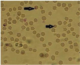
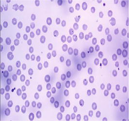
Diagnosis: Diagnosis can be based on history, age of kids, clinical signs especially of severe diarrhea, fecal examination and postmortem findings. Acute coccidiosis can be diagnosed by direct examination of feces but in chronic coccidiosis that have very low oocysts number are seen in feces (Figure 15, Table 1), direct examination of feces may not be adequate [46].
Figure 15.
Slide showing coccidiosis in goats.
Treatment and prevention: Sulfadimidine (sulfamethazine) injection (10–50 mg/kg) for five consecutive days is found effective. Vetcotrim bolus (10-30 m/kg Per Os once daily for 3 days) has a good result. Decoquinate (Deccox, 0.5 mg/kg) and Monensin 13-20grams/ton of feed can be used in non-lactating goats. Diclazuril (1 mg/kg, PO, once) and toltrazuril (20 mg/kg, PO, once) have been used successfully; doses may need to be repeated. A metabolite of toltrazuril, ponazuril (10 mg/kg, PO, once) reduced oocyst counts when used experimentally in goat kids. Ensure good nutrition program, Improved good hygiene in the house, minimize predisposing factors, avoid crowded pens and pastures, Feed and water troughs should be raised off the ground to prevent fecal contamination.
6.3 Theileriosis
Theileriosis is caused by Theileria spp. a genus comprising tick-borne transmitted protozoa of the family Theileridae, order Piroplasmida, subclass Piroplasmia, Phylum Apicomplexa. Theileria species affect domestic and wild ruminants, especially in Africa, Europe, Australia, and Asia [47].
Etiology: Theileriosis in sheep and goats is usually caused by T. lestoquardi (formerly T. hirci), T. uilenbergi or T. luwenshuni. Morbidity and mortality rates of up to 65% (T. luwenshuni) and 75% (T. uilenbergi) have been seen in susceptible animals introduced into endemic areas. Affected animals show sustained fever and anemia.
Clinical signs: The clinical signs of theileriosis in goats infected with T. lestoquardi are similar to other forms of theileriosis, which include anorexia, slight oculo-nasal discharges, fever, salivation, enlargement of superficial lymphnodes, weight loss, respiratory distress (Figure 16, Table 1), edema of the lungs, anemia, icterus and diarrhea, death may occur due to asphyxia. Abortions may also be seen. In most cases, experimental infection of sheep and goats with T. annulata resulted in only mild to moderate clinical signs of fever and lymphadenopathy [48].
Figure 16.
Respiratory distress and loss of weight by author.
Diagnosis: Theileriosis can be diagnosed on Giemsa-stained thin smears from blood or lymph node biopsies. At necropsy, impression smears can also be used to detect schizonts from many internal organs of infected animals such as the liver, spleen, lymph nodes and lungs. Other diagnostics tools like antigen- specific ELISAs and PCR are often used in diagnosis and can identify Theileria in the blood of both carriers and clinical cases. Some tests can differentiate the species of Theileria, while others are specific for the genus [48].
Treatment and prevention: Infected goats can be treated with antiparasitic drugs like Buparvaquone (2.5 mg/kg) is very effective in the early stage of the disease. Use of Oxytetracycline 10 mg/kg is found helpful. Antidiuretics and anti-inflammatory drugs may also be used when there is evidence of pulmonary edema. In advanced stage of the disease, treatment is less effective especially where there is extensive destruction of lymphoid and hematopoietic tissues [49]. Control of ticks by spraying or dipping of animals with acaracides is the most successful method used for the prevention of theileriosis, but this needs to be applied at regular intervals to be effective. Pyrethroid compounds are often used where animals are exposed to tickborne diseases.
6.4 Cowdriosis
Cowdriosis (Heartwater) is regarded as the most important infectious, noncontagious, tickborne rickettsial disease of ruminants that is clinically characterized by diarrhea, fever, hydropericardium, hydrothorax and edema of lung and brain. The disease is seen only in areas infested by ticks of the genus Amblyomma.
Etiology and transmission: The disease is caused by a pleomorphic ricketssia Ehrlichia ruminantium which is an obligate intracellular parasite previously known as Cowdria ruminantium.. Under natural conditions, E ruminantium is transmitted by Amblyomma ticks. Transmission occur mainly transstadially, but transovarial transmission rarely occurs.
Clinical signs: The incubation period of Heartwater in goats and sheep is 1–5 weeks and the course of acute disease takes 3–6 days. The clinical signs are dramatic in the peracute and acute forms. In peracute cases, animals may die suddenly without any premonitory signs; other animals developed dyspnea and intense convulsion. In the acute form, there is pale mucous membrane, anorexia and depression (Figure 17, Table 1). Other signs of hyperesthesia, nystagmus, chewing movements and a high-stepping stiff gait may be noticed. Terminally, prostration with bouts of opisthotonus, circling or galloping movement, and stiffening of the limbs, intermittent diarhea and convulsions are also seen. Subacute form is less pronounced, but there may be prolonged fever, mild incoordination and coughing. Nervous signs are inconsistent.
Figure 17.
Anorexia and depression by author.
Diagnosis: presumptive diagnosis is based on clinical, epidemiological and pathological features; E.ruminantium colonies can be identifiable in the brain or intima of blood vessels on staining with Giemsa or methylene blue; Molecular methods like real-time PCR has an advantage of being less time consuming and free of cross contamination; Serological tests such as indirect fluorescent antibody tests, enzyme linked immunosorbent assays (ELISA) and Western blot are used for definitive diagnosis.
Treatment and prevention: Sulfonamides and tetracyclines can be used in the early stage of the disease, when it advances the prognosis may be poor. Oxytetracycline 20%long acting at 10 mg/kg/day may be helpful or doxycycline at 2 mg/kg/day will be effective, if administered early in the course of heartwater infection. Diazepam may be required to control convulsions. Dexamethasone injection will be serve as supportive care, although the rationale behind its effectiveness is much more controversial. Control of tick infestation is a good preventive measure in some instances but may be difficult and expensive to maintain in others. In areas of endemicity, the use of dips against ticks of domestic animals is highly recommended. Affected animals must be confined in a quiet and cool areas that is devoid of any other disturbances because, any stimulation can cause a convulsion and abrupt death. Vaccination can be helpful in the prevention and control of cowdriosis.
6.5 Anaplasmosis
Anaplasmosis is a tickborne obligate intraerythrocytic bacteria of the order Rickettsiales, family Anaplasmataceae, genus Anaplasma, that infect red blood cells, causing fever and anemia.
Etiology and transmission:Anaplasma ovis may cause mild to severe disease in sheep, goats and deer which is typically transmitted by ticks or biting flies. Up to 17 different tick vector species (including Dermacentor, Rhipicephalus, Ixodes, Hyalomma, and Argas) have been reported to transmit Anaplasma spp. Iatrogenic transmission can occur when instruments are re-used without proper sanitation, including instruments used for dehorning, ear tagging, castrating, and vaccinating. In utero transmission has also been reported.
Clinical signs: Anaplasmosis is usually subclinical or less severe in young animals, it is severe and often fatal in older. Anaplasmosis is characterized by progressive anemia due to extravascular destruction of erythrocytes. Macrocytic anemia with circulating reticulocytes may be present late in the disease [50]. Animals with peracute infections succumb within a few hours of the onset of clinical signs. Acutely infected animals lose condition rapidly. Incoordination, loss of appetite, dyspnea, and a rapid pulse are usually evident in the late stages. The urine may be brown, but, contrary to babesiosis, with no hemoglobinuria. Mucous membranes appear pale and yellowish. There is abortion, hematologic parameters gradually return to normal after convalescence.
Diagnosis: Diagnosis based on clinical features, hematological changes, blood smears, and serologic testing. Microscopic examination of Giemsa-stained thin and thick blood films (Figure 18, Table 1) is critical to distinguish anaplasmosis from babesiosis and other conditions that result in anemia and jaundice, such as leptospirosis and theileriosis. Serological tests like ELISA, complement fixation, or card agglutination tests has been used to identify chronically infected carriers but with doubtful degree of accuracy. Nucleic acid-based detection methods can be used, but carrier level of infections may not be detected [50].
Figure 18.
Slide showing anaplsma spp.
Treatment and prevention: Oxytetracycline has been reported to reduce severity of the disease. The use of imidocarb has been shown to be very effective in the treatment of anaplasmosis. Good sanitary methods such as cleaning of stalls/pens regularly can help to reduce contamination. Avoid re-using of needles and disinfect medical equipments when use. Treatment with an effective acaricides to kill ticks may help to reduce the incidence of anaplasmosis.
Peste des petits ruminants is an acute, highly contagious transboundry viral disease primarily affecting goats and less commonly in sheep associated with high morbidity and mortality caused by PPR virus of the genus morbilliirus and family paramoviridae that closely resembles rinderpest virus.
Etiology and transmission: is caused by a PPR virus of the genus Morbillivirus of the family paramoviridae (sub family Paramixovirinae) under the order Mononegavirales which is related to but distinct from Rinderpest virus of cattle. The PPRV is genetically grouped into four genotypes (lineages) [51], based on the Fusion (F) and Nucleoprotein (N) gene sequences. Lineages I and II circulate mainly in West Africa, lineage III is mostly in Eastern part of Africa, while lineage IV is generally found in Asia, but has now spread to the African continent and become the most prevalent of all the lineages [52]. The disease is transmitted by infected aerosols in situation of close contact of animals and confinement seems to favor outreaks. Fomites like bedding, feed and water troughs also help in the transmission of PPR.
Clinical signs: In acute form, goats typically display an abrupt rise in temperature to 40–41°C. Within a few days, infected animals develop oculo-nasal discharges (Figure 19, Table 1), thirst, anorexia, depression and leukopenia [3].
Figure 19.
Purulent oculo-nasal discharges. By author.
Conjunctival mucous membranes may be congested, followed by serous and mucopurulent exudates. Affected animal develop necrotic oral erosions that produce a fetid smell. There is profuse diarrhea which develops within 2 to 3 days and is accompanied by abdominal pain, tachypnea, loss of weight and severe dehydration. There may be abortion 5 to 10 days after the onset of fever. The incSubation period is usually 4-5 days.
Diagnosis: History and clinical features give a presumptive diagnosis in endemic regions. The virus can also be detected in acute cases from various swabs and blood samples, using PCR and ELISA.
Treatment and prevention: PPRV infection has no specific treatment. Mortality can be reduced by supportive care, including the administration of Antibiotics such as chloramphenicol, penicillin, and streptomycin, inflammatory agents, as well as nutritional support. State and federal veterinarians should be notified if PPRV is suspected. Sheep and goats can be protected against PPR by immunization with rinderpest vaccines or by the simultaneous administration of PPR hyper immune bovine serum and virulent PPRV [52]. Premises should be decontaminated, and the area quarantined. Movement restrictions can also aid in the prevention of the disease.
7.2 Goat pox
The sheep pox virus (SPV) and goatpox virus (GPV) are serious acute, often fatal febrile contagious viral skin diseases of sheep and goats caused by the members of the Poxviridae, genus Capripoxvirus which is characterized by widespread skin eruptions. They are believed to be closely related antigenically and physicochemically, which are able to infect both sheep and goats.
Etiology and transmission: Sheep pox and goat pox is caused by the infection of sheep pox virus (SPV) or goat pox virus (GPV), closely related members of the genus Capripox virus in the family Poxviridae. SPV is mainly thought to affect sheep and GPV primarily to affect goats, but some isolates can cause mild to serious disease in both species. (CFSPH). Transmission is by direct contact, while indirect transmission by contaminated implements, vehicles or products such as litter or fodder. Indirect transmission by mechanical vectors like insects is also possible. Transmission by inhalation is important. SPV and GPV are shed in saliva, nasal and conjunctival secretions.
Clinical signs: Sheep pox and goat pox appear similar, with clinical signs that typically include: Fever, red spots that become blisters (Figure 20, Table 1) on the muzzle, eyelids, ears, udder or in severe cases all over the body [53]. Lesions can develop internally causing breathing difficulties, depression, lethargy, reluctant to feed, oculo-nasal discharges or swollen eyelids and enlarged superficial lymphnode.
Figure 20.
Clinical manifestation of capripoxvirus. By author.
Diagnosis:Capripoxviruses, their antigens and nucleic acids can be detected in skin lesions (e.g. biopsies, scrapings, vesicular fluid, scabs); oral, nasal and ocular secretions; blood; lymph node aspirates; and tissue samples from external or internal lesions collected at necropsy PCR can identify viral RNA directly in tissue samples, blood and secretions. Other tests to detect capripoxvirus antigens include enzyme-linked immunosorbent assays (ELISAs), immunostaining methods and agar gel immunodiffusion (AGID).
Treatment and prevention: There is no specific treatment for sheep pox or goat pox, but supportive treatment may reduce morbidity and other complications. Newly introduced animals should be quarantined. Other biosecurity measures, such as prevention of contact with other herds and disinfection of fomites are also helpful. Stringent cleaning and disinfection of farms and equipment, animal and vehicle movement controls within infected areas, Vaccination may be considered in the area where the disease is endemic.
7.3 Caprine arthritis encephalitis (CAE)
Is a viral disease of goats caused by a lentivirus called caprine arthritis encephalitis virus, which is an enveloped, single-stranded RNA in the family Retroviridae. The disease is found worldwide [54]. There are several genetically distinct isolates of the virus that differ in virulence.
Etiology and transmission: Caprine arthritis encephalitis virus is an enveloped, single-stranded RNA lentivirus in the family Retroviridae. There are several genetically distinct isolates of the virus that differ in virulence. Cross-species transmission is possible via feeding of colostrum or milk from infected goats. Therefore, the ovine and caprine lentiviruses are commonly referred to as small ruminant lentiviruses [55].
Clinical signs: the most common sign is polysynovitis-arthritis which includes joint swelling (Figure 21, Table 1) and lameness of varying severity. There is Stiffness, abnormal gait and posture, loss of weight and depression. In goats which develop the neurological form of the disease, show ataxia, incoordination, hypertonia and hyperreflexia are also common. The goat has increased difficulty standing and eventually is unable to stand [56]. Lameness may be sudden.
Figure 21.
Joint swelling seen in CAE. By author.
Diagnosis: Based on history and clinical manifestations. Serologic tests like agar gel immunodiffusion test and ELISA are useful to determine herd CAEV status.
Treatment and prevention: There is no specific treatments currently exist, likewise there is no vaccine but supportive care is indicated. To prevent spread of the disease, infected animals are separated from non-infected goats, or culled [55]. Separating goat kids from infected goats, and feeding the kids with cow’s milk, or pasteurized goat milk, will prevent infection [54]. Goats should also be quarantine and tested for CAE virus before introducing to the herd.
7.4 Contagious ecthyma (orf)
Orf is one of the most widespread highly infectious viral diseases of mostly small ruminants and sometimes other species, including wild animals. It occurs worldwide and characterized by scabby and pustular lesions on the muzzle, commissures of the lips and nostrils.
Etiology and transmission: is caused by Orf virus which is type species of the genus Parapoxvirus of subfamily Chordopoxvirinae of family Poxviridae. Natural transmission of disease occurs through direct or indirect contact with infected animals particularly with dried crusts that falls on the pastures during grazing.
Clinical signs: The primary lesion develops on the lips, muzzle and may extend to the mucosa of the oral cavity. Early in the infection, sores appear as blisters that develop into crusty scabs (Figure 22, Table 1). Lesions can sometimes be found on the feet and around the coronet, there is inappettance and loss of condition. During the course of the disease, there is presence of a nodules with red colorations in the center, a bluish white rings in the middle and peripheral erythematous. The lesions may progress to form papules and extends through vesicular and pustular stages to become encrusted. The Coalition of the numerous discrete lesions produces a large scab that result in the proliferation of dermal tissue that produces a verrucose mass beneath them [57].
Figure 22.
Crusty scab in Orf. By author.
Diagnosis: is based on clinical features and histopathology of skin lesions, transmission experiments and demonstration of a pox virus by electron microscopy of skin biopsies of infected skin.
Treatment and prevention: Antibiotics such as penicillin, chloramphenicol and10% Potassium permanganate solution is effective in case of lip lesions. In a situation complicated with stomatitis or enteritis, parenteral administration of antibiotics in conjunction with topical applications of salicylic acid ointment can give good result. Live vaccines should be used cautiously to avoid contaminating uninfected premises, and vaccinated animals should be separated from unprotected ones.
Parturient paresis is a non-febrile metabolism disturbance of pregnant and lactating ewes and does characterized by acute-onset of hypocalcemia, hyper excitability, circulatory collapse, flaccid paralysis, depression, recumbency, coma, and death.
Causes: Parturient paresis is as a result of decreased calcium intake on circumstance of high demand for calcium particularly during late gestation. This will result to low serum calcium concentration in pregnant animals with multiple fetuses. As fetuses mature and their bones mineralize, they require increasing amounts of calcium. Goats are required to mobilize stored calcium so as to increase calcium absorption, to meet up with the requirement. Failure to meet up with such calcium requirement especially during these periods put goats at significantly high risk of this condition.
Clinical signs: In early hypocalcemia, the most commonly noted clinical signs are lethargy and inappetence (Figure 23, Table 1), decreased body temperature, stiff gait, ataxia, salivation, depressed rumen motility, mild bloat or constipation, recumbency, weakened uterine contraction and if untreated, death.
Figure 23.
Lethargy and inappettance by author.
Diagnosis: A working diagnosis is based on history and clinical signs. In outbreaks occurring before parturition, pregnancy toxemia is the main differential diagnosis [58]. Diagnosis can be confirmed by testing serum calcium concentration before treatment. Urine ketone or serum beta-hydroxybutyrate concentrations should always be evaluated at the same time. Tentatively, diagnosis of acute hypocalcemia is aided by a rapid response to slow IV administration of calcium.
Treatment and prevention: Treatment is by IV administration of calcium borogluconate 50–150 milliliters of a 23% solution, which will elicit rapid response. Oral or subcutaneous administration of a calcium solution helps to prevent relapse [58]. Other treatment of Calcium-containing products that also contain phosphorus and magnesium, as well as dextrose, will have an additional therapeutic effect. Prevention is by supplementing adequate dietary requirement for calcium throughout gestation.
8.2 Pregnancy toxemia/ketosis
Is a metabolic often fatal disease that occurs in all parts of the world. It is most prevalent in very fat does or does carrying multiple fetuses. This is a condition of late pregnancy and early lactation most commonly occurring in the last month of gestation.
Causes: is caused by in adequate nutrition or disturbance in carbohydrate or sugar metabolism especially during last trimester of pregnancy in does. As the pregnancy progresses, the need for energy in the body increases, at the same time, the rumen capacity decreases as a result of fetal growth.
Clinical signs: Does with ketosis have poor appetite, lethargy and stress. They also tend to separate from the herd, lag behind and become depressed. There is abnormal gait and posture, apparent blindness with severe depression and frequent urination. A classic symptom is sweet-smelling (ketotic) breath. Goats may also grind their teeth and grunt, muscle tremors, opisthotonos, followed by recumbency (Figure 24, Table 1), coma and death usually follows within a few days.
Figure 24.
Goat with ketosis showing sternal recumbency. By author.
Diagnosis: To determine pregnancy toxemia in a flock. Blood glucose monitor should be used with a BHBA (beta-hydroxybutyrate) blood strip to test the glucose level. Does with a reading of 0.8 mmol/L or higher can be classified positive for pregnancy toxemia and should be treated accordingly.
Treatment and prevention: Administer a readily usable form of energy (especially glucose) and get the doe eating on her own usually with the help of anabolic steroids. Treatment is often ineffective if pregnancy toxemia is in advanced stages [59]. Oral administration of propylene glycol 1o0ml/day for three consecutive days may be helpful. Supplementation with calcium lactate may also be suggested. Subcutaneous administration of protamine zinc insulin at 0.4 U/kg daily may increase survival rate especially during the first half of pregnancy. Excessive fat should be reduced and weight gains should be allowed only during the 6 weeks before kidding will reduce the incidence of ketosis. Give high-quality forages in addition to supplements that are very palatable during the last 2 months of pregnancy. This allows does to receive adequate energy even though rumen volume is decreased.
8.3 Lactic acidosis (grain overload)
Is a carbohydrate fermentation disorder of the rumen that usually affect goats of all breeds and ages, as a result of feeding with large amount of highly fermentable concentrates, underfeeding of effective fiber and poor management practices.
Causes: occurs in goats that have been fed predominantly forage but abruptly introduced to a large of amount of readily digestible carbohydrates especially grain. It can be caused by abnormal feeding of an effective fiber and poor management practices.
Clinical signs: In acute acidosis, there will be sudden death. Other signs include an increased pulse and respiratory rates, decreased rumen motility, mild bloat, staggering, abnormal posture (Figure 25, Table 1) and even coma [60]. Subacute acidosis is difficult to recognize but occurs more frequently. Loss of appetite, panting, diarrhea, dehydration, lack of rumination and signs of discomfort may be noticed.
Figure 25.
Goat with grain overload.
Diagnosis: The diagnosis is obviously based on history and clinical findings. It can also be confirmed by, a low rumen fluid pH (less than 5.5), and the absence of live protozoa when rumen fluid is examined under the microscope.
Treatment and prevention: Treatment usually includes drenching with a solution of sodium bicarbonate, Magnessium sulphate, administration of an antibiotic to suppress the lactic acid-producing bacteria and a change in feeding practices. Prevention involves slowly introducing concentrate feeds to allow the adaptation of rumen microflora. Provision of a properly balanced diet and proper feed management practices.
Animal diseases are global challenges that are considered as a major impediment to livestock especially goat production. To establish an early warning and proper implementation of strategic preventive measures in a country prone to disease affliction, focus should be made on identifying principal epizootic diseases that have a strong impact on the public health, social and economic significance. This work clearly identified the commonest diseases ravaging goats from infectious: bacteria, fungal, parasitic, protozoa, rickettsia and viral, to non-infectious: metabolic disturbance/nutritional disorders and spells out the treatment regimen and the preventive measures that will remedy the predicament in goat production. It behooves on government, veterinarian, para-veterinarians and practioners to co-opt this idea and move forward to design a novel innovative policies and ideas that will help in the prevention, control and eradication of the diseases.
The authors are grateful to the staff and management of the Ministry of Agriculture, Yobe State for accessing their clinical records. The contributions of Prof. Lawal Said of Dept. of Vet. Medicine A.B.U Zaria and Dr. Adamu Kaikabo of NVRI Vom are also acknowledged.
Conflict of interest
The authors declare no conflict of interest.
References
1.Adebowale OAL. Dynamic of ruminant livestock Management in the Context of the Nigerian agricultural system. In: Javed K, editor. Livestock Production. London, UK, U.S.A: Intech. p. 61
2.Unigwe CR, Ogbu UM, Balogun FA, Orakwu OK, Nwafoh OC, Nwachukwu BC. Prevalence of small ruminant diseases/disorders at Makola veterinary hospital, Ibadan, Nigeria. Journal of Biology, Agriculture and Health Care. 2016;6(1):1-6
3.Bukar BA, El-Yuguda AD, Lawal S. The first Seroprevalence investigation of Peste des Petits ruminants virus among Sahel goat in Yobe state, Nigeria. Asian Journal of Medicine and Health. 2020;18(4):33-38
4.Lado MM, Salah KJ, Erneo BO. A case study of major constraints of small ruminants Management in Juba Country, central equatorial state, South Sudan. International journal of innovative science. Engineering and Technology. 2015;2(12):1-5
5.Fakoya EO, Oloruntoba A. Socio-economic determinants of small ruminant production among farmers in Osun state, Nigeria. Journal of Human Social Science Creativity. 2009;4(1):90-100
6.Kihu SM. Risk Factors and Socio-Economic Effects Associated with Spread of Peste Des Petits Ruminants (PPR) in Turkana County, Kenya [Thesis]. Kenya: University of Nairobi; 2014. pp. 4-38
7.Okoli IC. Incidence and modulating effect of environmental factors on Trypanosomoses, Peste des Petits ruminants and bronchopneumonia of west African dwarf goats in Imo state, Nigeria. Livestock Research for Rural Development. 2002;15(9):1-7
8.Nath TC, Bhuiyan MD, Mamun MA, Datta R, Chaudary SK, Hossain M, et al. Common infectious diseases of goats in Chitta District of Bangladesh. International Journal of Scientific Research in Agricultural Sciences. 2014;1(3):43-49
9.Lisa H, and Williamson MS. Caseous Lymphadenitis in Small Ruminants. Department of Large Animal Medicine, University of Georgia, College of Veterinary Medicine, Athens, Georgia, 2001; 17 (2): 359-371.
10.Baird GJ, Fontaine MC. Corynebacterium pseudotuberculosis and its role in ovine Caseous lymphadenitis. Journal of Comparative Pathology. 2007;137(4):179-210
11.Windsor PA. Control of Caseous lymphadenitis. Veterinary Clinics of North America: Food Animal Practice. 2011;27(1):193-202
12.Lignereux L, Chaber A, Saegerman C, Manso-Silvan L, Peyraud A, Apolloni A, et al. Unexpected field observations and transmission dynamics of contagious caprine Pleuro-pneumonia in a sand gazelle herd. Preventive Veterinary Medicine. 2018;157:70-77
13.OIE (World Organization for Animal Health). Manual of Diagnostic Tests and Vaccines for Terrestrial Animals. 2017. Available from: http://www.oie.int/en/international-standard-setting/terrestrial manual/access-online
14.More S, Botner A, Butterworth A, Calistri P, Depner K, Edwards S, et al. Assessment of listing and categorization of animal diseases within the framework of the animal health law (Regulation (EU) No. 2016/429): Contagious Caprine Pleuro-Pneumonia. EFSA AHAW Panel (European Food Safety Authority)-Panel on (Animal Health and Welfare). EFSA Journal. 2017;15(10):4996
15.Karen AM. Dermatophilosis in Animals. Wisconsin: Department of Medical Sciences, School of Veterinary Medicine, University of Wisconsin-Madison; 2019
16.Lughano K, Dominic K. Common Diseases of Sheep and Goats in Sub-Saharan Africa. Scotland: A Hand Book funded by Overseas Development Administration, Animal Health Program; 1996
17.Machado GP. Mastitis in small ruminants. In: Animal Husbandry Dairy Veterinary Science. Vol. 2. London, UK: IntechOpen; 2018. DOI: 10,15761/AHDVS,1000144
18.Blagitz MG, Souza FN, Batista CF, Diniz SA, Haddad JP, et al. Clinical findings related to Intramammary infections in meat-producing ewes. Tropical Animal Health Production. 2014;46:127-132
19.Kirk JH, Glenn JS, Maas JP. Mastitis in animal flock of milking sheep. Small Ruminant Research. 1996;22:187-191
20.Silva JG, Alves BHLS, Kung ES, Nascimento RB, Fernandes MFTS, et al. Etiology of mastitis in native goats and sheep born and raised in Brazilian semi-arid biom. Medicine Veterinary. 2013;7:26-31
21.Gelasakis AI, Mavrogianni VS, Petridis IG, Vasileiou NG, Fthenakis GC. Mastitis in sheep--the last 10 years and the future of research. Veterinary Microbiology. 2015;181:136-146
22.Valens N, Bobwealth O, Maria LB. Managing Foot Rot and Scald in Goats and Sheep; Peer Reviewed. Auburn, AL, USA: Alabama Extension; 2022
23.Lynn P, Mike N, Terry H. Footrot in Sheep and Goats. Expert Reviewed, Purdue Extension. AS-596-W. West Lafayette: Purdue University Cooperative Extension Service; 2011 IN 47907
24.Refai MK, El-Naggar AL, El-Mokhtar NM. Monograph on fungal diseases of sheep and goats. In: A Guide for Postgraduate Students in Developing Countries. Cairo, Egypt; 2017. p. 174. Available from: https://www.academia.edu/manuals
25.Stilwell G, Pissarra H. Cryptococcal meningitis in a goat: A case report. BMC Veterinary Research. 2014;10:84. DOI: 10.1186/1746-6148-10-84
26.Idnurm A, Lin X. Rising to the challenge of multiple Cryptococcus species and the diseases they cause. Fungal Genetic Biolology. 2015;78:1-6
27.Silva EC et al. Cryptococcus gattii molecular type VGII infection associated with lung disease in a goat. BMC Veterinary Research. 2017;13:41
28.Perfect JR. Cryptococcus neoformans: The yeast that likes it hot. FEMS Yeast Research. 2006;6:463-468
29.Silva STG et al. Nasal cryptococcosis in a sheep in Brazilian semi-arid. Brazillian Journal Veterinary Pathology. 2010;3:127-130
30.Rosa FB, Rubin MIB, Olinda RG, Paula VL, Stephanie CP, Rayane CG, et al. Granulomatous Leptomeningitis in a Goat Associated with Infection by Cryptococcus Neoformans. Vol. 9. Brazil: Biblioteca Virtual em Medicina Veterinária e Zootecnia; 2016. pp. 98-102
31.Berthelin CF, Legendre AM, Bailey CS, Kass PH, Wolf AM. Cryptococcosis of the nervous system in dogs, part 2: Diagnosis, treatment, monitoring, and prognosis. Prog Veterinary Neurolology. 1994;5:136-146
32.Villarroel A, Maggiulli TR. Rare Cryptococus gattii infection in an immunocompetent dairy goat following a cesarean section. Medical Mycolology. 2012;1:91-94
33.CFSPH. Technical Fact Sheets. Cryptococcosis. Available from: http://www.cfsph.iastate.edu/DiseaseInfo/
34.Chelsey K, Heidi W. Ringworm in Sheep and Goats. Little Rock, Arkansas: Division of Agriculture Research & Extension, University of Arkansas System; FSA3152
35.Bennett JW. An overview of the genus aspergillus. In: Machida M, Gomi K, editors. Aspergillus Molecular Biology and Genomics. United Kingdom: Caister Academic Press; 2010. pp. 1-17
36.Guarner J, Brandt ME. Histopathologic diagnosis of fungal infections In the 21st century. Clinical Microbiology Research. 2011;24:247-280
37.Elad D, Segal E. Diagnostic aspects of veterinary and human Aspergillosis. Front Microbiology. 2018;9:1303
38.Mahmoud MA et al. Prevalence of some respiratory diseases among sheep and goats in Shalateen, Halaieb and Abu-Ramad areas. Beni-Suef Veterinary Medicine Journal. 2005;15:196-202
39.Meason-Smith C et al. Panfungal PCR for identification of fungal pathogens in formalin-fixed animal tissues. Veterinary Patholology. 2017;54:640-648
40.Richardson M, Page I. Role of serological tests in the diagnosis of Mold infections. Curriculum Fungal Infection Research. 2018;12(3):127-136
41.Villarroel A. Internal Parasites in Sheep and Goats. USA, EM 9055: Cooperative Program of U.S. Department of Agriculture, and Oregon Counties. Oregon State University Extension Service; 2013
42.Lionel JD. Veterinary Viewpoints: Controlling Internal Parasites in Sheep and Goats. United State: Derinda Blakeney, APR, OSU College of Veterinary Medicine; 2021
43.Zauug JL. Abesiosis. In: Smith BP, editor. Large Animal Internal Medicine. St. Louis: Mosby, Elsevier; 2009. p. 1157
44.Radostits OM, Gay CC, Hinchcliff KW, Constable PD. Diseases associated with protozoa. In: Veterinary Medicine: A Textbook of Diseases of Cattle, Horses, Sheep, Pigs, and Goats. 10th ed. Vol. Pp. Amsterdam: Saunders Elsevier; 2008. pp. 1483-1540
45.Anthony A. Coccidiosis of goats. In: Professional Version MSD Veterinary Manual. Rahway, New Jersey, United State of America: Merck & Co., Inc.; 2022
46.Navarre CB, Pugh DG. Diseases of the gastrointestinal system. In: Pugh DG, editor. Sheep and Goat Medicine. Philadelphia, Pennsylvania: Saunders, An Imprint of Elsevier; 2002
47.Allison RW, Meinkoth JH. Anemia caused by rickettsia, mycoplasma, and protozoa. In: Weiss DJ, Wardrop KJ, editors. Schalm’s Veterinary Hematology. 6th ed. United State of America: Wiley-Blackwell; 2010
48.Spickler AR. Theileriosis in Cattle and Small Ruminants. 2019. Available from: http://www.cfsph.iastate.edu/DiseaseInfo/factsheets.php.
49.Ivan WM. Theileriousis in animals. In: MSD Veterinary Manual. Rahway, New Jersey, United State of America: Merck & Co., Inc.; 2022
50.Tabor AE. Anaplasmosis in Ruminants. In: MSD Veterinary Manual. Rahway, New Jersey, United State of America: Merck & Co., Inc.; 2022
51.Taylor W. The global eradication of Peste des Petits ruminants (PPR) within 15 years—Is this a pipe dream? Tropical Animal Health Production. 2015;48(3):559-667. DOI: 10.1007/s11250-016-0993-x
52.Kwiatek O, Hassan AY, Kamil SI, Ibrahim KA, Ishag MO, Aldi AO, et al. Asian lineage of Peste des Petits ruminants virus, Africa. Emerging Infectious Diseases. 2011;17(7):1223-1231. DOI: 10.3201/eid1707.101216
53.CFSPH. 2003-2017. Available from: www.cfsph.iastate.edu. Sheep Pox and Goat Pox.
55.Jeanne L, Emily J. Caprine arthritis and encephalitis. In: MSD Veterinary Manual. Rahway, New Jersey, United State of America: Merck & Co., Inc.; 2022
56.Smith MC, Sherman DM. Caprine arthritis encephalitis (CAE) and Maedi Visna (MV). In: Goat medicine. 2nd ed. New Jersey, United State of America: John Wiley & Sons; 2011
57.Philip RS. Overview of Contagious Ecthyma; Merck Veterinary Manual. 2014. Available from: https://www.merckvetmanual.com/contagious-ecthyma [Accessed: October, 2022]
58.George F. Parturient Paresis in Sheep and Goats Merck Veterinary Manual. 2022. Available from: https://www.merckvetmanual.com/ [Accessed: October, 2022]
59.Sweetlix. Ketosis or Pregnancy Toxemia in the Doe. 2006. Available from: www.sweetlix.com
 Open access peer-reviewed chapter