Open Access is an initiative that aims to make scientific research freely available to all. To date our community has made over 100 million downloads. It’s based on principles of collaboration, unobstructed discovery, and, most importantly, scientific progression. As PhD students, we found it difficult to access the research we needed, so we decided to create a new Open Access publisher that levels the playing field for scientists across the world. How? By making research easy to access, and puts the academic needs of the researchers before the business interests of publishers.
We are a community of more than 103,000 authors and editors from 3,291 institutions spanning 160 countries, including Nobel Prize winners and some of the world’s most-cited researchers. Publishing on IntechOpen allows authors to earn citations and find new collaborators, meaning more people see your work not only from your own field of study, but from other related fields too.
Hafiz Muhammad Rizwan, Muhammad Sohail Sajid, Faiza Bano, Urfa Bin Tahir, Aayesha Riaz, Muhammad Younus, Mahvish Maqbool, Ali Butt and Hafiz Muhammad Zohaib
Submitted: 10 February 2023Reviewed: 10 February 2023Published: 22 March 2023
To purchase hard copies of this book, please contact the representative in India:
CBS Publishers & Distributors Pvt. Ltd.
www.cbspd.com
|
customercare@cbspd.com
Small ruminants, especially goats, play an important role in the livestock economy of the world. Parasitism is one of the major problems facing goat populations around the world and is responsible for production losses. To control these kinds of losses and improve production, a timely diagnosis of parasitic infection is crucial. The improvement and development of conventional and molecular diagnostic tests help a lot with the early detection of parasitic infections. Strategies to use different control measures like chemotherapy, phytotherapy, pasture management, the use of bioactive crops, biological control measures, and the development of vaccines also help to improve the health and production of goats.
Section of Parasitology, Department of Pathobiology, KBCMA College of Veterinary and Animal Sciences, Sub-campus UVAS, Narowal, Lahore, Pakistan
Muhammad Sohail Sajid
Faculty of Veterinary Sciences, Department of Parasitology, University of Agriculture, Faisalabad, Pakistan
Faiza Bano
Faculty of Veterinary and Animal Sciences, Department of Parasitology and Microbiology, Pir Mehr Ali Shah Arid Agriculture University, Rawalpindi, Pakistan
Urfa Bin Tahir
Faculty of Veterinary Sciences, Department of Parasitology, University of Agriculture, Faisalabad, Pakistan
Aayesha Riaz
Faculty of Veterinary and Animal Sciences, Department of Parasitology and Microbiology, Pir Mehr Ali Shah Arid Agriculture University, Rawalpindi, Pakistan
Muhammad Younus
Section of Pathology, Department of Pathobiology, KBCMA College of Veterinary and Animal Sciences, Sub-campus UVAS, Narowal, Lahore, Pakistan
Mahvish Maqbool
Faculty of Veterinary Sciences, Department of Parasitology, University of Agriculture, Faisalabad, Pakistan
Ali Butt
Department of Livestock and Dairy Development, Punjab, Pakistan
Hafiz Muhammad Zohaib
Section of Parasitology, Department of Pathobiology, KBCMA College of Veterinary and Animal Sciences, Sub-campus UVAS, Narowal, Lahore, Pakistan
Faculty of Veterinary Sciences, Department of Parasitology, University of Agriculture, Faisalabad, Pakistan
*Address all correspondence to: hm.rizwan@uvas.edu.pk
1. Introduction
Goats are said to be the earliest tamed animals on human-made farms. Several ancient pieces of evidence show that they have been supporting people and their fields for around 10,000 years [1]. There are around 861.9 million goats in the world; among these, 59.7% of the goats are present in Asia, as compared to the worldwide ratio. China maintained its lead in terms of goat availability, followed by India, Pakistan, and Bangladesh, which account for about half of the world’s goat production load. Goats are commonly kept because they are less expensive to keep due to their small size, low food requirements, and lower care costs than cows [2]. Around 15.2 million metric tons of milk are produced by goats worldwide. In terms of goat milk output, India leads the way, with Sudan and Bangladesh trailing behind. Spain and France are the two largest producers of goat milk in Europe [3].
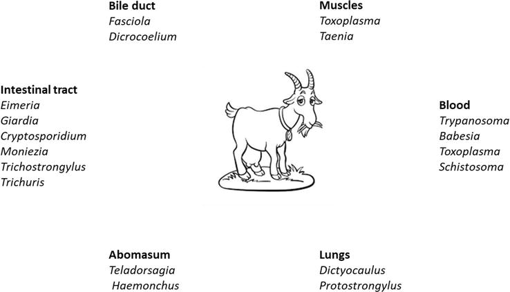
External and internal parasites significantly cause diseases in animals. These parasites cause permanent damage or death to animals, which can lead to economic losses and affect the performance of animals [4]. Overburdened parasites can impair reproductive capacity, limit growth rates, and make animals less productive for meat, fiber, or milk. Endo- and ectoparasites can infect goats and have been reported all over the world. Many nematodes and cestode species infect sheep and goats, causing parasitic gastritis and enteritis. Ostertagia circumcincta, Haemonchus contortus, Bunostomum trigonocephalum, Trichostrongylus axei, Oesophagostomum columbianum, and Nematodirus spp. are the most significant and prevalent around the world [5]. Parasitic species affecting the different organs of goats are given in Figure 1. Arthropod pests reduce goat production while also causing damage to body tissue, skin, hair, and blood loss. These parasites produce sores, cause skin irritation, and make the animal feel uneasy. Arthropods act as vectors and transmit diseases from a sick animal to a healthy animal. To get rid of these arthropods, animals move unintentionally and damage themselves and other animals, which can reduce production and cause weight loss [4].
Figure 1.
Parasitic species affecting the different organs of goats (made by the author).
Fecal examination is one of the best techniques for detecting parasites in parasitic infections, particularly endoparasites. Fecal examination can help identify parasite species and the severity of infection. However, the fecal examination methods failed to detect infection in its early stages. Some alternative methods are currently exploited for the parasitological examination of infections [6]. Antiparasitic drugs can be used to control parasitic infections. But due to the irrational use of existing drugs, parasites are becoming increasingly resistant to standard antiparasitic agents [7].
Goat raring and farming are the only sources of income for most farmers in the world, especially in developing countries like Pakistan, India, Bangladesh, etc. Most parasitic infections (especially chronic infections) are hidden killers for the economies of these farmers. Mostly, farmers are unaware of parasites, their diagnosis, their control, and their effect on the growth and production of goats. To obtain the related published research and data, we searched different databases (ScienceDirect, PubMed, ResearchGate, and available research papers with DOI). To search the data, the following keywords and subject terms were used: goat parasitism, protozoan infection, helminth infection, ectoparasitic infections, Eimeria, Trypanosoma, Giardia, Cryptosporidium, Toxoplasma, Babesia, Fasciloa, Paramphistomum, Schistosoma, Dicrocoelium, Taenia, Moniezia, Trichostrongylus. This chapter aims to compile all information on goat parasites, including pathogenesis, diagnosis, prevention, and control. Other than therapeutic control measures, alternative strategies (phytotherapy, biological control, grazing management, genetic management, and nutrition management) to control resistant parasites in goats are also discussed. Proper diagnosis of parasitic infections and strategic use of drugs can help reduce parasitic infections. The use of alternative parasite control strategies (pasture management, biological control, and phytotherapy) is crucial to reducing the production of resistant parasites.
Protozoans are single-celled parasites that mostly infect the gastrointestinal (GI) tract and circulatory system. Protozoans may reproduce sexually as well as asexually. In amoebas and flagellates, reproduction usually occurs through asexual multiplication in the form of binary fission. In Apicomplexa, the division occurs both sexually and asexually. Some protozoan species only need one host to complete their lifespans, and some of them have complex lifecycles that require two separate host species.
The trophozoite stage is linked to the pathogenesis of parasitic protozoans. In hemoflagellates, different terms such as epimastigote, amastigote, promastigote, and trypomastigote are designated as trophozoite stages based on the presence or absence of flagella. Trophozoite stages of Toxoplasma gondii are known by various names in Apicomplexa, including tachyzoite and bradyzoite. Some additional sexual and asexual stages that are included in the lifecycle of the species of these phyla include gametes, gametocytes, and merozoites, that is, the ones arising from a multinucleate schizont’s fission. Some protozoans also form cysts that have one or more contagious species. Upon multiplication in the cysts of some species, excystation occurs that releases more than one organism [8].
Protozoal parasites that cause coccidiosis to grow in the gut wall and can seriously harm the gut. Eimeria (E.) is a protozoon that causes coccidiosis. Only two (E. caprina and E. ninakohlyakimovae) of the nine different species of Eimeria found in numerous nations are capable of causing serious illness in goats. Other species do not infect goats or just show moderate symptoms [9]. Goat kids are most commonly infected, and this parasite has enormous potential for reproduction. Clinical symptoms may include a sudden onset of pasty diarrhea, abdominal pain, straining to pass feces and mucous, and blood-stained diarrhea [10].
Cryptosporidium is another protozoan parasite that can affect very young kids. This parasite causes diarrhea, which, if severe, can be fatal. Kids with compromised immune systems and insufficient colostrum intake are more likely to experience it. Moving young kids to the infected area, the use of artificial milk, and contaminated feed and water can also cause infection. The species of protozoa present in goat populations and their pathogenesis are given in Table 1.
Species
Predilection Site
Pathogenesis
Drug of Choice / Preventive Measure
References
Eimeria (E.) airlongi, E. caprina, E. ninakohlyakimvae, E. christenseni
Species of protozoa present in goat populations and their pathogenesis.
3.2 Helminths
Helminths are also referred to as parasitic worms and are primarily classified based on their external and internal morphology. Trematodes (flukes), cestodes (tapeworms), and nematodes (roundworms) are the three primary types of helminths. Except for blood flukes, helminths (tapeworms and flukes) have hermaphroditic species. Nematodes are the most prevalent class of helminths all around the world; they are bisexual and commonly called roundworms [16]. Nematodes are an important goat endoparasite and live in the gut and stomach.
Goats infected with nematodes shed parasitic eggs in their feces, which remain viable for a period of time, depending on temperature and humidity. During grazing, the infective stage (L3 on the grasses or L1 within the egg) is ingested by the goat and reaches its predilection side, where it causes damage. Nematodes, which belong to the genera Haemonchus, Ostertagia, Nematodirus, Cooperia, Oesophagostomum, and Trichostrongylus, are prevalent in goat populations. Diarrhea and weight loss are common signs in goats due to the presence of worms in the gut. Haemonchus (H.) contortus (Barber’s Pole Worm) is present in the abomasum (the fourth stomach) and causes anemia because they suck blood. Nematodes are important in goats as compared to sheep and cattle because they have low immunity and are susceptible to re-infection [17].
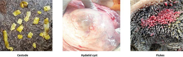
Cestodes are segmented, flat, and quite long worms present in the gut of the goat. One of the main species is Moniezia. The eggs or gravid segments are passed in the feces and ingested by oribatid mites (intermediate hosts). During grazing, goats ingest this mite along with grasses and get the infection. Most tapeworm infections are nonpathogenic, but heavy infections can lead to blockage of the intestine, GI disturbances, and thriftiness [18]. Flukes like Dicrocoelium dendriticum, Paramphistomum spp., Cotylophoron spp., and Fasciola spp. are commonly found in the goat population. Some of the helminths collected from slaughtered goats are given in Figure 2. All trematodes have an indirect life cycle, and the snail is one of the compulsory intermediate hosts. Some trematodes need another intermediate host other than a snail to complete their life cycle [19]. The species of helminths present in goat populations and their pathogenesis are given in Table 2.
Figure 2.
Helminths collected from slaughtered goats (made by the author).
Specie
Predilection site
Pathogenesis
Drug of Choice / Preventive Measure
References
Fasciloa (F.) hepatica, F. Magna
Bile duct
Progressive weight loss, anemia, edema (build of fluid, typically under the jaw), and sudden death
Species of helminths present in goat populations and their pathogenesis.
Worldwide, helminth infections in animals are the cause of many common and economically significant diseases. In essence, these parasites represent a risk to any livestock with outside access. Even though helminth infections can sometimes result in significant mortality rates, they typically influence overall health and welfare and lead to hidden productivity losses like lowered weight gain, milk supply, and wool and hair growth [22, 26, 30].
3.3 Arthropods
Arthropods are classified into insects and arachnids. Insects include lice, fleas, bugs, and flies, while arachnids include ticks and mites.
3.3.1 Insects
Goat lice are host-specific and only present on goats, or sometimes on sheep. Three biting species of lice, Bovicola (B.) caprae, B. limbata, and B. crassipes, are the main species of goat lice. These species feed on hair and skin and cause dermatitis. Three blood-sucking lice species of goats are Linognathus (L.) stenopsis, L. africanus, and L. pedalis. The L. stenopsis can be present all around the body, the L. africanus is usually found around the neck and head regions, and the L. africanus is found on the legs and feet of goats [31].
Itching, irritability, and scratching are all symptoms of pediculosis brought on by lice infestation. The lice infestation can be identified by the matted and dull coat of the animal, grooming behavior, and excessive scratching. Due to irritation caused by louse bites, animals scratch and rub with different objects, which can lead to skin damage and hair loss. Production losses and weight loss were reported due to improper nutrition, restlessness, and nervousness, while heavy infestation may lead to anemia. Within a herd, lice infestation can be transmitted through direct contact, while herd-to-herd infestation can be accomplished by introducing the infected animals. Lice can also move from one farm to another by clinging to flies [32].
Small (1 mm to 8 mm), wingless fleas with flattened sides and spines (combs) facing backward are known to infect goats. The majority of species move around a lot and only stay on the host for a portion of the time in order to feed on blood. The legs are strong and can be used to jump large distances. The Ctenocephalides felis (cat flea) and the Echidnophaga gallinacea (sticktight flea) are two species that frequently infest goats. Domestic goats have reported cases of severe anemia linked to a lot of cat flea bites. E. gallinacea tightly adheres to its host, typically near the ears and face. This species can stay attached to its host for 2–3 weeks and may spread in large populations, leading to ear and head ulcers. If fleas become an issue in a goat herd, extra precautions for monitoring herd dogs should be put in place because both of these flea types are quickly transferred to other animals [33].
Flies undergo a complete metamorphosis (eggs, larvae, pupae, and adult stage), and each stage inhabits a different habitat. Goats are particularly sensitive to species of flies like horn flies, stable flies, black flies, house flies, blow flies, mosquitoes, and horse flies. Flies can be extremely annoying and may have an impact on the goats’ performance. They prevent grazing and make goats group together or flee in order to escape their annoyance. Flies can bite (which is very painful) or suck blood, which can cause significant irritation to goats and transmit several diseases [34].
Horn flies are a common problem for cattle, but they can also occasionally be seen on goats, especially when goats and cattle use the same pasture. Both female and male horn flies feed on the blood of the host between 20 and 30 times a day. Horn flies remain on the animal constantly, only leaving to lay their eggs [35]. Stable flies (medium-sized flies) are similar to house flies and prefer to remain on goats’ feet and legs. Stable flies bite their hosts in a highly painful way and cause damage. On the biting side, blood may ooze out, which may be fatal in cases of heavy infestation. Goats on pasture frequently gather and mill around in large numbers due to stable fly infestations. Stable fly larvae grow on filthy hay, spilled feed, and straw bedding that is damp and decomposing organically [36]. Although some types of black flies and mosquitoes can feed on goats, they are typically not present in large enough numbers to cause huge damage. During the late spring season, the population of black flies and mosquitoes increases. Both of these flies require bodies of water to develop their larvae. Mosquito larvae grow in stagnant water, while black fly larvae develop in moving water. The greatest danger posed by mosquitoes is that they can spread disease [37].
House flies have a simple sponging mouthpart and do not bite goats. House flies do not directly harm animals but can irritate them and act as mechanical vectors for different pathogens. House flies can be a serious nuisance to confined animals, especially young goats. The attack of house flies increases when animals and sheds are not cleaned properly. Lacrimal, nasal, oral, and other goat secretions attract house flies. House flies rarely bother pastured goats unless they frequently visit loafing shelters. Although blowflies do not bite goats, they may be very annoying to both animals and livestock handlers, like house flies. The larvae of blowflies and houseflies grow in rotting organic waste, and decomposing animal remains. The best way to prevent these flies around barns is to practice good cleanliness. Premise sprays can be used inside buildings to suffocate adult flies. To expose flies to insecticide residue, surface areas in barns can be sprayed with premises sprays [38].
The adult nose-bot fly does not cause any harm to animals, but larvae live in goats’ nostrils. Larvae mature in the head sinuses before moving back down the nasal passages and finishing their development on the ground. Nasal membranes become inflamed as a result of migrating larvae (migrating to and from the head sinuses). Secondary infections can develop at the sites of migration. Animals that are infected show symptoms that include grinding of teeth, loss of appetite, excessive head shaking, and nasal discharge. A nose-bot infestation can also be identified by the presence of blood specks in the nasal discharge. When mature bot flies are present, goats exhibit unusually excitable behavior [39]. Ivermectin is very effective against larvae in all stages. Eprinomectin and doramectin are some other medications that have been said to be effective [40]. Keds are wingless flies that live their whole lives on goats or sheep. The transmission of keds from one to the other occurs through direct contact. Melophagus ovinus (sheep keds) is a problem in sheep but can also occur in goats. During her six-month life span, a female sheep kid gives birth to 10–15 young (one every eight days). The mouthparts of adult kids, which are sharp and resemble mosquito stingers, are used to feed. The feeding behavior of sheep kids causes severe irritation, and the animal rubs, bites, and scratches the body with other objects [41]. This condition is known as “cockle,” which decreases the market value of hide because it weakens and discolors it [42].
3.3.2 Arachnids
Several species of mites can infest the goat. The most common species of mites that infest goats are Demodex caprae (goat follicle mites), Psoroptes cuniculi (ear mites), Sarcoptes scabiei (scabies mites), and Chorioptes bovis (chorioptic scab mite). Demodectic mange is caused by Demodex caprae, which can produce cutaneous papules and nodules. These nodules or papules are caused by gland ducts or hair follicles becoming clogged and forming swellings. Demodectic mange mostly affects pregnant, dairy, and young goats. Nodules may burst and release mites, which could spread the mite to other animals. Close contact with infected animals, especially during mating or mingling, and the licking of infested animals are other means of mites’ transmission. Sarcoptic mange is caused by the scabies mite, which can burrow into the skin and cause varying degrees of dermatitis. In goats, a mild infestation of sarcoptic mange can be resolved without any severe clinical signs. However, in heavily infected goats, extensive hair loss (around the eyes, ears, and muzzle) and crusty lesions can be observed. Psoroptes cuniculi (ear mites) infestation causes lesions in or on the ear. These lesions result in the development of a crust and the discharge of an unpleasant odor into the external ear canal. The behavioral reactions due to ear mange include ear scratching, spasmodic contractions of neck muscles, loss of balance, and head shaking. Goats that are typically younger than a year typically have greater rates of infection than older animals. Weight loss and anemia are commonly seen in goats with chronic infestations of these mites [43]. In goats and domestic animals, chorioptic mange is caused by Chorioptes bovis. Common sites of infestation of this mite are the legs and feet of goats, especially the forefeet. These mites can also be found higher on the foot. Most lesions are small and barely noticeable. To get rid of mites’ infestation, treatment and control efforts should concentrate on all the animals in a herd. Retreatment of animals is necessary between 10 and 12 days to kill the newly hatching eggs. Isolating new animals should be performed to reduce the chances of mite introduction into herds [44].
Tick infestations not only damage the hide of animals but are also responsible for blood loss and the transmission of several pathogens. Ticks are divided into three groups (one-host, two-host, and three-host ticks) on the basis of the number of animals required to complete their life cycle. Ticks that parasitize goats are primarily members of the three-host tick family. Three-host ticks require three different animals to complete their whole life cycle, which can make their control challenging. There are three types of ticks known to parasitize goats, despite the fact that they are not frequently observed on goats. Dermacentor variabilis (American Dog Tick), Amblyomma maculatum (Gulf Coast Tick), and Amblyomma americanum (Lone Star Tick) are commonly found on goats [45].
The neck and withers are common A. americanum infestation sites on goats. Ticks can sometimes be found in the armpits and on the head. The lone mark on the back of adult females makes them easy to identify. The rear edge of adult males has non-connecting white marks. As compared to other ticks, the mouthparts of this tick are substantially longer. According to studies, goats can act as reservoirs for the bacteria Ehrlichia chaffeensis, which is transmitted by Lone Star ticks and causes the disease known as monocytic ehrlichiosis in humans. Due to this kind of zoonotic importance, care should be taken to handle goats infested with A. americanum [46]. According to a seasonal cycle, the infestation of goats by Gulf Coast ticks starts in the early spring and lasts until mid-summer. Throughout the summer, goats are infested with Lone Star ticks and American Dog ticks. All of these tick species should be controlled by targeted acaricidal applications; however, repetition may be necessary after three weeks. Extreme caution should be exercised when choosing an acaricide to treat the goats because there are very few approved acaricides for use in goats [47]. Species of arthropods infest the goat population, and their pathogenesis is given in Table 3.
Arthropod
Specie
Predilection site
Pathogenesis
Drug of Choice / Preventive Measure
References
Lice
Bovicola (B.) caprae, B. crassipes, B. Limbata, Linognathus (L.) stenopsis, L. africanus, Linognathus pedalis
Species of arthropods infesting the goat population and their pathogenesis.
3.4 Trends to diagnose goat parasitism
Infected animals’ clinical signs and symptoms vary depending on parasite predilection sites and feed requirements. For example, if the parasite is disrupting the digestive tract, the most common signs associated with this infection are diarrhea, weight loss, a rough coat, depression, and anorexia. If a parasite is infecting the stomach or liver and consuming blood, then the common signs are anemia, pale mucous membrane of the inner eyelid, and weight loss. Laboratory findings in such cases include plasma protein loss, decreased packed cell volume, and an increase in fecal egg count.
3.4.1 Conventional methods
The fecal egg count (FEC) is a technique used to assess how many parasite eggs are expelled per gram of feces (EPG). Although this is the best technique for use with live animals, there are some measuring challenges, such as the fact that the EPG does not indicate the number of worms because adult worms of each parasite species lay a different number of eggs. Identification of eggs does not mean the exact identification of parasitic species, and some EPG determination procedures could be less accurate than others.
It has been demonstrated that the FEC, in particular for H. contortus (the barber pole worm), primarily reflects the animal’s worm burden and acts as a seasonal signal of variations in the degree of infection. The relative direction of infection can be seen by looking at trends in FEC over time. When worms other than H. contortus are prevalent, FEC is a less reliable indicator of adult worm loads [48]. Some diagnostic laboratories may also offer services for the culture of feces in order to hatch worm eggs and collect larvae. This makes it possible to recognize the specific species of intestinal or abdominal parasites present in the herd. To determine the degree of resistance against anthelmintics, “drench-rite tests” can be performed to choose the best effective drug [49].
The maintenance of an animal’s “erythropoiesis,” or capacity to produce red blood cells, can be impacted by nematode parasites. The percentage of red blood cells in the blood is known as the packed cell volume (PCV). The normal value of PCV typically exceeds 30%. Anemia usually manifests itself when PCV falls below 20%. Nematode infection may lead to chronic anemia, which means red blood cell production is not as fast as needed to meet demand. Notably, H. contortus can cause severe acute blood loss and mortality. The PCV readings are not always used as a “stand-alone” diagnostic tool; they have been used to complement other response criteria [50].
To determine the level of anemia, the color of the mucous membrane (where several capillaries are close to the surface) of animals like the gums, the vulva, and the eyelid can be observed. If these membranes are extremely pale, deworming is necessary right away since death is imminent. In South Africa, the FAMACHA eye color chart method was created to assist farmers in monitoring and assessing the degree of anemia without the need for laboratory testing. This method involves looking at the lower eyelid mucous membranes and comparing them to a laminated color chart with sheep eyes at five various levels: 1) Red means non-anemic, 2) red-pink means non-anemic, 3) pink means mildly anemic, 4) pink-white means anemic, and 5) white means severely anemic.
This approach can be useful for identifying animals that need therapy. But this technique is only valid for blood-sucking nematodes like H. contortus, as anemia is the main pathologic consequence of infection. The comprehensive testing of FAMACHA in South Africa and the US produced remarkable results. It has been shown that if animals were checked weekly and only salvage treatments were given, up to 70% of adult animals would not require deworming, and only a small number would require more than one treatment. The overall number of treatments may be reduced by up to 90% as compared to earlier treatment plans. This prevents the development of dewormer resistance because the majority of the worms would not be exposed to anthelmintics [51, 52].
The direct and absolute method for identifying and determining the exact number of parasites is to examine the animals immediately after slaughter or death to collect, count, and identify the parasites present. Only a veterinarian or other qualified specialist can perform this with high precision. Haemonchus contortus, which is present in the lining of the abomasum, can help determine the severity of the infection. It should be noted that the animal must not have been dead for very long. The likelihood of discovering worms increases with how recently the animal died. This is due to the fact that the worms will eventually travel as deep down the intestine as they can after dying. Telodorsagia and Trichostrongylus must be noted as being too small to be seen without a microscope. Even if thousands of these worms are present, they are hidden by the contents of the stomach and are invisible to the naked eye [49].
3.4.2 Serology-based assays
In live animals, where tissue or biologic samples are inaccessible, the identification of parasitic infection through a serology-based assay is the gold standard. Antibody and antigen detection are the two categories of serology-based diagnostic assays. Enzyme-linked immunosorbent assay (ELISA) is an important serology-based assay with different types like the Falcon assay screening test, ELISA, and dot-ELISA. Other serology-based tests used for identification of parasites include the hemagglutination (HA) test, the complement fixation (CF) test, the indirect immunofluorescent antibody (IFA) test, the direct immunofluorescent antibody (DFA) test, the immunoblotting test, and quick diagnostic procedures [53].
Serology-based tests are more specific and sensitive, even though they are easier to use and take less time to complete. It is crucial for differential diagnosis in animals where blood smears fail to identify the parasite, for example, Babesia and Plasmodium infections, or in animals with low parasitemia or asymptomatic infections [54]. It is possible for the parasite to spread after organ or blood transplants if an infected patient is asymptomatic and declared negative. When egg production is low or irregular, serology testing for the parasitic infection has been demonstrated to be helpful in confirming the chronic infection [55]. Due to antigenic variation, the sensitivity and specificity of a serology assay can vary in different regions. A serology assay of one parasitic infection can also show cross-reactivity with other related parasites due to the presence of the same antigens.
3.4.3 Nucleic acid-based approaches
Due to the limitations of the above-mentioned assays (microscopy and serology assays), scientists developed the polymerase chain reaction (PCR), which amplifies the specific genes of a specific parasite. With the advancement in diagnosis, traditional PCR is updated to nested PCR (improving sensitivity and specificity), multiplexed PCR (amplification of several different DNA sequences at the same time), and real-time PCR (quantification of the product after each cycle). Luminex-based assays and loop-mediated isothermal amplification are new emerging techniques for the identification of parasitic infections [53, 56]. The sensitivity and specificity of nucleic acid-based techniques are much higher than those of existing diagnostic assays. In addition, nucleic acid-based techniques can detect infection in animals with low parasitemia or even asymptomatic infection [57].
3.5 Prevention and control of parasitic infections
Parasitic protozoan infections have long been regarded as one of the world’s most serious unresolved livestock and public health issues, particularly in most tropical regions. Leishmaniasis, coccidiosis, and American and African trypanosomiasis account for the highest mortality rates in developing countries because of a lack of hygienic practices and inefficient prophylactic measures [58]. Most of these diseases were thought to be extinct in developed countries, but they are now resurfacing as opportunistic infections, typically in immunocompromised people. The emerging of these diseases may be a result of tourism, immigration flows (particularly from endemic areas), and global warming brought on by dramatic climatic changes [59].
The development of protective vaccinations has been the subject of parasitology, but their effectiveness is still not progressive. Chemotherapy is still the mainstay of care for the majority of parasite infections. The drugs that are currently being used were first launched decades ago, and some of them even have significant effects. But due to the irrational use of most of the anti-protozoal drugs, their efficacy has decreased. In the past few years, there has been little success reported in the development of new anti-protozoal drugs. However, due to recent technological advancements, research has been done to develop new antiparasitic drugs. The establishment of public-private partnerships that concentrate on tropical parasitic diseases and the sequencing of parasitic genomes are the two most significant developments [60]. The control of parasites, especially helminths, relies on pasture management practices, routine checkups of animals, efficient use of chemotherapeutics, and strict adoption of hygienic measures.
Arthropods may be a problem for man because they contaminate his food and other resources. Arthropods spread several infectious diseases to animals and irritate or harm them directly. For these reasons, man may need to control, eliminate, or repel them. When arthropods are present around goats, they can cause a significant problem. Insecticide-based control should be required to reduce damage and production losses. Permethrin- or pyrethroid-based products are the only ones that have been allowed to treat goats. Those that contain piperonyl butoxide deliver the best results when combined with pyrethroid products. When only treating the barns, goat farmers have more alternatives, which are frequently referred to as “premise sprays.” As opposed to a non-residual substance like pyrethrum, a residual spray that lasts for a while is the best choice for a premises spray. Make sure to treat vertical arthropod-resting places, such as barn walls, while using residual sprays. When applying the product, make sure the surface is neither greasy nor moist. Automated misting systems are now being used by more livestock owners who keep their animals mostly in barns. These can be useful, but caution should be exercised when using them, especially if there is hay or animal feed nearby. Insecticide resistance might also develop as a result of excessive use of these systems [38]. Other means to control arthropods are physical measures (such as heat or cold), biological interference, the use of chemical repellents, trapping of arthropods, and destruction of their habitat.
3.6 Alternatives to antiparasitic drug treatment
3.6.1 Use of herbal anthelmintics
In animals, parasites can cause acute and chronic infections, which ultimately reduce production (growth rate, milk production, and meat production). Along with the systematic use of antiparasitic agents, research has shown the positive role of complementary and alternative medicine in treating parasitic infections. Various plants or plant products have been traditionally used in veterinary medicine for their antiparasitic activity. The need of the hour is to standardize the use of these phytogenic substances for parasite control at the farm level. Indigenous plants that can be used for their antiparasitic effects include Heyysarvum coronarium, Heracleum spp., Allium sativum, Artemisia maritime, Melia azadarach, Artemisia vulgaris, Chrysanthemum spp., Areca catechu, Calotropis procera, Carica papaya, and Azadirachta indica [61, 62].
The juice of crushed leaves of Aloe ferox can be applied to the skin to treat tick and mite infections, and if this juice is used with drinking water, it can control the helminth infection. Elephantorrhiza elephantina root boiling water can be used to treat arachnid (spray on skin) and helminth (drinking water) infections. Boiling the leaves of Acokanthera oppositifolia and Bulbine latifolia is very useful to control all kinds of parasitic infections. An extract of the bark of Centella coriacea and Cussonia spicata and an extract of the leaves of Agapanthus praecox and Albuca setosa have shown anthelmintic effects [63].
3.6.2 Biological control
Biological control of parasitic infections can be done either by using agents of plant origin, like grasses, or agents of zoological origin, such as bacteria, viruses, fungi, predators, and parasites. Fungi having antiparasitic activity have been known for a long time. Fungi are divided into three groups based on their morphology, spectrum, and efficacy. One is the predacious fungi, which have specialized structures to trap nematodes on their mycelium, such as adhesive knobs, rings, and networks. Important predatory fungi include Monacrosporium spp. and Arthobotrys spp. [64]. The second group consists of the endoparasitic fungi, which produce spores. Examples include Harposporium anguillulae and Drechmeria coniospora [65]. The third group includes egg parasitic fungi that have ovicidal activity and can be pivotal in the control of those nematodes that are capable of surviving for longer durations in the environment in the egg stage. Verticillium chlamydosporium has been shown to be effective against Ascaris lumbricoides eggs from naturally infected pigs [66].
3.6.3 Grazing strategies
Stocking density is an important aspect of controlling parasitic infections, as it is directly linked to the exposure of animals to infective larvae as well as pasture contamination. A general guideline considers five sheep or five to seven goats equal to one cow, and it is suggested to graze five to seven goats per acre of pasture. Sheep and goats have different grazing habits. Sheep prefer grazing on the ground, while goats prefer to browse on trees and bushes. Pasture management also includes careful inspection of the grazing area to avoid overgrazing and sustain productive pastures. It includes the use of a grazing area that is not contaminated with parasitic larvae and has not been used for grazing sheep and goats over the last 6 to 12 months, but it could be used for grazing cattle or horses. It also includes the removal of silage crops or hay. The burning pasture will eliminate parasitic larvae [67].
3.6.4 Genetics management
It is the best and most long-term remedy to control parasitic infections, as some breeds of animals are more resistant to parasitic infections. Selecting animals for parasitic resistance can be an important tool in this regard, and it does not have any bad effects on the animal’s growth. Nowadays, breeding policies are concentrating on the development of breeds that are resistant to parasitic infections. This trait has a heritability of 20–40%. Grazing resistant animals with susceptible animals on the same pasture can reduce the challenge to susceptible animals as resistant animals may act to sweep the pasture [68].
3.6.5 Nutritional management
Nutritional management plays a key role in controlling parasitic infections. The animals’ protein intake is directly related to their ability to resist nematode infections. Protein-deficient animals have poor immunity due to lower levels of IgA and are thus less resistant to parasitic infections. Vitamins such as A, B12, and E and minerals like Fe, Cu, Co, Zn, and P also provide a better immunity against parasitic infections. The addition of molybdenum at 6–10 mg/day has resulted in a decreased worm burden in lambs, which is not attributable to copper deficiency. A probable reason could be an increase in blood eosinophil levels and jejunal mast cells due to molybdenum administration [69].
3.7 Conclusions
The goat population contributes greatly to the world economy and plays a role in food security. Goat raring necessitates minimal infrastructure and investment. Like other animals, goats are also susceptible to both endo- and ectoparasites. Parasitic infection reduces the production (of milk, meat, and hair) and is responsible for economic losses. Internal parasites (protozoa and helminths) damage the organs, especially the gastrointestinal tract, and reduce the absorption of digested food. External parasites (lice, fleas, flies, ticks, and mites) damage the hide, are responsible for blood loss, and transmit several pathogens. The timely diagnosis of parasitic infections reduces the damage and production losses. With the advancement of diagnosis, parasitic infections can be detected easily and more accurately. Several antiparasitic drugs are available to treat parasitic infections, but irrational uses of these drugs can develop resistant parasites. Alternative techniques like phytotherapy, biological control, grazing management, bioactive crops, and nutrition are in practice to reduce the chances of parasitic resistance.
References
1.Ensminger ME, Parker RO. Sheep and Goat Science. 5th ed. Danville, Illinois: The Interstate Printers and Publishers Inc.; 1986. DOI: 10.1007/s11882-014-0421-0
2.Monteiro A, Costa JM, Lima MJ. Goat System productions: Advantages and disadvantages to the animal, environment and farmer. Kukovics S, editors. Goat Science. UK: IntechOpen; 2018. DOI: 10.5772/intechopen.70002
3.Faostat. 2008. Available from: http://faostat.fao.org/default.aspx. 10.1016/j.appet. 2017.08.21
4.Batool A, Sajid MS, Rizwan HM, Iqbal A, Rashid I, Jan I, Bano F, Ahmad F, Ahmad W, Khan MN. Association of various risk factors with the distribution of gastrointestinal, haemo and ectoparasites in small ruminants. Journal of Animal Health and Production. 2022;10(2):204-213. DOI: http://dx.doi.org/10.17582/journal.jahp/2022/10.2.204.213
5.Jas R, Kumar D, Bhandari A, Pandit S. Seasonal alteration in prevalence and intensity of naturally occurring gastrointestinal helminth infection in goats of new alluvial zone of West Bengal, India. Biological Rhythm Research. 2017;48:867-876
6.Jas R, Ghosh JD, Das K. Polyclonal antibody based coproantigen detection immunoassay for diagnosis of Oesophagostomum columbianum infection in goats. Veterinary Parasitology. 2010;170:262-267
7.Datta S, Dandapat P, Jas R. Diagnosis of mixed gastrointestinal nematode infection in goat by an indirect-ELISA. Iranian Journal of Veterinary Research. 2018;19(3):189-193
8.Hill DE, Dubey JP. Toxoplasma gondii. In: Ortega YR, Sterling CR, editors. Foodborne Parasites. Cham: Springer; 2018. pp. 119-138
9.Etsay K, Megbey S, Yohannes H. Prevalence of sheep and goat coccidiosis in different districts of Tigray region, Ethiopia. Nigerian Journal of Animal Science. 2020;22(3):61-69
10.Wang CR, Xiao JY, Chen AH, Chen J, Wang Y, Gao JF, et al. Prevalence of coccidial infection in sheep and goats in northeastern China. Veterinary Parasitology. 2010;174(3-4):213-217
11.Mohamaden WI, Sallam NH, Abouelhassan EM. Prevalence of Eimeria species among sheep and goats in Suez governorate, Egypt. International Journal of Veterinary Science and Medicine. 2018;6(1):65-72. DOI: 10.1016/j.ijvsm.2018.02.004
12.Batista JS, Oliveira AF, Rodrigues CM, Damasceno CA, Oliveira IR, Alves HM, et al. Infection by Trypanosoma vivax in goats and sheep in the Brazilian semiarid region: From acute disease outbreak to chronic cryptic infection. Veterinary Parasitology. 2009;165(1-2):131-135. DOI: 10.1016/j.vetpar.2009.07.005
13.Jafari H, Jalali MH, Shapouri MS, Hajikolaii MR. Determination of Giardia duodenalis genotypes in sheep and goat from Iran. Journal of Parasitic Diseases. 2014;38(1):81-84. DOI: 10.1007/s12639-012-0199-8
14.Paraud C, Chartier C. Cryptosporidiosis in small ruminants. Small Ruminant Research. 2012;103(1):93-97. DOI: 10.1016/j.smallrumres.2011.10.023
15.Kage S, Mamatha GS, Lakkundi JN, Shivashankar BP, D'Souza PE. Detection of incidence of Babesia spp. in sheep and goats by parasitological diagnostic techniques. Journal of Parasitic Diseases. 2019;43(3):452-457. DOI: 10.1007/s12639-019-01109-3
16.Cattadori I, Haydon D, and Hudson P. Parasites and climate synchronize red grouse populations. Nature 2005;433:737-741. https://doi.org/10.1038/nature03276
17.Mickiewicz M, Czopowicz M, Moroz A, Potărniche AV, Szaluś-Jordanow O, Spinu M, et al. Prevalence of anthelmintic resistance of gastrointestinal nematodes in polish goat herds assessed by the larval development test. BMC Veterinary Research. 2021;17(1):1-12
18.Zvinorova PI, Halimani TE, Muchadeyi FC, Matika O, Riggio V, Dzama K. Prevalence and risk factors of gastrointestinal parasitic infections in goats in low-input low-output farming systems in Zimbabwe. Small Ruminant Research. 2016;143:75-83
19.Domke AVM, Chartier C, Gjerde B, Leine N, Vatn S, Stuen S. Prevalence of gastrointestinal helminths, lungworms and liver fluke in sheep and goats in Norway. Veterinary Parasitology. 2013;194(1):40-48
20.Hashemnia M, Rezaei F, Nikousefat Z, Ghashghaii A. Acute caprine fasciolosis: A case with unusual migration to lung. Journal of Parasitic Diseases. 2015;39(3):514-517. DOI: 10.1007/s12639-013-0387-1
21.Godara R, Katoch R, Yadav A, Rastogi A. Epidemiology of paramphistomosis in sheep and goats in Jammu, India. Journal of Parasitic Diseases. 2014;38(4):423-428. DOI: 10.1007/s12639-013-0264-y
22.Shen XH, Li YF, Wang L, Yang WH. Investigation on Schistosoma japonicum infection in goats in Dantu District, Zhenjiang City from 2004 to 2019. Zhongguo Xue Xi Chong Bing Fang Zhi Za Zhi. 2020;32(4):393-396. DOI: 10.16250/j.32.1374.2020008
23.Godara R, Katoch R, Yadav A, Borah MK. Dicrocoeliosis in goats in Jammu, India. Journal of Parasitic Diseases. 2014;38(2):201-204. DOI: 10.1007/s12639-012-0212-2
24.Hajipour N, Allah Rashidzadeh H, Ketzis J, Esmaeili Seraji R, Azizi H, Karimi I, et al. Taenia ovis in small ruminants in Iran: Prevalence, pathology, and economic loss. Veterinary Science. 2020;7(1):34. DOI: 10.3390/vetsci7010034
25.Iacob OC, El-Deeb WM, Paşca SA, Turtoi AI. Uncommon co-infection due to Moniezia expansa and Moniezia benedeni in young goats from Romania: Morphological and histopathological analysis. Annals of Parasitology. 2020;66(4):501-507. DOI: 10.17420/ap6604.291
26.Macaldowie C, Jackson F, Huntley J, McKellar A, Jackson E. A comparison of larval development and mucosal mast cell responses in worm-naïve goat yearlings, kids and lambs undergoing primary and secondary challenge with Teladorsagia circumcincta. Veterinary Parasitology. 2003;114:1-13
27.Elseadawy R, Abbas I, Al-Araby M, Abu-Elwafa S. Molecular identification of different Trichostrongylus species infecting sheep and goats from Dakahlia governorate, Egypt. Journal of Parasitic Diseases. 2021;45(1):218-227. DOI: 10.1007/s12639-020-01299-1
28.Gul N, Tak H. Prevalence of Trichuris spp. in small ruminants slaughtered in Srinagar District (J&K). Journal of Parasitic Diseases. 2016;40(3):741-744. DOI: 10.1007/s12639-014-0570-z
29.Zafari S, Mohtasebi S, Sazmand A, Bahari A, Sargison ND, Verocai GG. The prevalence and control of lungworms of pastoral ruminants in Iran. Pathogens. 2022;11:1392. DOI: 10.3390/pathogens11121392
30.Arsenopoulos KV, Fthenakis GC, Katsarou EI, Papadopoulos E. Haemonchosis: A challenging parasitic infection of sheep and goats. Animals (Basel). 2021;11(2):363. DOI: 10.3390/ani11020363
31.González-Álvarez VH. Presence of two lice species (Insecta: Phthiraptera) in a goat (Capra hircus) from La Comarca Lagunera, Mexico: A case report. International Journal for Research in Applied Sciences and Biotechnology. 2020;7(5):152-155
32.Fentahun T, Woldemariam F, Chanie M, Berhan M. Prevalence of ectoparasites on small ruminants in and around Gondar town. American-Eurasian Journal of Scientific Research. 2012;7(3):106-111
33.Ajith Y, Dimri U, Gopalakrishnan A, Devi G. A study on prevalence and factors associated with ectoparasitism in goats of two agro-climatic regions in India. Journal of Parasitic Diseases. 2017;41(3):739-746
34.Yadav A, Khajuria JK, Soodan JS. Warble fly infestation in goats of Jammu. Journal of Veterinary Parasitology. 2006;20(2):149-152
35.Brewer GJ, Boxler DJ, Domingues LN, Fryxell RT, Holderman C, Loftin KM, et al. Horn fly (Diptera: Muscidae) biology, management, and future research directions. Journal of Integrated Pest Management. 2021;12(1):42
36.Sinshaw A, Abébé G, Desquesnes M, Yoni W. Biting flies and Trypanosoma vivax infection in three highland districts bordering lake Tana, Ethiopia. Veterinary Parasitology. 2006;142(1-2):35-46
37.Bashar A, Hossen M, Chowdhury SR, Hossain M, Rahman M, Rahman M. Prevalence of haemoprotozoan diseases in Black Bengal goats of Sylhet region of Bangladesh. Alexandria Journal for Veterinary Sciences. 2020;65(1):70-80
38.Kangume M, Muhangi D, Byaruhanga J, Agaba A, Sserunkuma J, Kisembo SJ, et al. Tsetse Fly Distribution and Occurrence of Trypanosoma Species among Cattle and Goats around Queen. Uganda: Elizabeth National Park; 2020. DOI: 10.21203/rs.3.rs-32266/v2
39.Sotiraki S, Hall MJ. A review of comparative aspects of myiasis in goats and sheep in Europe. Small Ruminant Research. 2012;103(1):75-83
40.Ahaduzzaman M. The global and regional prevalence of oestrosis in sheep and goats: A systematic review of articles and meta-analysis. Parasites and Vectors. 2019;12(1):1-17
41.Kassayeq E, Kebede E. Epidemiological study on manage mite, lice and sheep keds of small ruminants in Tigray region, northern Ethiopia. Ethiopian Veterinary Journal. 2010;14(2):51-66
42.Mwaki DM, Kidambasi KO, Kinyua J, Ogila K, Kigen C, Getange D, et al. Molecular detection of novel Anaplasma sp. and zoonotic hemopathogens in livestock and their hematophagous biting keds (genus Hippobosca) from Laisamis, northern Kenya. Open Research Africa. 2022;5(23):1-13
43.Zeryehun T, Mengesha L. Prevalence of mange mites of goats in and around Kombolcha, South Wollo, Amhara National Regional state, Northeastern Ethiopia. World Applied Sciences Journal. 2012;19:106-111
44.Yasine A, Kumsa B, Hailu Y, Ayana D. Mites of sheep and goats in Oromia zone of Amhara region, north eastern Ethiopia: Species, prevalence and farmers awareness. BMC Veterinary Research. 2015;11(1):1-6
45.Kifle T, Mathewos M, Fesseha H, Abate A, Wolde A. Study on prevalence of ixodid ticks of goats and acaricide utilization practices of herd owners in Benatsemay District, south Omo zone, South-Western Ethiopia. Veterinary Medicine: Research and Reports. 2021;12:225
46.Cisak E, Wójcik-Fatla A, Zajac V, Sroka J, Buczek A, Dutkiewicz J. Prevalence of tick-borne encephalitis virus [TBEV] in samples of raw milk taken randomly from cows, goats and sheep in eastern Poland. Annals of Agricultural and Environmental Medicine. 2010;17(2):283-286
47.Johns JL, Higgins B, Schroller SG, Flanders MM, Heller MC. Test comparison for the detection of Anaplasma phagocytophilum antibodies in goats, and prevalence of granulocytic anaplasmosis in goats from Northern California and Southern Oregon. Small Ruminant Research. 2022;207:106608
48.Sargison ND. Understanding the epidemiology of gastrointestinal parasitic infections in sheep: What does a faecal helminth egg count tell us? Small Ruminant Research. 2013;110(2-3):78-81
49.Sandoval E, Morales G, Ybarra N, Barrios M, Borges J. Comparison between two McMaster egg counting slide used for the diagnostic of gastrointestinal nematode infection in ruminants. Zootecnia Tropical. 2013;29(4):495-501
50.Luethy D, Stefanovski D, Salber R, Sweeney RW. Prediction of packed cell volume after whole blood transfusion in small ruminants and South American Camelids: 80 cases (2006-2016). Journal of Veterinary Internal Medicine. 2017;31(6):1900-1904
53.Ndao M. Diagnosis of parasitic diseases: Old and new approaches. Interdisciplinary Perspectives on Infectious Diseases. 2009;2009:278246
54.Carter WJ, Yan Z, Cassai ND, Sidhu GS. Detection of extracellular forms of babesia in the blood by electron microscopy: A diagnostic method for differentiation from Plasmodium falciparum. Ultrastructural Pathology. 2003;27(4):211-216
55.Hillyer GV, de Galanes MS, Rodriguez-Perez J, Bjorland J, Silva de Lagrava M, Ramirez Guzman S, et al. Use of the FalconTM assay screening test-enzyme-linked immunosorbent assay (FAST-ELISA) and the enzyme-linked immunoelectrotransfer blot (EITB) to determine the prevalence of human fascioliasis in the Bolivian Altiplano. The American Journal of Tropical Medicine and Hygiene. 1992;46(5):603-609
56.Zarlenga DS, Higgins J. PCR as a diagnostic and quantitative technique in veterinary parasitology. Veterinary Parasitology. 2001;101(3-4):215-230
57.Mens P, Spieker N, Omar S, Heijnen M, Schallig H, Kager PA. Is molecular biology the best alternative for diagnosis of malaria to microscopy? A comparison between microscopy, antigen detection and molecular tests in rural Kenya and urban Tanzania. Tropical Medicine and International Health. 2007;12(2):238-244
58.Alemayehu B, Alemayehu M. Leishmaniasis: A review on parasite, vector, and reservoir host. Health Science Journal. 2017;11(4):519
59.Scott ME. The impact of infection and disease on animal populations: Implications for conservation biology. Conservatory Biology. 1988;2(1):40-56
60.Monge-Maillo B, López-Vélez R. Therapeutic options for old world cutaneous leishmaniasis and new world cutaneous and mucocutaneous leishmaniasis. Drugs. 2013;73(17):1889-1920
61.Jalil PJ, Shnawa BH, Hammad SM. Silver nanoparticles: Green synthesis, characterization, blood compatibility and protoscolicidal efficacy against Echinococcus granulosus. Pakistan Veterinary Journal. 2021;41:393-399
62.Wajiha I, Qureshi NA. In vitro anticoccidial, antioxidant activities and biochemical screening of methanolic and aqueous leaves extracts of selected plants. Pakistan Veterinary Journal. 2021;41:57-63
63.Sanhokwe M, Mupangwa J, Masika PJ, Maphosa V, Muchenje V. Medicinal plants used to control internal and external parasites in goats. Onderstepoort Journal of Veterinary Research. 2016;83(1):a1016
64.Grønvold J, Henriksen SA, Larsen M, Nansen P, Wolstrup J. Biological control aspects of biological control with special reference to arthropods, protozoans and helminths of domesticated animals. Veterinary Parasitology. 1996;64:47-64
65.Santos CDP, Charles TP. Efeito da aplicaçäo de conídios de Drechmeria coniospora em cultivos de fezes contendo ovos de Haemonchus contortus. Arquivo Brasilerio de Medicina Veterinaria e Zootecnia. 1995;47:123-128
66.Lysek H, Sterba J. Colonization of Ascaris lumbricoides eggs by the fungus Verticillium chlamydosporium Goddard. Folia Parasitologica. 1991;38:255-259
67.Dida FM. Strategies for goat feeding and management during drought. In: Kukovics S, editor. Goat Science. UK: IntechOpen; 2018. DOI: 10.5772/intechopen.101161
68.Leboeuf B, Delgadillo JA, Manfredi E, Piacère A, Clément V, Martin P, et al. Management of goat reproduction and insemination for genetic improvement in France. Reproduction in Domestic Animals. 2008;43(2):379-385. DOI: 10.1111/j.1439-0531.2008.01188.x
69.Rizwan HM, Abbas H, Sajid MS, Rashid MI, Jones MK. Preventive management of the parasitic diseases through trace elements. In: Abbas RZ, Ahmad A, editors. Veterinary Pathobiology and Public Health. Pakistan: Unique Scientific Publishers; 2021. pp. 190-202. DOI: 10.47278/book.vpph/2021.016
Written By
Hafiz Muhammad Rizwan, Muhammad Sohail Sajid, Faiza Bano, Urfa Bin Tahir, Aayesha Riaz, Muhammad Younus, Mahvish Maqbool, Ali Butt and Hafiz Muhammad Zohaib
Submitted: 10 February 2023Reviewed: 10 February 2023Published: 22 March 2023
 Open access peer-reviewed chapter