Open Access is an initiative that aims to make scientific research freely available to all. To date our community has made over 100 million downloads. It’s based on principles of collaboration, unobstructed discovery, and, most importantly, scientific progression. As PhD students, we found it difficult to access the research we needed, so we decided to create a new Open Access publisher that levels the playing field for scientists across the world. How? By making research easy to access, and puts the academic needs of the researchers before the business interests of publishers.
We are a community of more than 103,000 authors and editors from 3,291 institutions spanning 160 countries, including Nobel Prize winners and some of the world’s most-cited researchers. Publishing on IntechOpen allows authors to earn citations and find new collaborators, meaning more people see your work not only from your own field of study, but from other related fields too.
Nickel-titanium alloys are the most common shape memory alloys (SMA) widely used in dentistry. The attractive properties of Ni-Ti BioSMAs (shape memory effect, superelasticity, good corrosion, wear resistance, and good biocompatibility) make them highly desirable candidates for the design of implants, prosthetic components, orthodontic wires, and endodontic files. The aim of this chapter is to present the advantages of Ni-Ti alloy in dentistry through a selection of optimal chemical composition and various surface treatments (mechanical polishing, electrochemical polishing, chemical etching in acid solutions, heat treatment). The osteoconductivity of the Ni-Ti alloy on human bone cells and the anticorrosive and antibacterial effects of nanocoating orthodontic wires were tested. The results on human bone cells suggested good adhesion and proliferation of osteoblast-like cells to nickel-titanium surfaces, which could improve osseointegration of the potential dental implants. A study of magnetron sputtering method (MS) with TiN-Cu-nanocoatings on orthodontic archwires showed favorable results in corrosion resistance and antibacterial properties. Future improvement of mechanical properties is focused on applying various methods of surface modifications, especially applying coatings with antibacterial properties.
Department of Prosthodontics, School of Dental Medicine, University of Belgrade, Serbia
Marijana Popović Bajić*
Department of Restorative Odontology and Endodontics, School of Dental Medicine, University of Belgrade, Serbia
Igor Đorđević
Department of Prosthodontics, School of Dental Medicine, University of Belgrade, Serbia
Marija Živković
Department of Orthodontics, School of Dental Medicine, University of Belgrade, Serbia
Vojkan Lazić
Department of Prosthodontics, School of Dental Medicine, University of Belgrade, Serbia
Vukoman Jokanović
Institute for Nuclear Science, Vinča, Belgrade, Serbia
Ilija Nasov
Plasma Center for Plasma Technology, Skopje, North Macedonia
Slavoljub Živković
Department of Restorative Odontology and Endodontics, School of Dental Medicine, University of Belgrade, Serbia
*Address all correspondence to: dr.marijanapopovic@gmail.com
1. Introduction
Nickel-titanium is the only SMA with valuable biomedical applications among the different SMAs. Properties of nitinol that suit its application in dentistry are good biocompatibility, corrosion resistance, superelasticity, and fatigue resistance. This biomaterial is widely used in all fields of dentistry: prosthetics, orthodontics, and endodontics.
Even though Ni-Ti medical devices were first introduced in the early 70s, it passed 20 years since that discovery and wider commercial use was established [1]. During the 80s, among various experimental efforts, the scientific community was full of skepticism about the biological safety of these alloys, considering the fact that equiatomic nickel-titanium alloys consist of around 50 weight percent of nickel. At that time, nickel sensitivity, toxicity, and carcinogenicity were widely discussed [2].
With further understanding and improvement of the technological production processes and the corrosion resistance of these alloys, a more comprehensive commercial application began. Yet, many studies conducted during the mid-1990s indicated good biocompatibility, thanks to encouraging findings of corrosion resistance of nitinol equiatomic or near equiatomic alloy. The same decade was known for the advent of nitinol stent [3, 4]. It was reported that formation of the protective surface layer could prevent nickel ion release [5]. The nickel-free zone within the upper surface and the presence of homogenous titanium oxide are the main factors defining good biocompatibility of this alloy.
Besides inevitable biological safety, any material considered for biomedical use must possess biofunctional properties, also known as biomechanical compatibility with the surrounding environment in the human body. Two unique functional properties, pseudoelasticity and shape memory effect, are considered to be responsible for this alloy’s biomechanical compatibility. Also, favorable properties are compressive strength (higher than human bone) and elastic modulus (almost similar to osseous tissue) [6].
Due to thermoelastic martensitic transformation, this alloy can adapt to the conditions of the environment. Crystal state defines the physical properties of the material. The austenitic form is rigid, contrary to the ductile martensitic, which is recognized as a distinctive feature, i.e., the alloy’s capability to be adjusted to the desired application. Phase transformations are characterized by diffusion-less, coordinated movement of the atoms [7]. It occurs while receiving or releasing minimum energy for a given temperature or mechanical load. This property of the Ni-Ti alloy enables its applications in orthodontics and endodontics.
Bearing in mind that the functional properties of these alloys are sensitive and highly dependent on chemical composition changes, the fabrication of this device for biomedical use must fulfill requirements based on Ni/Ti ratio. Consequently, by changing the alloy composition to 0.1%, the transformation temperature can be changed to 10°C [8]. More precisely, higher Ti content increases the transformation temperature. Oppositely, increasing the Ni content by 1%, Ms temperature decreases significantly, at a rate of about 100°C [9]. Also, these minor changes in composition make considerable changes in performance characteristics. Titanium-rich alloys are known for their excellent shape recovery (i.e., thermal phase transformation phenomenon).
In contrast, nickel-rich Ni-Ti alloys gained attention for their superelastic behavior (i.e., stress-triggered phase transformation), which is why most Ni-Ti biomedical applications rely on this feature. Moreover, higher nickel content has been reported to improve mechanical properties [10], and yield strength is the most important for biomedical purposes. The addition of heavy metals, like nickel, can reduce grain size during solidification. Besides improving mechanical properties, grain refining enhances surface characteristics and consequently improves tear resistance.
This unique property was discovered in the early 60s by Buehler and his co-workers at the U.S. Naval Ordnance Laboratory. This feature is recognized as the ability of a material to transform its macroscopic shape upon temperature changes. Depending on the reversibility, the shape memory phenomenon can exist as a one-way or two-way SME. If the alloy is permanently mechanically deformed at low temperatures (cold state), it returns to its original shape upon heating [11]. In this one-way phenomenon, the material can be deformed to its original shape upon heating. However, the two-way shape memory effect is characterized by reversible transformations from low to high temperatures. TWSME is not an ordinary material property, as alloy must undergo specific training to obtain this property [12].
In both cases, OWSME or TWSME alloy starts to deform below the martensite start temperature, Ms.
2.2 Superelasticity (pseudoelasticity)
The pseudoelastic feature of nitinol was discovered in the early 1970s and represented materials’ properties to reach elastic deformation up to 8% [6]. Even though the term superelastic is commonly used in literature, the pseudoelastic phenomenon is more convenient when explaining this functional property. When the material is exposed to stress at temperatures above Af, nonlinear deformation without residual strain is developed.
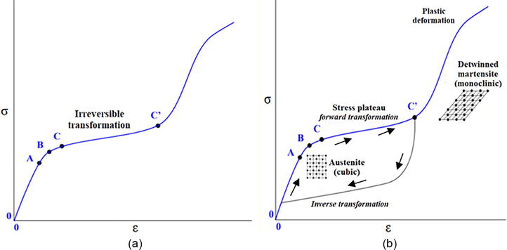
Figure 1a represents a typical stress–strain diagram for conventional metallic materials, and it is visible that upon loading, stress increases linearly. Contrary to this conventional behavior, the stress-strain diagram for pseudoelastic Ni-Ti alloy is shown in Figure 1b. The interval from 0 to A represents behavior when stress increases linearly with a strain upon loading. Point B is the yield point. The interval from C to C′ is a loading plateau (i.e., stress plateau) where large strains are developed with a slight stress increase. At point C, the Ni-Ti alloy can reach an 8% strain. Concurrently, the structure lattice (point C′) transforms in stress-induced martensite (monoclinic crystal lattice), and the material possesses that crystallographic form until the stress is removed. After unloading, it reverts into parent austenite because the inverse transformation occurs. The strain is recovered at the lower point of the unloading plateau, and the material exhibits a body-centered cubic lattice (austenite phase). In conclusion, the uniquenesses of the pseudoelastic Ni-Ti alloy diagram is a hysteresis loop (also known as a stress-strain flag), and the size of this loop can vary regarding the variation in chemical composition or thermomechanical treatment of the material.
Figure 1.
Stress-strain diagram: (a) typical curve for conventional metallic materials, A-Linear elasticity, B-Elastic limit, C-Yield point, C’-Fracture point; (b) Ni-Ti alloy with inverse transformation behavior, that is, “flag” of a diagram, A-Austenite phase, B-Martensite twinned phase, C-C’-Non-linear deformation i.e. non-linear behavior of stress and strain, C’-Martensite detwinned phase.
2.3 Corrosion resistance
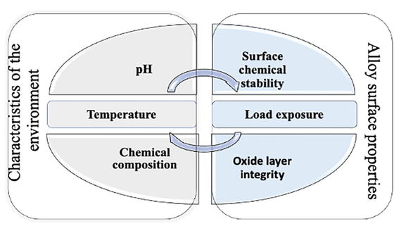
In terms of a wide array of medical applications, the corrosion resistance of nitinol medical devices is a crucial factor in defining their biocompatibility. Corrosion of nickel-titanium alloys is a contextual phenomenon (Figure 2), depending on surface treatments and the nature of the environment [4]. The investigation of the influence of pH change on corrosion is well documented. These alloys are corrosion-resistant in pH-neutral conditions [13].
Figure 2.
Corrosion dependent variables.
Nickel-titanium alloys are known for their passivization state. Still, in order to explain the overall mechanisms of corrosion resistance of nitinol alloy, the most important fact is that the human body presents very challenging surroundings. The reason for that is the presence of dissolved oxygen, chlorides, and changes in pH levels (acid-base disbalance). Moreover, different parts of the human body vary significantly in oxygen concentrations and pH values. Various studies have demonstrated lower corrosion resistance of these alloys in acidic solutions and chloride-containing environments [14, 15, 16]. In general, it is well documented that the corrosion potential increases as the pH value decreases [17]. Also, an increase in the environment temperature decreases surfaces’ ability to repassivate.
On the contrary, Montero-Ocampo and his co-workers reported the beneficial effects of heat treatment (annealing) on corrosion resistance [6]. Results of their study showed that microstructure evolution generated corrosion density hysteresis of almost zero during thermomechanical processing and a pitting potential of >1 [18].
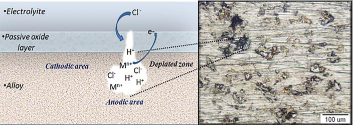
According to the attack mechanism, Ni-Ti devices are more prone to localized forms of corrosion and pitting. When exposed to aggressive anionic species, local breakdown occurs [19, 20]. The localized surface area becomes anodic, while the rest of the alloy surface is cathodic (Figure 3). Further, pit propagation is driven by an autocatalytic process, which means that once the aggressive anion penetrates the surface, the surroundings become depleted in oxygen [21, 22]. Pits grow in the direction of gravity, producing an excess of positive charges inside the pit. These conditions initiate more migration of anions into the localized defect, causing its propagation.
Figure 3.
Mechanism of pitting corrosion in Cl−-containing environment.
Numerous studies point out chloride ions as one of the most aggressive agents responsible for the degradation of passive alloys, such as nickel-titanium, from the aspect of the individual effect of elements within corrosion compounds. The reason for this is, above all, the small size of this molecule that allows it to penetrate more easily into the surface oxide layer. In addition, the absorption of Cl− ions is easy as it has a negative charge on the positively charged oxide layer [23, 24]. After the initial penetration of chloride into the surface layer, further outcomes can go in two directions, repassivation and complete or partial recovery, or further propagation.
The possibility of repassivation, that is, the ability of the oxide layer to perform self-healing after surface damage, is crucial for its long-term stability. This ability depends on the quality of the oxide film. The homogeneity of the oxide will ensure continuity and prevent metal leakage ions. Otherwise, the inhomogeneous oxide on the surface will allow the accumulation of hydrogen ions inside the initially formed crack, which creates a more positive potential value (Figure 3).
2.4 Surface chemical stability
The surface characteristics of Ni-Ti alloy are highly dependent on the preparation method. Literature-based evidence is coherent about the statement that the Ni/Ti ratio on the surface can vary significantly [25]. Also, during exposure to the corrosive environment, the thickness of the oxide on the surface of the nickel-titanium alloy can be in a wide range of nanometer scale, from 4 to 3500 nm [26]. Several studies have analyzed the oxide layer growth potential on the sample’s surface. Results showed that untreated samples developed a thicker oxide on the surface (120–340 nm), while on mechanically polished or etched samples, the film was up to 10 times thinner (11–16 nm) [27]. However, the protective role of the surface oxide layer against corrosive agents is determined by the ability of the alloy to develop a homogeneous and compact oxide. The integrity of this layer is much more important than its thickness [28].
To date, various surface treatments have been investigated. The most common are traditional methods, which include mechanical polishing, electrochemical polishing, chemical etching in acid solutions, and heat treatment (treatment in an autoclave) [29]. In addition, there are plasma ion implantation methods and bioactive coatings formation [25].
Untreated samples develop a surface layer of oxygen, carbon, titanium oxide, and small amounts of nickel. Conventional preparation, such as mechanical polishing, can increase titanium concentrations up to 5 times [4].
Also, polished surfaces consist of a combination of titanium, titanium oxide, a small amount of nickel oxide, and elemental nickel. In contrast, the oxygen concentration is significantly lower on chemically etched surfaces. Previous studies [30, 31] indicated multiple advantages of polishing over chemical etching. It was concluded that etching with various acids can create pitting defects up to 8 μm deep. Such roughness can favor local corrosion.
Besides, if the alloy is autoclaved in water, the titanium ratio increases 20 to 30 times more than nickel. As the immersion duration increases, more Ti develops on the surface. Consequently, nickel concentrations decrease [4].
The process of spontaneous formation of transition metal oxides is measured by the Gibbs free energy [32]. The more significant presence of Ti oxide on the surface of nickel-titanium alloys is explained by its formation requiring four times less energy than Ni oxide.
Given that in vivo implantation is accompanied by severe environmental factors (body fluids- blood, saliva, acid-base imbalance, body salts, etc.), the surface structure may differ significantly from that in vitro. In line with that, Hanawa and his co-workers [33] showed that nitinol devices implanted in human bone develop a thin calcium phosphate (Ca/P) layer in combination with titanium oxide. Results of further investigations confirmed that this layer is considered responsible for nitinol’s good biocompatibility.
To join the advantages and overcome the limitations of the screw and cement-retained prosthetic components, a new retention system based on the two-way shape memory effect (TWSME) has been developed lately, etc. nitinol sleeve attachment component.
To be able to solve the problem of inadequate dental implant position and to achieve adequate angulation of the superstructure in implant-prosthodontics, nickel-titanium was used for the first time to manufacture a memory abutment in the 90s of the last century (Dyna memory abutment) [34]. But, this abutment was compatible only with Dyna implants. Due to limited application and high production costs, this system did not achieve wide commercial use.
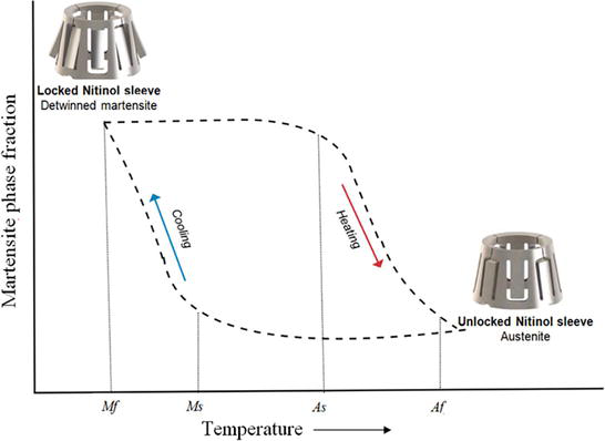
Contemporary product design (Rodo abutment, Smileloc® Abutment System, Nitinol sleeve) and machine-precise components lead to widespread application. This component is compatible with different implant systems (Straumann, Neodent, Nobel Biocare, Biomet, and Biohorizons). The transition to the Rodo abutment (Smileloc® Abutment System) led to the appearance of a slightly more advanced abutment system with a memory shape (Figure 4). As part of a crown on implants, a nitinol sleeve over the abutment has recently been used as an alternative to screw-retained or cement-retained crowns to overcome technical and biological complications in implant prosthetics. This system, familiar to clinicians for the last 10 years, is characterized by a retention nitinol component placed between the implant superstructure and the crown. The nitinol component is the precision-machined sleeve [35]. The retention component can persist in two forms, i.e., austenite cubic structure (at high temperatures) and martensitic body-centered tetragonal crystal structure (at low temperatures). Figure 4 represents the working principle of the shape memory alloy sleeve. The temperature change leads to changes within the crystal lattice resulting in a macroscopic change in the shape of the component from an “unlocked” to a “locked” form.
Figure 4.
Two-way shape memory mechanism of nitinol abutment sleeve.
According to the data from X-ray images taken after 6 months of follow-up, crowns retained on the nitinol sleeve were kept in the same position without any restoration movement [36]. Although these results suggested stable retention, evidence of long-term clinical studies is needed to evaluate optimal retention force, especially in pro arch implant prosthodontics restorations.
When martensite form is heated, it begins to change into austenite (Figure 4). The temperature at which this phenomenon starts is called austenite start temperature (As). The temperature at which this phenomenon is complete is called austenite finish temperature (Af). When the austenite form of the sleeve is cooled, it begins to change into martensite. The temperature at which this phenomenon starts is called martensite start temperature (Ms). The temperature at which martensite is again completely reverted is called martensite finish temperature (Mf). Hysteresis is generally defined as the difference between the temperatures at which the material is 50% transformed to austenite upon heating and 50% transformed to martensite upon cooling. This difference can be up to 20–30°C [37].
The uniqueness of nickel-titanium alloy was used to develop a second type of abutment, the EZ Crown system. Even though this abutment consists of a nitinol component, its retention is based on features different from the shape memory sleeve abutment.
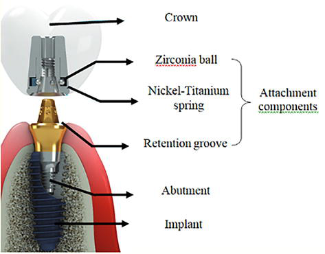
Namely, the superstructure of the EZ Crown internal component (attachment system) consists of a nitinol retention spring, the zirconia ball, and the cylindrical suprastructure (Figure 5) [39].
Figure 5.
Retention system of EZ crown [38].
In this complex configuration, the nitinol spring is in a martensitic stabilized configuration, meaning that the product will not develop either pseudoelasticity or the shape memory effect. As seen from Figure 5, Zr balls are positioned into the undercut region of the abutment retention groove.
Once the Ni-Ti is placed over the Zr ball, it develops a stable retention force.
Thanks to the low modulus of elasticity and good springback capacity of the martensitic stabilized nickel-titanium component, the nitinol spring will provide a continuous force on the zirconia ball [40].
The first dental Ni-Ti implants were introduced in the 80s of the last century [41].
Important properties of the metallic biomaterials are that they exhibit similar elastic deformation behavior as local tissue in human environment. It is known that elastic deformation of Co-Cr alloys and stainless steel is limited to 1%. Contrary, human osseous tissue can be elastically deformed up to 10% strein. Nitinol is only commercially available metallic material that can behave in a similar way when exposed to mechanical deformation.
Regarding the desired properties of the materials for implant manufacture, preference should be given to alloys with a high damping capacity and a low modulus of elasticity. High damping capacity is the desired property when it comes to orthopedic materials. This feature represents materials’ ability to absorb and evenly transfer mechanical forces, eliminating sudden shocks and oscillations.
Thanks to good flexibility, nitinol alloy can convert accepted mechanical energy into thermal energy, making the implant resistant to shocks caused by external loading. Therefore, metallic materials with high damping capacity values will result in a well-balanced stress-strain ratio within the alloy [42].
Regarding the modulus of elasticity, the desired property of the alloys is that these values should be as low as possible and close to those of Young’s modulus of bone. The Ti6Al4V titanium alloy used for making implants has a modulus of elasticity of 110 GPa, and for pure titanium, that value is 116 GPa. The Co-Cr alloy has a modulus of elasticity of 210–235 GPa, and the nickel-titanium is 80 GPa for the austenitic and 30 GPa for the martensitic form of the alloy. Young’s modulus for human bone is 20 GPa [43].
If the material’s physical properties for making implants are not aligned with the purpose of these devices, it can lead to technical and biological complications. The low damping capacity of the alloy more often leads to problems with the implant itself (fracture), and differences in the values of the modulus of elasticity of the material for making the implant and the surrounding bone can develop unfavorable effects on the supporting tissue and decrease bone density [38].
Regarding the mechanical properties of Ni-Ti, this material is biomechanically compatible with the surrounding environment. In comparison to Ti-base implants, it was reported that Ni-Ti alloy exhibits higher biomechanical compatibility with human bone under extension flexion in the sagittal plane [11].
Lately, in bioengineering science, considerable efforts have been made to develop devices that mimic the natural human environment. Additive manufacturing enables the production of devices with desired porosity. This design is suitable for the production of scaffolds as a porous matrix allows for osteoblasts to grow.
However, data in the literature indicate the importance of precise defining the size and distribution of these pores because, in the case of the presence of irregular porosity of the material, the strength could be because, with the increase in pores, the sensitivity of the metal structure to cracks propagation also increase. The suggested application requires far more preclinical and clinical investigation of the osteoconductivity of Ni-Ti.
3.1.1 Experimental procedure
The aim was to evaluate osteoblast-like cell adhesion and proliferation in the presence of Ni-Ti alloy.
The study was carried out in accordance with the Declaration of Helsinki and approved by the Institutional Ethics Committee (School of Dental Medicine, University of Belgrade, approval no. 36/7).
Ni-Ti alloy was produced by a standard casting process, i.e. vacuum remelting. To be able to obtain sample dimensions for experimental testing, electro-erosion cutting was performed. The final experimental sample dimensions were 2r = 11 mm, thickness 1.7 mm, n = 5. Round-over glass with the same dimensions as Ni-Ti samples served as control. All of the samples were cleaned with acetone, alcohol, and deionized water for 90 seconds, blow-dried, sterilized with UV-C light for 1 hour, and transferred to a culture flask with 12 wells.
3.1.1.1 Cell culture
Osteoblast-like cells ([MG-63] ATCC® CRL-1427™, USA) were incubated under strictly controlled conditions (at 37°C in a humidified 5% CO2 atmosphere) in a complete growth medium (Dulbecco’s modified Eagle’s medium (DMEM) with 4 mM L-glutamine, 10% Foetal bovine serum (FBS), and 1% ABAM, Sigma-Aldrich, Steinheim, Germany), and passaged every 3 days.
3.1.1.2 Adhesion pattern and cell viability
For the assessment of biocompatibility after direct exposure, osteoblast-like cells were seeded into each well at a density of 2 × 10⁴ cells cm−2. Human bone cells were plated on Ni-Ti samples and control cover glass. The incubation period was 24 h.
The number and the viability of attached osteoblast-like cells were evaluated using differential staining. In order to remove partially attached cells, the samples were washed with Phosphate-Buffered Saline. Then, samples were stained with 2 μg/mL Hoechst 33342 dye for 20 min, which bound to the DNA of live cells. The number of viable cells was assessed by emission of blue fluorescence and counted using ImageJ (NIH).
3.1.1.3 Preparation for scanning electron microscopy
After incubation, samples with attached cells were fixed, contrasted and submerged into 2.5% glutaraldehyde (SPI Supplies, West Chester, PA, USA) and 0.4% paraformaldehyde (Merck KGaA, Darmstadt, Germany) in 1 M Na-phosphate buffer (NaH2PO4·2H2O and Na2HPO4·2H2O; Merck KGaA, Darmstadt, Germany). Samples were gold-dusted using a Precision Etching Coating System (682 PECS, Gatan, Pleasanton, USA). The morphology and the attachment pattern of osteoblast-like cells were examined using a scanning electron microscope (JOEL JSM-6500F).
3.1.2 Results
3.1.2.1 Cell attachment and proliferation in the presence of Ni-Ti
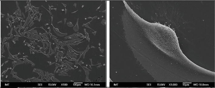
The cell cultures showed that the cells had grown very close to the Ni-Ti surfaces (Figure 6). The shape of the attached cells was predominately elongated, with clearly visible large cell extensions suggesting good adhesion. Cells with membrane outgrowths predominate (pseudopodia and philopodia).
Figure 6.
Scanning electron microscopy of osteoblast-like cells cultured on Ni-Ti after 24 h.
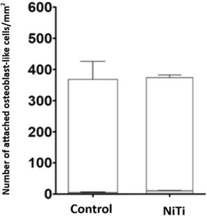
The results of fluorescence microscopy showed that after 24 h of exposure, tested Ni-Ti samples used were not cytotoxic, as the density of attached cells was almost comparable to the control indicating normal cell growth, and the results of staining cells with propidium iodide showed that the viability of attached cells in all samples was higher than 95% (98.6 ± 0.5% in control; 96.6 ± 0, 1% in Ni-Ti (Figure 7).
Figure 7.
Mean number of viable attached cells after 24 h.
This study conducted on human bone cells suggested good susceptibility of osteoblast-like cells to nickel-titanium surfaces.
3.2 Application of Ni-Ti alloy in orthodontics
In dentistry, the Ni-Ti alloy has the widest application for the production of orthodontic wires. In modern orthodontics, the choice of materials plays a significant role in achieving optimal outcomes. Among these materials, Ni-Ti archwires have gained prominence due to their unique mechanical properties.
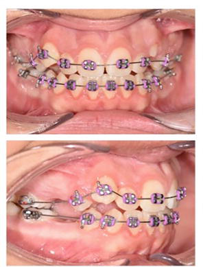
Since they were originally introduced, the development of orthodontic archwires has continued, enhancing their properties in a range of clinical settings. The orthodontic archwires exert gentle and continual forces on teeth, which leads to tooth repositioning. When a force is applied over several weeks to months, the orthodontic archwire elastic properties of the archwire are needed. Furthermore, distinct orthodontic archwires are required for different stages of the orthodontic treatment. Due to their unique properties—the shape memory effect and superelasticity, Ni-Ti archwires can effectively correct the misalignment of teeth, which is extremely important in the initial phase of orthodontic treatment (leveling and alignment). When the force is transferred to the bone, the bone responds with resorption and apposition, allowing the tooth movement (Figure 8) [44].
Figure 8.
The initial phase of orthodontic treatment (leveling and alignment) with Ni-Ti 0.012 inch archwire. Due to its superelastic properties, this archwire can tolerate a great degree of teeth misalignment.
Due to superelasticity, on unloading, Ni-Ti archwires may return to their original shape before loading. It is possible for the alloy to be deformed until 7–8% strain, which is almost forty times the capacity of the stainless steel wire. The loading leads to periodontal ligament deformation, and mobilization begins at a plateau of light forces recognized to provide the best biological tooth movement while the crystalline structure of the Ni-Ti alloy reverts to its initial configuration [45].
Since Ni-Ti alloy is a material with a shape memory effect, it has the property to “remember” its original shape after being elastically or pseudoplastically deformed by increasing its temperature. This effect results from thermoelastic martensitic transformation, where a body-centered cubic phase (austenitic phase) transforms into an orthorhombic or monoclinic martensite phase [46]. Therefore, in orthodontics, when the orthodontic archwire is deformed, it aligns and moves the teeth by returning to their natural shape. The stress remains almost at a constant level during the range of archwire activation, generating a low level of force, producing a good response from surrounding tissues, more comfort for the patient, and more physiological tooth movement. High orthodontic forces can produce root resorption, which is undesirable because it can affect the tooth’s long-term viability [47]. Apart from using Ni-Ti alloys in archwires, they can be used in open coil springs, as well as closing loops. NiTi retraction loops for canine distalization are not widely used in everyday practice since their forming is not as easy as with other alloys. Open coil springs are used most often for molar distalization, space opening, or tooth uprighting, delivering light and continuous forces. However, these forces are very often below the optimal range, so the authors advise more activation of the Ni-Ti coils to take greater advantage of their superelastic properties [48]. The effectiveness of space closure with Ni-Ti coils compared to elastomeric chains is likely to be similar in clinical investigation [49]. However, the moderate quality evidence of a metanalysis suggests a faster space closure with Ni-Ti closing springs compared to elastomeric chains [50]. Force degradation of both elastomeric chains and Ni-Ti coils was noticed and should be considered by clinicians when choosing the manner of space closure [50].
Fixed appliances consist of orthodontic brackets, which are bonded to labial or lingual and orthodontic archwires, placed into the slots of orthodontic brackets to exert a force on the teeth. Therefore, oral hygiene is compromised due to greater adhesion of plaque and bacteria, and the risk of enamel decay and gingivitis is increased during the orthodontic treatment [51, 52]. Since silver nanoparticles are known for their antibacterial properties in numerous studies [53, 54], it is possible to design a bactericide archwire to help with dental plaque control. The electrodeposition of silver nanoparticles onto Ni-Ti orthodontic archwires can significantly reduce oral bacteria in bacterial culture by more than 90% without changing their calorimetric and mechanical properties and release of nickel; however, further clinical investigation of these findings is needed to prove these results [55].
The conventional Ni-Ti archwire shows a homogenous composition along its length. Functionally graded Ni-Ti shows varying composition along its length, allowing for tailored mechanical properties at different segments of the wire, which can provide customized force levels required for different teeth. The manufacturing of functionally graded Ni-Ti wires involves various methods. Microstructurally graded Ni-Ti can be created by variations in heat treatment conditions (annealing or aging temperature) along the length of the Ni-Ti wire or Ni-Ti plates across their thickness. Compositionally graded Ni-Ti has a variation in composition (contents of Ni and Ti) within the body of the alloy. Also, the structure geometry can be purposely graded in the case of geometrically graded Ni-Ti archwires [56].
Also, two major problems in using NitTi alloys in orthodontics - friction and corrosion which lead to Ni-Ti release and mechanical weakening, can be encountered with different surface modifications of the archwire [57]. The results of a clinical study revealed that TiO2 coating on the archwire surface decreased the surface roughness and adhesion of Streptococcus mutans. However, the advantage of coated archwire on surface roughness was lost after 1 month [58].
Future investigation should be focused on enhancing mechanical properties using different methods of surface modifications, preferably creating a coating with antibacterial properties.
3.2.1 Magnetron sputtering method
The Magnetron sputtering (MS) method is a state-of-the-art coating technology that relies on physical vapor deposition (PVD) in vacuum conditions [59]. MS enables rapid deposition of diverse metal/alloy coatings on the surface of various materials with improved adhesion, high purity of the obtained films and excellent coverage of the substrate features [60]. Overall, the use of this outstanding technology provides very high hardness, abrasion resistance, fine microstructure, as well as long-term chemical, thermal, and environmental stability, which is essential for materials sustainability and safe clinical use [59]. By applying MS, modification of the material’s surface in single-layer, multilayer, or graded thin-layer coatings for obtaining systems with superior functional characteristics could be achieved.
In order to improve the properties of the Ni-Ti alloy in dentistry, this MS method was applied with protective coatings onto orthodontic archwires. The greatest challenge for safe orthodontic treatment is corrosion, chemically or microbiologically induced, resulting in the release of nickel (Ni). The aim of this investigation was to enhance resistance to corrosion and introduce antibacterial properties to Ni-Ti orthodontic archwires by coating them with copper (Cu) doper titanium nitride (TiN-Cu). Using cathodic arc evaporation (CAE), Ni-Ti archwires were coated with TiN-Cu and direct current magnetron sputtering (DC-MS). The morphology of the sample has been examined using field emission scanning electron microscopy (FESEM) whereas chemical composition was analyzed using energy-dispersive X-ray spectroscopy (EDS), X-ray diffraction (XRD) and Fourier transformed infrared spectroscopy (FTIR). To estimate the ion release, inductively coupled plasma optical emission spectrometry (ICP-OES) was used. The biocompatibility of samples was examined using 3-(4,5-dimethylthiazol-2-yl)-2,5-diphenyl tetrazolium bromide (MTT) assay. Antibacterial activity was tested for Streptococcus mutans and Streptococcus mitis.
The physicochemical characterization results showed that the coatings with the presence of TiN phase and incorporated Cu were well-designed. Such topography encourages corrosion resistance and consequently increases biocompatibility due to decreased surface roughness. EDS spectrum demonstrated the existence of Cu, Ti, and N in these samples, whereas XRD and FTIR analyses showed the presence of the TiN phase with incorporated Cu, indicating that the desired phases were obtained. TiN-Cu nanocoated archwires demonstrated a significantly reduced Ni release (p < 0.05). The 28-day eluates of TiN-Cu-nanocoated archwires had the highest relative cell viability (p < 0.05). Antibacterial tests showed a significant decrease in Streptococcus mutans and Streptococcus mitis counts, regarding TiN-Cu-nanocoated archwires. In contrast, TiN coatings showed no reduction in Streptococcus mutans, indicating the importance of copper in the obtained results.
The most notable decrease in concentrations of Streptococcus mitis was found in the case of TiN-Cu-coated archwires (p < 0.05) (Table 1). Considering antibacterial tests and biocompatibility, TiN-Cu-nanocoated archwires may be a promising candidate for further clinical research [61].
Streptococcus mutans
Streptococcus mitis
TiN-Cu Nanocoated Archwires
Ni-Ti Archwires
SS Archwires
TiN-Cu-Nanocoated Archwires
Ni-Ti Archwires
SS Archwires
Log CFU ± SD
4.19 ± 0.22
4.29 ± 0.24
5.87 ± 1.10
3.08 ± 0.99
3.75 ± 0.82
5.54 ± 0.60
Table 1.
Final bacterial count for Streptococcus mutans and Streptococcus mitis.
3.3 Application of Ni-Ti alloy in endodontics
Thanks to the high flexibility of the alloy, nickel-titanium endodontic instruments represent the gold standard in root canal therapy. In order to ensure the possibility of simultaneous cleaning and shaping of complex morphology of the canal, the alloy must possess the property of superelasticity, and the characteristic unique only for binary alloys with a higher proportion of nickel (56 wt.%) compared to titanium (44 wt.%) [62]. Good flexibility and the possibility to be elastically deformed up to 8% are the main characteristics of endodontic instruments. Those properties ensure high fatigue resistance of endodontic files, thus preventing instrument breakage during the mechanical shaping of the root canal. Compared to stainless steel and cobalt alloys, good mechanical properties, favorable axial and torsional resistance, and lower values of the elastic modulus are all very important in the shaping of the root canal [14].
A big problem in endodontics is cleaning and shaping of the narrow and curved root canals. The intricate interplay of temperature, heating duration, and cooling rates during alloy production has been established to influence the superelasticity and shape memory of Ni-Ti files [63]. The application of heat treatment preserves the crystallographic structure of the alloy, imparting endodontic files with heightened flexibility and resistance to fractures [64].
Ni-Ti endodontic files predominantly exhibit either an austenitic phase (conventional Ni-Ti, M-wire, R-phase) or a martensitic phase (controlled memory (CM) wire, gold, and blue heat-treated Ni-Ti alloys) [45]. Specialized heat treatment has led to the development of superelastic alloys with a stable martensitic phase, showcasing a lower modulus of elasticity (30–40 GPa) compared to austenitic (80–90 GPa). Furthermore, the modulus in the R-phase is lower than in martensite [65]. The martensitic phase, with its double-phase structure reorientation, offers superior resistance to cyclic fatigue compared to austenitic phases [65, 66].
The production of conventional Ni-Ti wires involves a cold drawing process, yielding a microstructure that incorporates martensite residues within the austenitic matrix. To reduce internal stresses and drawbacks associated with the rearrangement of the crystal grid, a crucial step involves subjecting the alloy to heat treatment within the temperature range of 450–550°C [67]. Heat treatment enables endodontic files to be more resistant to breakage during root canal treatment.
Specific surface treatments play a pivotal role in enhancing the physical and mechanical properties of endodontic files. These treatments, such as electropolishing, electric discharge machining, ionic implementation, cryogenic treatment, and nitriding, contribute to the overall performance and longevity of endodontic files [14, 63].
Electropolishing is an electrochemical process for surface finishing Ni-Ti files. This method removes surface irregularities by dissolving metal ions in an electrolyte bath, creating a thin passive layer that enhances resistance to cyclic fatigue, torsional load, and corrosion [63, 64, 65]. RaCe systems and EndoSequence files are examples of electropolished files, offering improved cutting, reduced screwing inside the canal, and enhanced apical penetration [63].
In the ever-evolving landscape of endodontics, Electric Discharge Machining (EDM) technology has emerged as a transformative force, offering a non-contact thermal erosion process to produce electrically conductive materials. This cutting-edge method involves controlled electrical discharge in the presence of an insulating fluid, presenting a revolutionary approach to shaping Ni-Ti alloys. EDM technology, represented by systems like Hyflex EDM and Neoniti, stands at the forefront of endodontic innovation. These systems showcase not only enhanced durability and performance but also a commitment to precision and efficiency in root canal procedures. As endodontics continues to advance, EDM technology promises to play a pivotal role in shaping the future of root canal treatments.
In the late 1980s, ion implantation in plasma emerged as a groundbreaking technique. Applying a highly negative pulsating voltage to the plasma-submerged file in a vacuum chamber allowed ions (argon, boron, and nitrogen) to penetrate the file’s surface without compromising its superelastic properties. Studies have demonstrated that ionic nitrogen implantation improves resistance to cyclic fatigue and enhances cutting efficiency, leading to improved wear resistance [63].
The immersion sol-gel method provides another avenue for enhancing file surfaces. By coating endodontic files with a protective layer of flexible TiO2 through this method, a surface porous oxide film is formed. This film increases the stability of the surface layers, offering protection against corrosion [68].
The thermal nitriding method introduces titanium nitride (TiN) to the file’s surface, consisting of a thin outer layer of TiN and a thicker inner layer of Ti2Ni. This process significantly increases corrosion resistance, particularly in contact with sodium hypochlorite (NaOCl) which is used as an irrigant [68, 69].
Endodontic files subjected to these advanced surface treatments exhibit greater resistance to corrosive defects and improved overall performance. While some debate exists regarding the impact of file design versus finishing on cyclic fatigue resistance, it is evident that these surface treatments contribute significantly to the evolution of endodontic technology [69, 70].
Thanks to its biocompatibility, corrosion resistance, superelasticity, and fatigue resistance, Ni-Ti alloys are widely used in all areas of dentistry: prosthetics, orthodontics, and endodontics. The favorable characteristics include high compressive strength, similar to human bone, as well as an elasticity modulus nearly equivalent to bone tissue. These attributes are essential for the fabrication of dental implants. Pseudoelasticity and shape memory effect are responsible for biomechanical compatibility. Thanks to its mechanical, antibacterial, and anticorrosive effect, this alloy is very successfully used for the production of orthodontic wires. Nickel-rich Ni-Ti alloys have attracted attention due to the possibility of stress-induced phase transformation, which is why most endodontic files rely on this feature. As technology moves forward, the integration of advanced surface treatments promises to further improve the efficiency and longevity of dental supplies, ultimately benefiting dental medicine as a whole.
1.Castleman LS, Motzkin SM, Alicandri FP, Bonawit VL, Johnson AA. Biocompatibility of nitinol alloy as an implant material. Journal of Biomedical Materials Research. 1976;10:695-731. DOI: 10.1002/jbm.820100505
2.Genchi G, Carocci A, Lauria G, Sinicropi MS, Nickel CA. Human health and environmental toxicology. International Journal of Environmental Research and Public Health. 2020;17:679. DOI: 10.3390/ijerph17030679
3.Shabalovskaya SA. On the nature of the biocompatibility and on medical applications of NiTi shape memory and superelastic alloys. Bio-medical Materials and Engineering. 1996;6:267-289
4.Ryhänen J, Niemi E, Serlo W, Niemelä E, Sandvik P, Pernu H, et al. Biocompatibility of nickel-titanium shape memory metal and its corrosion behavior in human cell cultures. Journal of Biomedical Materials Research. 1997;35:451-457. DOI: 10.1002/(sici)1097-4636(19970615)35:4<451::aid-jbm5>3.0.co;2-g
5.Nasakina EO, Sudarchikova MA, Sergienko KV, Konushkin SV, Sevost’yanov MA. Ion release and surface characterization of nanostructured nitinol during long-term testing. Nanomaterials (Basel). 2019;9:1569. DOI: 10.3390/nano9111569
6.Duerig T, Pelton A, Stöckel D. An overview of nitinol medical applications. Materials Science and Engineering: A. 1999;273-275:149-160. DOI: 10.1016/S0921-5093(99)00294-4
7.Soffa WA, Laughlin DE. Diffusional phase transformations in the solid state. In: Physical Metallurgy. Oxford, UK: Elsevier; 2014. pp. 851-1020. ISBN 978-0-444-53770-6
8.Li G, Yu T, Zhang N, Chen M. The effect of Ni content on phase transformation behavior of NiTi alloys: An atomistic modeling study. Computational Materials Science. 2022;215:111804. DOI: 10.1016/j.commatsci.2022.111804
9.Chekotu J, Groarke R, O’Toole K, Brabazon D. Advances in selective laser melting of nitinol shape memory alloy part production. Materials. 2019;12:809. DOI: 10.3390/ma12050809
10.Sui S, Chew Y, Weng F, Tan C, Du Z, Bi G. Achieving grain refinement and ultrahigh yield strength in laser aided additive manufacturing of Ti−6Al−4V alloy by trace Ni addition. Virtual and Physical Prototyping. 2021;16:417-427. DOI: 10.1080/17452759.2021.1949091
11.One Way and Two Way Shape Memory Effect: Thermo Mechanical Characterization of Ni-Ti Wires - PDF Free. Download Available from: https://docplayer.net/263607-One-way-and-two-way-shape-memory-effect-thermo-mechanical-characterization-of-ni-ti-wires.html [Accessed: July 2, 2023]
12.Huang W, Toh W. Training two-way shape memory alloy by reheat treatment. Journal of Materials Science Letters. 2000;19:1549-1550. DOI: 10.1023/A:1006721022185
13.Davis JR. Corrosion: Understanding the Basic (06691G). Philadelphia, USA: ASM International; 2020. ISBN 13: 9780871706416
14.Cioffi M, Gilliland D, Ceccone G, Chiesa R, Cigada A. Electrochemical release testing of nickel-titanium orthodontic wires in artificial saliva using thin layer activation. Acta Biomaterialia. 2005;1:717-724. DOI: 10.1016/j.actbio.2005.07.008
15.Huang H-H, Chiu Y-H, Lee T-H, Wu S-C, Yang H-W, Su K-H, et al. Ion release from Ni-Ti orthodontic wires in artificial saliva with various acidities. Biomaterials. 2003;24:3585-3592. DOI: 10.1016/S0142-9612(03)00188-1
16.Taqa A, Fathi W, Mohammed R. Evaluation of nickel ion release from orthodontic wires in different types of artificial saliva. RDENTJ. 2014;14:182-188. DOI: 10.33899/rden.2014.160888
17.Lee T-H, Huang T-K, Lin S-Y, Chen L-K, Chou M-Y, Huang H-H. Corrosion resistance of different nickel-titanium archwires in acidic fluoride-containing artificial saliva. The Angle Orthodontist. 2010;80:547-553. DOI: 10.2319/042909-235.1
18.O’Brien B, Carroll WM, Kelly MJ. Passivation of nitinol wire for vascular implants—A demonstration of the benefits. Biomaterials. 2002;23:1739-1748. DOI: 10.1016/S0142-9612(01)00299-X
19.Eliaz N. Corrosion of metallic biomaterials: A review. Materials (Basel). 2019;12:E407. DOI: 10.3390/ma12030407
20.Seo D-I, Lee J-B. Localized corrosion and repassivation behaviors of additively manufactured titanium alloys in simulated biomedical solutions. npj Materials Degradation. 2023;7:1-12. DOI: 10.1038/s41529-023-00363-4
21.Bhandari J, Khan F, Abbassi R, Garaniya V, Ojeda R. Modelling of pitting corrosion in marine and offshore steel structures – A technical review. Journal of Loss Prevention in the Process Industries. 2015;37:39-62. DOI: 10.1016/j.jlp.2015.06.008
22.Bae I, Kim B-H, Kim D-G, Sohn I-B, Yang S-W. Salt heat treatment and passivation to improve the corrosion resistance of nitinol (Ni-Ti). Materials (Basel). 2021;14:7789. DOI: 10.3390/ma14247789
23.Modelling of Pitting Corrosion in Marine and Offshore Steel Structures – A Technical Review - ScienceDirect Available from: https://www.sciencedirect.com/science/article/abs/pii/S0950423015300024 [Accessed: January 17, 2024]
24.Grgur B. Korozija I Zaštita. Belgrade, Serbia: Faculty of technology and metallurgy; 2020. ISBN: 978-86-7401-365-6
25.Mohammadi Z, Soltani MK, Shalavi S, Asgary S. A review of the various surface treatments of Ni-Ti instruments. Iranian Endodontic Journal. 2014;9:235-240
26.Sullivan SJL, Dreher ML, Zheng J, Chen L, Madamba D, Miyashiro K, et al. Effects of oxide layer composition and radial compression on nickel release in nitinol stents. Shape Memory and Superelasticity. 2015;1:319-327. DOI: 10.1007/s40830-015-0028-x
27.Clarke B, Carroll W, Rochev Y, Hynes M, Bradley D, Plumley D. Influence of nitinol wire surface treatment on oxide thickness and composition and its subsequent effect on corrosion resistance and nickel ion release. Journal of Biomedical Materials Research. 2006;79A:61-70. DOI: 10.1002/jbm.a.30720
28.Trepanier C, Venugopalan R, Pelton AR. Corrosion resistance and biocompatibility of passivated Ni-Ti. In: Yahia L, editor. Shape Memory Implants. Berlin, Heidelberg: Springer Berlin Heidelberg; 2000. pp. 35-45, ISBN 978-3-642-64118-3
29.Shabalovskaya S, Anderegg J, Van Humbeeck J. Critical overview of nitinol surfaces and their modifications for medical applications. Acta Biomaterialia. 2008;4:447-467. DOI: 10.1016/j.actbio.2008.01.013
30.Arjmand F, Zhang L. Mechanical polishing, surface roughness, near-surface deformation, and electrochemical corrosion of alloy 690TT. Surface and Interface Analysis. 2015;47:1120-1126. DOI: 10.1002/sia.5858
31.Milošev I, Kapun B. The corrosion resistance of nitinol alloy in simulated physiological solutions: Part 1: The effect of surface preparation. Materials Science and Engineering: C. 2012;32:1087-1096. DOI: 10.1016/j.msec.2011.11.007
32.Rudolf R, Stambolić A, Kocijan A. Atomic layer deposition of ATiO2 layer on Nitinol and its corrosion resistance in a simulated body fluid. Metals. 2021;11:659. DOI: 10.3390/met11040659
33.Hanawa T, Ota M. Calcium phosphate naturally formed on titanium in electrolyte solution. Biomaterials. 1991;12:767-774. DOI: 10.1016/0142-9612(91)90028-9
34.Metals as Biomaterials. Wiley. Available from: https://www.wiley.com/en-us/Metals+as+ Biomaterials-p-9780471969358 [Accessed: April 8, 2023]
35.Shah KC, Chao D, Wu BM, Jensen OT. Shape-memory retained complete arch guided implant treatment using nitinol (Smileloc) abutments. Oral and Maxillofacial Surgery Clinics of North America. 2019;31:427-435. DOI: 10.1016/j.coms.2019.03.005
36.Shah KC, Linsley CS, Wu BM. Evaluation of a shape memory implant abutment system: An up to 6-month pilot clinical study. The Journal of Prosthetic Dentistry. 2020;123:257-263. DOI: 10.1016/j.prosdent.2018.11.012
37.Jokanović V, Petrović B, Zivkovic M. The main characteristics and application of the shape memory alloys in orthodontics and endodontics. Serbian Dental Journal. 2019;66:29-35. DOI: 10.2478/sdj-2019-0004
38.Bahraminasab M, Sahari B. Ni-Ti Shape Memory Alloys, Promising Materials in Orthopedic Applications. London, UK: IntechOpen; 2013. pp. 261-278. ISBN 978-953-51-1084-2
39.Choi J-W, Song C-H, Huh J-B. Implant-supported fixed dental prostheses with new retention type using zirconia ball and nickel-titanium spring. The Korean Academy of Oral & Maxillofacial Implantology. 2019;23:16-24. DOI: 10.32542/implantology.2019002
40.Shah K, Seo Y, Wu B. Clinical application of a shape memory implant abutment system. The Journal of Prosthetic Dentistry. 2016;117:8-12. DOI: 10.1016/j.prosdent.2016.06.007
41.Sachdeva R, Fukuyo S, Suzuki K, Oshida Y, Miyazaki S. Shape memory Ni-Ti alloys - applications in dentistry. Materials Science Forum. 1990;56-58:693-698. DOI: 10.4028/www.scientific.net/MSF.56-58.693
42.Senthilnathan K, Shamimi A, Bonsignore C, Paranjape H, Duerig T. Effect of prestrain on the fatigue life of superelastic nitinol. Journal of Materials Engineering and Performance. 2019;28:5946-5958. DOI: 10.1007/s11665-019-04334-2
43.Niinomi M, Liu Y, Nakai M, Liu H, Li H. Biomedical titanium alloys with Young’s moduli close to that of cortical bone. Regen Biomater. 2016;3:173-185. DOI: 10.1093/rb/rbw016
44.Melsen B, Cattaneo PM, Dalstra M, Kraft DC. The importance of force levels in relation to tooth movement. Seminars in Orthodontics. 2007;13:220-233
45.Fernandes DJ, Peres RV, Mendes AM, Elias CN. Understanding the shape-memory alloys used in orthodontics. ISRN Dentistry. 2011;2011:132408
46.Otsuka K, Wayman CM, Nakai K, Sakamoto H, Shimizu K. Superelasticity effects and stress- induced martensitic transformations in CuAlNi alloys. Acta Metallurgica. 1976;24(3):207-226
47.Harris DA, Jones AS, Darendeliler MA. Physical properties of root cementum: Part 8. Volumetric analysis of root resorption craters after application of controlled intrusive light and heavy orthodontic forces: A microcomputed tomography scan study. American Journal of Orthodontics and Dentofacial Orthopedics. 2006;130:639-647
48.Sifakakis I, Bourauel C. Nickel–titanium products in daily orthodontic practice. In: Orthodontic Applications of Biomaterials. Cambridge, United States: Woodhead Publishing; 2017. pp. 107-127
49.Bokas J, Woods M. A clinical comparison between nickel titanium springs and elastomeric chains. Australian Orthodontic Journal. 2006;22(1):39-46
50.Mohammed H, Rizk MZ, Wafaie K, Almuzian M. Effectiveness of nickel-titanium springs vs elastomeric chains in orthodontic space closure: A systematic review and meta-analysis. Orthodontics & Craniofacial Research. 2018;21(1):12-19
51.Shokeen B, Viloria E, Duong E, Rizvi M, Murillo G, Mullen J, et al. The impact of fixed orthodontic appliances and clear aligners on the oral microbiome and the association with clinical parameters: A longitudinal comparative study. American Journal of Orthodontics and Dentofacial Orthopedics. 2022;161(5):475-485
52.Živkovic Sandic M, Popovic B, Carkic J, Nikolic N, Glisic B. Changes in subgingival microflora after placement and removal of fixed orthodontic appliances. Srpski Arhiv za Celokupno Lekarstvo. 2014;142(5-6):301-305
53.Espinosa-Cristóbal LF, Holguín-Meráz C, Zaragoza-Contreras EA, Martínez-Martínez RE, Donohue-Cornejo A, Loyola-Rodríguez JP, et al. Antimicrobial and Substantivity properties of silver nanoparticles against oral microbiomes clinically isolated from young and young-adult patients. Journal of Nanomaterials. 2019;2019:3205971
54.Jokanović V, Živković M, Zdravković N. A new approach to extraordinary efficient protection against COVID 19 based on nanotechnology. Serbia Dental Journal. 2020;67(2):100-109
55.Gil FJ, Espinar-Escalona E, Clusellas N, Fernandez-Bozal J, Artes-Ribas M, Puigdollers A. New bactericide orthodonthic archwire: Ni-Ti with silver nanoparticles. Metals. 2020;10(6):702
56.Shariat BS, Meng Q , Mahmud AS, Wu Z, Bakhtiari R, Zhang J, et al. Experiments on deformation behaviour of functionally graded Ni-Ti structures. Data in Brief. 2017;13:562-568
57.Uysal I, Yilmaz B, Atilla AO, Evis Z. Nickel titanium alloys as orthodontic archwires: A narrative review. Engineering Science and Technology, an International Journal. 2022;36:101277
58.Venkatesan K, Kailasam V, Padmanabhan S. Evaluation of titanium dioxide coating on surface roughness of nickel-titanium archwires and its influence on Streptococcus mutans adhesion and enamel mineralization: A prospective clinical study. American Journal of Orthodontics and Dentofacial Orthopedics. 2020;158:199-208
59.Sarakinos K, Alami J, Konstantinidis S. High power pulsed magnetron sputtering: A review on scientific and engineering state of the art. Surface and Coatings Technology. 2010;204:1661
60.Tudose IV, Comanescu F, Pascariu P, Bucur S, Rusen L, Iacomi F, et al. Chapter 2 - chemical and physical methods for multifunctional nanostructured interface fabrication. In: Dinca V, Suchea MP, editors. Micro and Nano Technologies, Functional Nanostructured Interfaces for Environmental and Biomedical Applications. Elsevier; 2019. pp. 15-26. eBook ISBN: 9780128144022
61.Ilić B, Petrović B, Marinković J, Miletić Vukajlović J, Stevanović M, Potočnik J, et al. Investigation of ion release and antibacterial properties of TiN-Cu-nanocoated nitinol archwires. Coatings. 2023;13:1587
62.Zupanc J, Vahdat-Pajouh N, Schäfer E. New thermomechanically treated Ni-Ti alloys – A review. International Endodontic Journal. 2018;51:1088-1103. DOI: 10.1111/iej.12924
63.Gavini G, Santos MD, Caldeira CL, Machado MEL, Freire LG, Iglecias EF. Nickel-titanium instruments in endodontics: A concise review of the state of the art. Brazilian Oral Research. 2018;32(Suppl. 1):e67
64.Gambarini G, Cicconetti A, Di Nardo D, Miccoli G, Zanza A, Testarelli L. Influence of different heat treatments on torsional and cyclic fatigue resistance of nickel-titanium rotary files: A comparative study. Applied Science (Basel). 2020;10(16):5604-5611
65.Gambarini G, Galli M, Di Nardo D, Seracchiani M, Donfrancesco O, Testarelli L. Differences in cyclic fatigue lifespan between two different heat treated Ni-Ti endodontic rotary instruments: Wave one gold vs edge one fire. Journal of Clinical and Experimental Dentistry. 2019;11(7):e609-e613
66.Jamleh A, Alghaihab A, Alfadley A, Alfawaz H, Alqedairi A, Alfouzan K. Cyclic fatigue and torsional failure of edge taper platinum endodontic files at simulated body temperature. Journal of Endodontia. 2019;45(5):611-614
67.Zinelis S, Eliades T, Eliades G. A metallurgical characterization of ten endodontic Ni-Ti instruments: Assessing the clinical relevance of shape memory and superelastic properties of Ni-Ti endodontic instruments. International Endodontic Journal. 2010;43(2):125-134
68.Srivastava S, Alghadouni MA, Alotheem HS. Current strategies in metallurgical advances of rotary Ni-Ti instruments: A review. Journal of Dental Health, Oral Disorders & Therapy. 2018;9(1):72-77
69.Han-Hsing Lin J, Karabucak B, Lee SM. Effect of sodium hypochlorite on conventional and heat-treated nickel-titanium endodontic rotary instruments - an in vitro study. Journal of Dental Science. 2021;16(2):738-743
70.Huang X, Shen Y, Wei X, Haapasalo M. Fatigue resistance of nickel-titanium instruments exposed to high-concentration hypochlorite. Journal of Endodontia. 2017;43(11):1847-1851
Written By
Minja Miličić Lazić, Marijana Popović Bajić, Igor Đorđević, Marija Živković, Vojkan Lazić, Vukoman Jokanović, Ilija Nasov and Slavoljub Živković
Submitted: 29 January 2024Reviewed: 08 February 2024Published: 21 March 2024