
Abstract
Tissue engineering focuses on developing replacement tissues and organs to maintain, restore, or improve their function. To achieve this goal, an optimal scaffold is required to promote cell growth and biomolecules release involved in the repair process. In tissues, the extracellular matrix (ECM) provides spatial and mechanical cues to cells and physical support. Therefore, creating a scaffold that mimics the ECM of a tissue or organ of interest to facilitate its repair represents an urgent need. Collagen is the most abundant protein in the ECM and is essential for maintaining the biological and structural integrity of the tissue as well as providing physical support. Collagen-based scaffolds can be obtained from a decellularized collagen matrix, preserving the original tissue shape and ECM structure, or by extracting, purifying, and polymerizing collagen alone or with other natural or biosynthetic polymers and ceramics, which can be chemically or physically cross-linked, modified with natural/synthetic polymers or inorganic materials, or supplemented with biochemical factors. The properties of collagen for obtaining tissue engineering products and the intellectual property of collagen-based scaffolds in clinical trials and patents are discussed. Here, we described the importance of collagen for tissue and organ repair.
Keywords
- tissue engineering
- collagen
- biological scaffold
- decellularized
- biosynthetic scaffold
- repair
1. Introduction
Collagen represents the primary structural protein in the body, comprising approximately 30 wt% of the body’s total protein content [1]. Its different types sustain mechanical and bodily stresses, support various cell types, and anchor many growth factors [1, 2]. Besides, this protein effectively modulates the biological behavior of cells to stimulate native tissue repair [3]. To date, about 28 distinct types of collagens have been identified in vertebrates [4]. Of which, the most abundant fibrillar protein is Type I collagen.
As well known, collagen is a key component of the extracellular matrix (ECM), which strongly regulates the extracellular scaffolding, maintains the biological and structural integrity of ECM, and plays critical roles in the regulation of the phases of wound healing [5]. This protein induces platelet activation and aggregation during the early phase to induce homeostasis. Besides, collagen degradation releases fragments that promote fibroblast proliferation and synthesis of growth factors that regulate angiogenesis and reepithelialization [5].
Tissue engineering is an emergent strategy that combines cells, biomaterial scaffolds, and biologically active molecules to repair a specific damaged tissue. When these products are implanted at the lesion, the scaffold will be replaced by the cells’ own matrices during the regeneration. Therefore, the scaffolds should be biocompatible and biodegradable and provide appropriate signals for the seeded cells that regulate the process [6].
Biological scaffolds are structures used in tissue engineering to support cells and bioactive molecules required to repair or replace injured tissues. These scaffolds are fabricated from natural materials by removing the cellular content from source tissues while preserving the structural and molecular composition of the remaining ECM [7]. This complex is crucial in tissue engineering because the ECM provides physical support to tissues, maintains their architecture, and can regulate cellular functions, providing spatial and mechanical signals to cells [6, 8]. The microstructure of the ECM varies depending on the type of tissue. However, their proteins, such as collagen, fibronectin, laminin, and glycosaminoglycans (GAGs), are responsible for maintaining the 3D structure, mechanical support and favorably influence the mitogenesis, chemotaxis, and cell differentiation fate [9, 10]. Each of these processes is key to promoting the formation of new tissues and organs [6].
The decellularization process is highly relevant in tissue engineering due to its efficient removal of the cellular and nucleic components while preserving the structural and functional proteins of the extracellular matrix (ECM) [11, 12], especially the collagen fibers, which are the main component of ECM, therefore of the acellular matrices [3, 6]. During a procedure to obtain an acellular matrix, the spatial arrangement of collagen fibers within the ECM represents an important structural aspect due to the mechanical properties associated with fiber direction. In addition, the presence and integrity of ECM proteins and their three-dimensional organization strongly affect the quality and downstream clinical outcome when the scaffolds are used for tissue repair and reconstruction (10). The ECM regulates cell behavior and phenotype while cells, in turn, continuously produce, degrade, and remodel the ECM. This reciprocal process is crucial to tissue development, homeostasis, and wound healing [9].
The decellularized matrices have been widely used as biological scaffolds to repair various tissues and organs such as cartilage, skin, bone, bladder, blood vessels, heart, liver, and lung, among others [6, 13, 14]. It has been reported that collagen strongly regulates the microstructure of decellularized matrices for repairing tissue. To date, a decellularized matrix from porcine cornea was used for
This chapter focuses on the importance of collagen for tissue engineering products because the protein has garnered broad interest as a biomedical material. In particular, collagen is a highly biocompatible macromolecule with potent biological functions for tissue repair and regeneration [1]. One of the objectives of this chapter is to offer a complete overview of the use of acellular matrices as biological scaffolds because they are mainly used for tissue repair, considering key factors such as biocompatibility and biodegradability attributable to ECM composition, especially collagen content.
2. Structure and function of collagen
Collagen consists of a triple helix chain formed by α chains. Each alpha (α) chain contains about 1000 amino acids with a molecular weight of approximately 100 kDa. It is composed of a specific set of amino acids repeating sequence (Gly-Xaa-Yaa)n [3]. In this case, glycine residues are located in every third position, and the amino acids in the Xaa and Yaa positions are often proline and hydroxyproline. It should be noted that collagen is cleaved from a precursor molecule known as procollagen by the C-terminal proteolytic processing of soluble procollagen precursors [3].
Collagen is a crucial regulator of the structural integrity of tissues and organs. In particular, regulates the structure of connective tissues, such as bone and cartilage [8]. Moreover, collagen interacts with diverse types of cells through its specific peptide repeat unit and triple helix structure, promoting their adhesion, proliferation, and differentiation by interactions with specific receptors such as integrins, glycoproteins, and proteoglycan receptors [3, 16]. Another feature that ratifies the importance of collagen is its capacity to store and deliver endogenous growth factors and cytokines, providing a suitable microenvironment for cells that participate in tissue and organ development and wound repair [3].
Using acellular matrices naturally provides collagen, an essential tool for producing tissue engineering technologies (Figure 1). Collagen offers low immunogenicity, a porous structure, permeability, good biocompatibility, and biodegradability [8, 17, 18]. Each property of collagen represents an opportunity to explore the diverse processes in which this protein participates, especially in tissue maintenance and repair. Here, we mentioned the collagen properties in decellularized matrices widely used in tissue engineering.
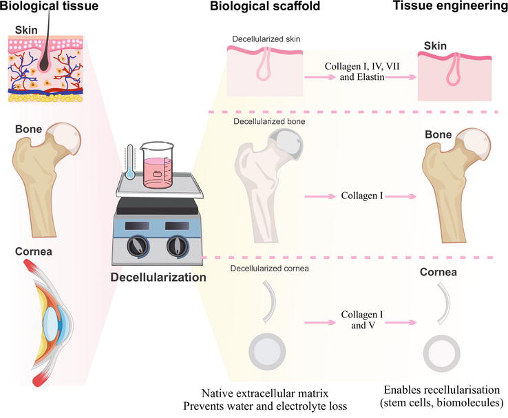
Figure 1.
Decellularized scaffolds with application in tissue engineering. Acellular scaffolds from different tissues can be obtaining by physicochemical and enzymatic process that preserve the native collagen content.
3. Decellularized scaffolds
3.1 Corneal decellularized matrix
Decellularized corneas have been reported as a viable alternative that acts as a scaffold for corneal engineering. Several strategies have been used to lyse cells and remove cellular material from the cornea [12]. Acellular corneal matrices (ACM) have been proposed as an alternative to mimic the native structure and composition of the human cornea. These matrices contain collagen I and V [19] and can be recellularized with corneal cells due to their ability to provide a structure similar to the native cornea [20].
Preclinical studies and human clinical applications of decellularized corneas have been successfully described. In animal lamellar keratoplasty experiments, porcine ACM was shown to integrate into the corneal wound bed and improve the reconstruction of the lamellar integrity of the corneal stroma, suggesting that acellular bioengineered cornea may be a good alternative for the treatment of feline corneal sequestrum [21]. Another
Regarding the use of ACM as a scaffold for tissue engineering constructs, one study reported the
A comprehensive evaluation of decellularized porcine corneas after clinical transplantation has been reported. In this study, porcine ACM was transplanted in the center of the left cornea of a 40-year-old male patient with a 1.5-mm-diameter corneal ulcer caused by bacterial keratitis, whose vision had rapidly deteriorated. Porcine ACM (Acornea, Product, 350 μm thick) and a lamellar graft were customized to the patient’s corneal defect size. Within the 2-month follow-up period, the corneal ulcer graft became transparent and his visual acuity improved from 0.01 to 0.1. However, the patient’s visual improvement with porcine ACM was insufficient. Therefore, to improve his vision, the patient underwent a second deep lamellar keratoplasty using a human donor cornea. It is important to note that the porcine ACM showed good integration with the host cornea, keratocytes had grown within the ACM, and no inflammatory cells were detected [24].
3.2 Bone repair/regeneration
The natural matrix of bone is highly composed of collagen fibrils, specifically type I collagen. Therefore, this type of collagen is widely used in bone repair [3]. However, the poor mechanical properties offered by collagen in bone tissue require the preservation of the inherent collagen and inorganic components within the bone. This condition is one of the more relevant parameters during the bone decellularization process [10].
3.3 Acellular dermal matrix
Acellular dermal matrix (ADM) is a biological scaffold that provides skin-native tissue biochemical properties and ultrastructural architecture to support tissue repair [27]. These matrices have high collagen types I, IV, and VII and elastin content, promoting the graft’s favorable elastic properties [14]. In a preclinical context, ADM has been used to treat chronic skin wounds because it provides molecules that improve intercellular communication and neovascularization in wound surface repair [28].
Numerous preclinical evidence has demonstrated the repair effect of ADM, especially on skin wounds. Construct based on ADM recellularized with mesenchymal stromal cells from Wharton jelly (hWJ-MSC) promoted the close of porcine full-thickness excisional wounds through the formation of stratified epithelium, basement membrane, and dermal papillae, improving the appearance of the repaired tissue at 30 days posttreatment (Figure 2) [29]. In addition, an experimental study about human ADM seeded with adipose-derived stem cells was reported. The construct enhanced wound healing in a murine model demonstrated by increased reepithelialization in 12 days, synthesis of granulation tissue, and vascularization during early wound healing [30].
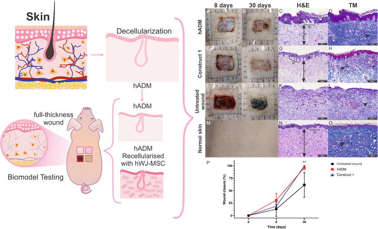
Figure 2.
4. Collagen and natural biopolymers scaffolds
Natural polymers have biocompatibility and bioactivity but are challenging to engineer due to limited processability, high contamination risk, and poor mechanical properties. Examples include bioactive proteins such as silk, collagen, gelatin, fibrinogen, and polysaccharides such as cellulose, chitosan, and alginate, and glycosaminoglycans, such as hyaluronic acid.
4.1 Chitosan
Chitosan is a polysaccharide found in the exoskeleton of crustaceans, mollusks, and insects. Chitosan has low toxicity, is non-immunogenic, and has antimicrobial activity, making it an excellent choice for tissue engineering applications. Its use as a biomaterial is justified by its biodegradability and biocompatibility. In addition, it is inexpensive and can be obtained by partial deacetylation of chitin [31, 32]. Additionally, chitosan is the only positively charged biopolymer that can interact with the ECM’s structural molecules [33]. This unique cationic biopolymer can be combined with another anionic biopolymer to form a two-component scaffold with optimal mechanical and biological properties [34]. The combination of collagen with chitosan is a promising alternative for tissue healing purposes, given the antimicrobial and hemostatic properties of this polysaccharide; in addition, chitosan improves the structure of collagen fibers, and it is used to try to control the degradation time of the scaffold and improve scaffold mechanical properties.
The main application forms of collagen/chitosan scaffolds in tissue engineering for skin and bone repair are membrane [35, 36], hydrogel [37], hydrogel 3D-bioprinter [38], and sponge [39].
On the other hand, a freeze-dried chitosan-collagen sponge for wound dressing was found to be nontoxic and biocompatible in an animal model. In addition to excellent water retention properties, which are critical for reducing the risk of dehydration at the wound site, it also showed excellent neovascularization and fibroblast proliferation, which were shown to promote substantial full-thickness skin wound healing [39]. An
4.2 Alginate
Alginate is a naturally occurring anionic and hydrophilic polysaccharide. It is one of the most abundant biosynthesized materials, mainly derived from brown algae and bacteria [42]. Alginate is particularly interesting for a wide range of applications as a biomaterial, especially as a support matrix or delivery system for tissue repair and regeneration [43]. Due to its outstanding properties in biocompatibility, biodegradability, non-antigenicity, and chelating ability, alginate has been widely used in various biomedical applications, including tissue engineering and drug delivery [44, 45]. Chelation with divalent cations is the easiest way to prepare alginate hydrogels from an aqueous solution under mild conditions. As a result of the natural polysaccharide, alginate has a pH-dependent anionic nature and can interact with cationic polyelectrolytes and proteoglycans. Therefore, cationic drug and molecule delivery systems can be obtained through simple electrostatic interactions [44]. However, alginate lacks cell-binding sites, limiting long-term cell survival and viability. Collagen (Col) contains cell-binding motifs, facilitating cell attachment, interaction, and spreading, consequently maintaining cell viability and promoting cell proliferation. In particular, collagen–alginate hydrogel has attracted much attention due to its excellent biocompatibility, gelling under mild conditions, low cytotoxicity, controllable mechanical properties, wide availability, and easy incorporation of other biomaterials and bioactive agents [46].
It has been reported that collagen, in combination with alginate, improves the hydrogel’s mechanical properties and tunes the stiffness simply by the concentration of Ca2+ [47]. Moreover, due to the collagen-containing cell-binding ligands and the inherent lack of cell-binding motifs in alginate, increasing the amount of collagen in the collagen/alginate hydrogel improves the cell-binding ligands, allowing more cell adhesion and attachment, thereby maintaining cell viability and promoting cell proliferation.
On the other hand, alginate is considered a nondegradable biomaterial because the human body lacks a specific enzyme called alginase to cleave alginate polymer chains. However, covalently crosslinked alginate can be gradually degraded by ion exchange, in which covalent ions, such as Ca2+, are constantly released from alginate to replace monovalent cations, such as Na+, from the environment [46]. Currently, sodium alginate and collagen, combined with other biomaterials or alone, are widely used hydrogels in cartilage, skin, intervertebral disc, and bone tissue engineering.
A promising approach for bone tissue engineering is the
On the other hand, 3D bioprinting is an innovative technology for cartilage tissue engineering. Collagen type I (COL) mixed with sodium alginate (SA) to serve as 3D bioprinting bioinks and incorporated chondrocytes to construct
Collagen and alginate wound dressings already on the market include DermaCol/Ag (advanced wound dressing containing collagen, sodium alginate, carboxymethylcellulose, ethylenediaminetetraacetic acid (EDTA), and silver chloride); ColActive® Plus (collagen, sodium alginate, carboxymethylcellulose and ethylenediaminetetraacetic acid dressing); and FIBRACOLTM plus (a soft, absorbent and conformable 90% collagen, 10% calcium alginate dressing).
4.3 Hyaluronic acid
Hyaluronic acid (HA) is a natural glycosaminoglycan found in the extracellular matrix of most connective tissues and is composed of repeating disaccharide units of N-acetylglucosamine and glucuronic acid. There are several ways in which HA can mimic the ECM: (1) Like other ECM components, it can provide a physical scaffold for cells to attach and migrate. (2) It can regulate cell behavior and signaling pathways by interacting with cell surface receptors. (3) It can serve as a reservoir for growth factors and other signaling molecules that can be released in a controlled manner to regulate cell behavior and tissue repair processes. Other known capabilities of this material include immune regulation and regeneration induction [53].
HA, due to its chemical composition, is a polymer that has a strong affinity for water and breaks down quickly. HA-based scaffolds have been extensively studied in the field of tissue engineering because they are highly biocompatible, biodegradable, and can be easily modified chemically. Hydrogels, sponges, injectable hydrogels, and electrospun scaffolds are all possible forms of HA-collagen scaffolds [54, 55]. The collagen-HA substrate is cell-compatible
Otherwise, the layer-by-layer (LBL) self-assembly technique is a highly effective method for immobilizing the major components of the extracellular matrix, such as collagen and hyaluronic acid, on titanium-based implants and forming a polyelectrolyte multilayer (PEM) film by electrostatic interaction, and this covalently immobilized film may be beneficial for early osseointegration of implants [62]. Likewise, multilayer coatings on Ti64 substrates based on jellyfish collagen and HA polyelectrolytes with incorporation of methylglyoxal as antimicrobial agent are surface coatings that are promising candidates for future bone tissue engineering applications to provide antimicrobial activity with bone-inducing functions [63].
5. Collagen and synthetic biopolymers scaffolds
Synthetic biopolymers are preferred scaffolding materials for their defined chemistries, cost-effectiveness, ability to tailor physicochemical properties, and longer shelf life. However, they are not bioactive and can induce inflammatory responses. Examples include polylactic acid (PLA), polyglycolic acid (PGA), poly-L-lactide (PLLA), polyε-caprolactone (PCL), polylactic-glycolic acid (PLGA) copolymers, and polyhydroxyalkanoates (PHA).
5.1 Polycaprolactone
Polycaprolactone (PCL) is a biodegradable polyester composed of a sequence of methylene units between which ester groups are formed. This semicrystalline polymer has a melting point of 58–60°C, low viscosity, and easy processability. At room temperature, short-chain polycaprolactone is amorphous, soft, and rubbery. Due to the uniform structure, however, it crystallizes easily, resulting in material reinforcement. PCL is degraded by hydrolysis of its ester bonds under physiological conditions (such as in the human body) and has, therefore, received much attention for its use as an implantable biomaterial. In particular, it is especially interesting to prepare long-term implantable devices due to their degradation, which is even slower than that of polylactide. PCL has been approved by the U.S. FDA in specific applications used in the human body, such as a drug delivery device, suture (sold under the brand name Monocryl), or adhesion barrier.
PCL is a candidate material for bone defect repair because of its favorable properties, such as biocompatibility, biodegradability, non-toxicity, low degradation rate, and good mechanical properties. However, PCL’s surface is hydrophobic and does not promote cell adhesion and proliferation [64]. Therefore, combining PCL and natural polymer can improve the hydrophilicity, substrate softness, and biological properties of cell adhesion and proliferation. Gun Woo et al. used a novel method that combines PCL 3D scaffolds with fish collagen (Col) and the osteogenic abalone intestine gastrointestinal digests (AIGIDs) from
Otherwise, the development and fabrication of biocompatible synthetic vascular grafts with hybrid structures using biopolymers are highly needed. The poly(ε-caprolactone)/collagen/heparin composite vascular graft is the main advantage of this composite: it not only has mechanical properties similar to autologous blood vessels and good biocompatibility. A synergistic effect between collagen and heparin released upon degradation promoted proper tissue regeneration when used as a vascular graft [68]. Also, Park et al. reported the fabrication of PCL-based scaffolds (D = 3 mm) with collagen (lined inner layer) and silica (random outer layer) to enhance vascular cell responses. Human umbilical vein endothelial cells (HUVEC) and mouse fibroblasts were seeded into the inner and outer layers, respectively. Collagen in the lined inner layer is essential for the antithrombogenic design as it prevents platelet adhesion and activation. The seeding of mouse fibroblast cells allows the maintenance of vascular tone by improving the surface hydrophilicity of the PCL stimulating the secretion of angiogenic and epithelial growth factors involved in wound healing [69].
In skin scaffolds using polycaprolactone and collagen, Lizarazo et al. reported that PCL/Col stimulates the secretion of angiogenic and epithelial growth factors involved in wound healing in human Wharton’s jelly mesenchymal stromal cells [70]. Jouibary et al. demonstrated that electrospun polycaprolactone/collagen/graphene oxide scaffolds had mechanical properties similar to those of normal skin [71]. Li et al. integrated antibacterial ZnO quantum dots into the biocompatible PCL/Col fibrous scaffolds by electrospining method to achieve synergistic wound-healing effect and the PCL-Col/ZnO fibrous scaffolds containing vascular endothelial growth factor (VEGF) also promoted wound-healing effect through promoting expression of transforming growth factor-β (TGF-β) and the vascular factor (CD31) in tissues in the early stages of wound healing [72].
5.2 Polylactic acid
Polylactic acid (PLA) can be divided into three different sub-families, namely: PDLLA (poly DL-lactic acid), PLLA (poly(L-lactic acid), and PDLA (poly(D-lactic acid)). These three subgroups of PLA have the same chemical makeup but differ in their three-dimensional molecular structure. PLA is a biodegradable polymer produced from renewable resources, including corn and potato starch, sugar beet sugar, and sugar cane. Polylactic acid and its copolymers have attracted considerable attention in environmental, biomedical, and pharmaceutical applications [73]. Because it is a bioresorbable material, it can be used in medical applications to make hybrid scaffolds for mesenchymal cell cultures. Among the properties of PLA is its degradation process, which begins with hydrolysis of the polymer chain and ends with the production of CO2 and H2O, which are incorporated into the Krebs cycle [74]. PLA has been used and evaluated in fixation devices such as screws, pins, and washers to promote healing of thoracic, hand, leg, finger, and toe fractures; ligament reconstruction procedures; soft and hard tissue fixation; bone and osteochondral fragment alignment; meniscus repair; and hyaline cartilage fixation [75, 76].
Hybrid scaffolds are being used to improve bone repair therapies. Some examples of these are: tissue-engineered bone was constructed by combining bone marrow mesenchymal stem cells with nano-hydroxyapatite/collagen I/poly-L-lactic acid scaffolds and implanted into the bone tunnel of core decompression (CD) to treat early avascular necrosis of the femoral head. The construct showed promising results in enhancing the curative effect of CD and may provide a strategy for the treatment of this condition [77]. A nano-HAP/collagen/PLA composite scaffold was developed using biomimetic synthesis. Osteoblasts adhered, spread, and proliferated in the scaffold’s pores within one week
Researchers used 3D printing to create PLA scaffolds that were treated with collagen, minocycline, and citrate-hydroxyapatite nanoparticles. The resulting scaffolds have enhanced osteogenic activity and antibiofilm properties, making them suitable for bone repair [81]. Likewise, porcine skin collagen(PSColl)or tilapia fish scale collagen(TFColl)was immobilized onto PLA. PSColl-PLA or TFColl-PLA film showed significantly enhanced mineralization of osteoblast-like cells [82]. Similarly, electrospun scaffolds composed of PLA/Col/ amorphous calcium phosphate show significant production of bone-repair-related growth factors [83] and PLA 3D printed loaded with SDF-1–collagen support cell growth of endothelial cells and induce neo-vessel formation [84].
In cartilage tissue engineering, significant innovation is the fabrication of chitosan/collagen hydrogel scaffolds from 3D-printed PLA struts and cellulose nanofibers, which showed no cytotoxic effect on mesenchymal stem cells and allowed cell growth, attachment, proliferation, and migration through the scaffolds [85] PLLA-collagen hybrid sponge cell seeding prevented scaffold collapse and promoted cartilage tissue formation
For tendon and ligament regeneration, PLA-Pluronic® (PLA-P) and PLA-Tetronic® (PLA-T) copolymers formed into knitted patches and associated with collagen I/chondroitin sulfate (Coll CS) 3-dimensional matrices. PLA-based copolymers associated with collagen and CS sponge showed perfect tissue integration and allowed neotissue synthesis after 12 weeks
6. Acellular matrix and polymer scaffold technology search
Acellular matrices and polymeric scaffolds are two approaches in regenerative medicine that provide structural support for cell growth, differentiation, and adhesion. They both aim to regenerate damaged tissues. Patent searches are a valuable tool for research related to tissue engineering, acellular matrices, and biosynthetic scaffolds as they contain detailed information about research, processes, and technological advances not published in scientific articles. Patents help us understand the current state of the art, including existing technologies, research, and active areas of work. They also provide ideas for new techniques and process improvements to enhance acellular matrices and biosynthetic scaffolds. Considering this information, a technological search has been carried out in relation to these two types of technologies.
Similarly, patent documents often provide details about the composition of a product by describing the materials used, proportions, and processing techniques, which allows one to focus and customize the matrix or scaffold in specific tissue engineering research. On the other hand, continuous monitoring of emerging technologies, trends, inventors, companies, or institutions can be performed, which allows the identification of behaviors that influence research decision-making.
A search for patents related to acellular matrices and biosynthetic scaffolds was carried out using a strategy that involved selecting keywords, search fields, and relevant technologies. The goal was to obtain information on existing patents [88]. For the selection of keywords and search fields, a previous bibliographic review was carried out, from which the following keywords were selected:
Acellula*, decellulariz*, extracellula*, graft*, matrix, bone*, dermal*, corneal, collagen*, repa*, regenerativ*, and biosyntheti* scaffol*.
A search equation was designed with limited keywords related to tissue engineering, including bone, dermis, and cornea. Then, a robust search equation was designed that included the International Patent Classification (IPC) codes and the cooperative patent classification (CPC) codes as a filter, excluding words that did not contribute to the terminology, such as “cosmeti*” and other types of acellular membranes, in order to obtain precise information.
There were 140 search results, including 67 relevant results related to acellular matrices and 42 patent documents related to synthetic polymers and synthetic matrices, including collagen. Of the 67 biological acellular matrices, 43% of the relevant results are related to bone acellular matrices, 31% are related to skin acellular membranes, 18% do not specify the type of acellular matrix, and 7% are related to acellular cornea. Of the 42 results for biosynthetic matrices, 50% are related to scaffolds for bone regeneration, 21% are related to skin, 19% do not specify, and 10% are related to cornea. Similarly, 31 of the 42 technologies include collagen. It should be noted that in cases where the type of repair is not specified, the acellular matrix or biosynthetic scaffold may be used in several types of tissues. No related technologies have an FDA application number. On the other hand, it was found that the publication dates of the 140 technologies found ranged from 1995 to 2023 (Figure 3).
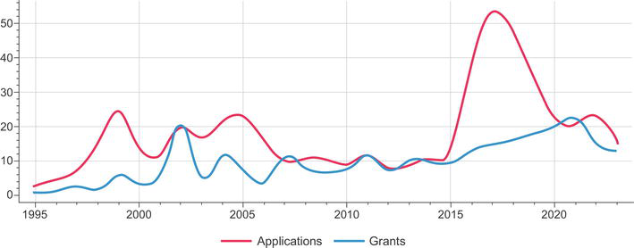
Figure 3.
Advances on the development of related technologies on decellularized matrices and biosynthetic scaffolds used in tissue engineering. Patent applications (red line) and granted technologies (blue line) from 1995 to 2023.
In conclusion, it was observed that most of the technologies of both acellular matrices and biosynthetic scaffolds (unlike clinical trials) are related to the engineering of bone tissues, followed by dermal matrices; taking into account this, it was deduced that it is necessary to increase the technological production in corneal tissues. It is noteworthy that from 2015 to 2019 more patent applications were registered related to acellular matrix technologies and biosynthetic scaffolds for bone, skin, and cornea. Their production decreased for 2020, possibly due to the global contingency of COVID-19. However, the year in which more patents were conceived was 2021 (Figure 3). Finally, the information contained in these documents can be used as inspiration to create new technologies or improve existing methods.
7. Clinical trials
Several clinical trials have been conducted with favorable results using collagen matrices, acellular membranes of the skin, cornea, and acellular bone matrices for their used in tissue repair or regeneration.
Strategies based on biopolymers combined with collagen for tissue treatment have been described. Since collagen is an abundant protein in connective tissue and an essential component of acellular matrices, collagen-based materials are indispensable in regenerative medicine [89], and collagen membranes have been studied in the regeneration of various tissues. For example, the use of a collagen membrane in bone repair has been reported with results comparable to endoprostheses, in this case in guided bone regeneration surgery in dentistry [89, 90, 91]. Results related to using a hydroxyapatite-collagen sponge biomaterial enriched with stem cells have been reported. These technologies were implanted in patients with alveolar cleft defects. The results showed reduced morbidity and decreased intensity and frequency of donor site pain in the group treated with the resorbable collagen sponge and stem cells at six months [92, 93]. Furthermore, in the case of articular cartilage regeneration, collagen membranes have been used in conjunction with autologous chondrocytes to form a construct capable of repairing articular defects [94].
On the other hand, corneal tissue engineering uses corneal tissue with cells; these corneal substitutes have been evaluated for viability and compatibility in patients and have been shown to promote healing and regeneration in corneal wounds. However, it has also been shown to have the possibility of improving corneal wound healing and regeneration [95, 96]. Similarly, clinical trials using acellular dermal matrices obtained from human skin [97, 98, 99] have demonstrated the efficacy of acellular dermal matrix in treating chronic wounds, diabetic foot ulcers, and other complex skin defects [100, 101]. In one clinical trial, 86 patients with diabetic foot ulcers were randomized into the study, with 47 patients receiving acellular matrix (study group) and 39 patients receiving standard-of-care therapy (control group). The proportion of healed ulcers between the groups was statistically significant (P = 0.0289), with the odds of healing being 2,7 times higher in the study group than in the control group, which indicated the potential of ADM by single application as an effective treatment of chronic diabetic ulcers [102].
On the other hand, a clinical study described using a human dermal matrix in combination with a skin graft as an alternative reconstructive solution for treating three different clinical cases related to full-thickness skin wounds. According to histological and ultrastructural analysis from repaired skin biopsies one year after the treatment with ADM, regenerative healing of the wound area with well-organized/oriented connective tissue, as well as cellular infiltration and blood vessel formation was observed in all clinical cases [103]. Several commercially available ADMs have shown significant repair effects for treating these diseases [104]. In addition, the extracellular matrix obtained from the dermis provides a support structure and growth factors that allow the expansion and proliferation of cells. This has great potential in tissue engineering and regenerative medicine [98, 105]. Human application of different acellular scaffolds is the ultimate and most important goal of tissue engineering and regenerative medicine [106]. Due to their biological composition and natural origin, these matrices promote biocompatibility, integrity, and cell and molecular movements crucial during tissue repair.
Finally, collagen-elastin matrices have been used as skin substitutes in treating burns hand and skin reconstruction; grafting with the matrix and unmeshed skin was carried out in a one-stage procedure. After three months, the pliability of the grafted area was excellent. A full range of motion was achieved in all hands, and no blisters or unstable or hypertrophic scars occurred [107]. we found more studies related to the use of mesenchymal stem cells [108], adipose-derived cells, and xenogeneic grafts based on ultrapure collagen [109]; all this for the repair of different tissues; however, these trials do not yet have specific results. Clinical studies that do not present results may be because most of the clinical study still needs to be completed, or it was not possible to complete it due to failures in its planning [110].
8. Conclusion
Numerous techniques for obtaining collagen-based scaffolds, their preclinical and clinical application, and technology watch related to technologies based on this protein have been reported in this manuscript, highlighting the property of collagen as the main component of ECM. In tissue and organ repair, each process undoubtedly requires the participation of collagen because it predominantly forms the structure of the tissue and contains cellular and protein binding sites. These features confirm the importance of collagen in all sceneries of applications in tissue engineering, from obtaining acellular biological matrices to generating biosynthetic scaffolds for treating damaged organs and tissues.
Acknowledgments
The authors want to acknowledge Instituto Distrital de Ciencia Biotecnología e Innovación en Salud for its valuable help with the provision of financial and technical resources to draft the manuscript.
References
- 1.
Bauer AJP, Liu J, Windsor LJ, Song F, Li B. Current development of collagen-based biomaterials for tissue repair and regeneration. Soft Materials. 2014; 12 :359-370. DOI: 10.1080/1539445X.2014.925474 - 2.
Kular JK, Basu S, Sharma RI. The extracellular matrix: Structure, composition, age-related differences, tools for analysis and applications for tissue engineering. Journal of Tissue Engineering. 2014; 5 :2041731414557112. DOI: 10.1177/2041731414557112 - 3.
Zhu J, Li Z, Zou Y, Lu G, Ronca A, D’Amora U, et al. Advanced application of collagen-based biomaterials in tissue repair and restoration. Journal of Leather Science and Engineering. 2022; 4 :30. DOI: 10.1186/s42825-022-00102-6 - 4.
Exposito J-Y, Valcourt U, Cluzel C, Lethias C. The fibrillar collagen family. International Journal of Molecular Sciences. 2010; 11 :407-426. DOI: 10.3390/ijms11020407 - 5.
Mathew-Steiner SS, Roy S, Sen CK. Collagen in wound healing. Bioengineering. 2021; 8 :1-15 - 6.
Hoshiba T, Lu H, Kawazoe N, Chen G. Decellularized matrices for tissue engineering. Expert Opinion on Biological Therapy. 2010; 10 :1717-1728. DOI: 10.1517/14712598.2010.534079 - 7.
Keane TJ, Swinehart IT, Badylak SF. Methods of tissue decellularization used for preparation of biologic scaffolds and in vivo relevance. Methods. 2015; 84 :25-34. DOI: 10.1016/j.ymeth.2015.03.005 - 8.
Dong C, Lv Y. Application of collagen scaffold in tissue engineering: Recent advances and new perspectives. Polymers (Basel). Feb 2016; 8 (2):1-20 - 9.
Costa A, Naranjo JD, Londono R, Badylak SF. Biologic scaffolds. Cold Spring Harbor Perspectives in Medicine. Sep 2017; 7 (9):1-23 - 10.
Xiong C, Yao W, Tao R, et al. Application of decellularized adipose matrix as a bioscaffold in different tissue engineering. Aesthetic Plastic Surgery. 2023. DOI: 10.1007/s00266-023-03608-4 - 11.
Chen K, Lin X, Zhang Q , Ni J, Li J, Xiao J, et al. Decellularized periosteum as a potential biologic scaffold for bone tissue engineering. Acta Biomaterialia. 2015; 19 :46-55. DOI: 10.1016/j.actbio.2015.02.020 - 12.
Lynch AP, Ahearne M. Strategies for developing decellularized corneal scaffolds. Experimental Eye Research. 2013; 108 :42-47. DOI: 10.1016/j.exer.2012.12.012 - 13.
Belviso I, Romano V, Sacco AM, Ricci G, Massai D, Cammarota M, et al. Decellularized human dermal matrix as a biological scaffold for cardiac repair and regeneration. Frontiers in Bioengineering and Biotechnology. 2020; 8 :1-18. [Internet] Available from:https://www.frontiersin.org/articles/10.3389/fbioe.2020.00229 - 14.
Zhang X, Chen X, Hong H, Hu R, Liu J, Liu C. Decellularized extracellular matrix scaffolds: Recent trends and emerging strategies in tissue engineering. Bioactive Materials. 2022; 10 :15-31. DOI: 10.1016/j.bioactmat.2021.09.014 - 15.
Hashimoto Y, Funamoto S, Sasaki S, Honda T, Hattori S, Nam K, et al. Preparation and characterization of decellularized cornea using high-hydrostatic pressurization for corneal tissue engineering. Biomaterials. 2010; 31 :3941-3948. DOI: 10.1016/j.biomaterials.2010.01.122 - 16.
An B, Lin Y-S, Brodsky B. Collagen interactions: Drug design and delivery. Advanced Drug Delivery Reviews. Feb 2016; 97 :69-84 - 17.
Chevallay B, Herbage D. Collagen-based biomaterials as 3D scaffold for cell cultures: Applications for tissue engineering and gene therapy. Medical & Biological Engineering & Computing. 2000; 38 :211-218. DOI: 10.1007/BF02344779 - 18.
Wolf K, Alexander S, Schacht V, Coussens LM, von Andrian UH, van Rheenen J, et al. Collagen-based cell migration models in vitro and in vivo. Seminars in Cell & Developmental Biology. 2009; 20 :931-941. DOI: 10.1016/j.semcdb.2009.08.005 - 19.
Ricard-Blum S. The collagen family. Cold Spring Harbor Perspectives in Biology. 2011; 3 :a004978. DOI: 10.1101/cshperspect.a004978 - 20.
Shafiq MA, Gemeinhart RA, Yue BYJT, Djalilian AR. Decellularized human cornea for reconstructing the corneal epithelium and anterior stroma. Tissue Engineering. Part C, Methods. 2012; 18 :340-348. DOI: 10.1089/ten.TEC.2011.0072 - 21.
Xu H, Sapienza JS, Jin Y, Lin J, Zheng X, Dong H, et al. Lamellar keratoplasty using acellular bioengineering cornea. Animals. 2022; 12 (1016):1-13 - 22.
Hashimoto Y, Funamoto S, Sasaki S, Negishi J, Honda T, Hattori S, et al. Corneal regeneration by deep anterior lamellar keratoplasty (DALK) using decellularized corneal matrix. PLoS One. 2015; 10 :e0131989. DOI: 10.1371/journal.pone.0131989 - 23.
Alio del Barrio JL, Chiesa M, Garagorri N, Garcia-Urquia N, Fernandez-Delgado J, Bataille L, et al. Acellular human corneal matrix sheets seeded with human adipose-derived mesenchymal stem cells integrate functionally in an experimental animal model. Experimental Eye Research. 2015; 132 :91-100. DOI: 10.1016/j.exer.2015.01.020 - 24.
Shi Y, Bikkuzin T, Song Z, Jin X, Jin H, Li X, et al. Comprehensive evaluation of decellularized porcine corneal after clinical transplantation. Xenotransplantation. Nov 2017; 24 (6):1-7 - 25.
Li P, Feng M, Hu X, Zhang C, Zhu J, Xu G, et al. Biological evaluation of acellular bovine bone matrix treated with NaOH. Journal of Materials Science. Materials in Medicine. 2022; 33 :58. DOI: 10.1007/s10856-022-06678-z - 26.
Emami A, Talaei-Khozani T, Tavanafar S, Zareifard N, Azarpira N, Vojdani Z. Synergic effects of decellularized bone matrix, hydroxyapatite, and extracellular vesicles on repairing of the rabbit mandibular bone defect model. Journal of Translational Medicine. 2020; 18 :361. DOI: 10.1186/s12967-020-02525-3 - 27.
Dussoyer M, Michopoulou A, Rousselle P. Applied sciences decellularized scaffolds for skin repair and regeneration. Applied Sciences. 2020; 10 :1-24 - 28.
Wu X, Kathuria N, Patrick CW, Reece GP. Quantitative analysis of the microvasculature growing in the fibrin Interface between a skin graft and the recipient site. Microvascular Research. 2008; 75 :119-129. DOI: 10.1016/j.mvr.2007.04.012 - 29.
Correa-Araujo L, Prieto-Abello L, Lara-Bertrand A, Medina-Solano MM, Guerrero L, Camacho B, et al. Bioengineered skin constructs based on mesenchymal stromal cells and acellular dermal matrix exposed to inflammatory microenvironment releasing growth factors involved in skin repair. Stem Cell Research & Therapy. 2023; 14 :1-18. DOI: 10.1186/s13287-023-03535-w - 30.
Doornaert M, Depypere B, Creytens D, Declercq H, Taminau J, Lemeire K, et al. Human decellularized dermal matrix seeded with adipose-derived stem cells enhances wound healing in a murine model: Experimental study. Annals of Medicine and Surgery. 2019; 46 :4-11. DOI: 10.1016/j.amsu.2019.07.033 - 31.
Rico-Llanos GA, Borrego-González S, Moncayo-Donoso M, Becerra J, Visser R. Collagen type I biomaterials as scaffolds for bone tissue engineering. Polymers. 2021; 13 :599. DOI: 10.3390/POLYM13040599 - 32.
Munhoz MAS, Hirata HH, Plepis AMG, Martins VCA, Cunha MR. Use of collagen/chitosan sponges mineralized with hydroxyapatite for the repair of cranial defects in rats. Injury. 2018; 49 :2154-2160. DOI: 10.1016/j.injury.2018.09.018 - 33.
Battafarano G, Rossi M, De Martino V, Marampon F, Borro L, Secinaro A, et al. Strategies for bone regeneration: From graft to tissue engineering. International Journal of Molecular Sciences. Jan 2021; 22 (3):1-22 - 34.
Horn MM, Martins VCA, de Guzzi Plepis AM. Interaction of anionic collagen with chitosan: Effect on thermal and morphological characteristics. Carbohydrate Polymers. 2009; 77 :239-243. DOI: 10.1016/j.carbpol.2008.12.039 - 35.
Soares DG, Rosseto HL, Basso FG, Scheffel DS, Hebling J, Costa CA de S. Chitosan-collagen biomembrane embedded with calcium-aluminate enhances dentinogenic potential of pulp cells. Brazilian Oral Research. scielo. 2016; 30 :1-10 - 36.
Becerra J, Rodriguez M, Leal D, Noris-Suarez K, Gonzalez G. Chitosan-collagen-hydroxyapatite membranes for tissue engineering. Journal of Materials Science. Materials in Medicine. 2022; 33 :18. DOI: 10.1007/s10856-022-06643-w - 37.
Grabska-Zielińska S, Sionkowska A, Coelho CC, Monteiro FJ. Silk fibroin/collagen/chitosan scaffolds cross-linked by a glyoxal solution as biomaterials toward bone tissue regeneration. Materials. 2020; 13 :1-20 - 38.
Heidenreich AC, Pérez-Recalde M, González Wusener A, Hermida ÉB. Collagen and chitosan blends for 3D bioprinting: A rheological and printability approach. Polymer Testing. 2020; 82 :106297. DOI: 10.1016/j.polymertesting.2019.106297 - 39.
Zhang M-X, Zhao W-Y, Fang Q-Q , Wang X-F, Chen C-Y, Shi B-H, et al. Effects of chitosan-collagen dressing on wound healing in vitro and in vivo assays. Journal of Applied Biomaterials & Functional Materials. 2021; 19 :2280800021989698. DOI: 10.1177/2280800021989698 - 40.
Da Cunha MR, Maia FLM, Iatecola A, Massimino LC, Plepis AM de G, Martins V da CA, et al. In vivo evaluation of collagen and chitosan scaffold, associated or not with stem cells, in bone repair. Journal of Functional Biomaterials. Jul 2023; 14 (7):1-19 - 41.
Jirofti N, Golandi M, Movaffagh J, Ahmadi FS, Kalalinia F. Improvement of the wound-healing process by curcumin-loaded chitosan/collagen blend electrospun nanofibers: In vitro and In vivo studies. ACS Biomaterials Science & Engineering. 2021; 7 :3886-3897. DOI: 10.1021/acsbiomaterials.1c00131 - 42.
Gombotz WR, Wee S. Protein release from alginate matrices. Advanced Drug Delivery Reviews. 1998; 31 :267-285. DOI: 10.1016/S0169-409X(97)00124-5 - 43.
Orive G, Tam SK, Pedraz JL, Hallé J-P. Biocompatibility of alginate-poly-L-lysine microcapsules for cell therapy. Biomaterials. 2006; 27 :3691-3700. DOI: 10.1016/j.biomaterials.2006.02.048 - 44.
Qin Y. Alginate fibres: An overview of the production processes and applications in wound management. Polymer International. 2008; 57 :171-180. DOI: 10.1002/pi.2296 - 45.
Rinaudo M. Main properties and current applications of some polysaccharides as biomaterials. Polymer International. 2008; 57 :397-430. DOI: 10.1002/pi.2378 - 46.
Hu T, Lo ACY. Collagen–alginate composite hydrogel: Application in tissue engineering and biomedical sciences. Polymers (Basel). 2021; 13 :1-26 - 47.
Ledo AM, Vining KH, Alonso MJ, Garcia-Fuentes M, Mooney DJ. Extracellular matrix mechanics regulate transfection and SOX9-directed differentiation of mesenchymal stem cells. Acta Biomaterialia. 2020; 110 :153-163. DOI: 10.1016/j.actbio.2020.04.027 - 48.
Diogo GS, Marques CF, Sotelo CG, Pérez-Martín RI, Pirraco RP, Reis RL, et al. Cell-laden biomimetically mineralized shark-skin-collagen-based 3D printed hydrogels for the engineering of hard tissues. ACS Biomaterials Science & Engineering. 2020; 6 :3664-3672. DOI: 10.1021/acsbiomaterials.0c00436 - 49.
Sotome S, Uemura T, Kikuchi M, Chen J, Itoh S, Tanaka J, et al. Synthesis and in vivo evaluation of a novel hydroxyapatite/collagen–alginate as a bone filler and a drug delivery carrier of bone morphogenetic protein. Materials Science and Engineering: C. 2004; 24 :341-347. DOI: 10.1016/j.msec.2003.12.003 - 50.
Yang X, Lu Z, Wu H, Li W, Zheng L, Zhao J. Collagen-alginate as bioink for three-dimensional (3D) cell printing based cartilage tissue engineering. Materials Science and Engineering: C. 2018; 83 :195-201. DOI: 10.1016/j.msec.2017.09.002 - 51.
Guillaume O, Naqvi SM, Lennon K, Buckley CT. Enhancing cell migration in shape-memory alginate-collagen composite scaffolds: In vitro and ex vivo assessment for intervertebral disc repair. Journal of Biomaterials Applications. 2015; 29 :1230-1246. DOI: 10.1177/0885328214557905 - 52.
Bowles RD, Williams RM, Zipfel WR, Bonassar LJ. Self-assembly of aligned tissue-engineered annulus fibrosus and intervertebral disc composite via collagen gel contraction. Tissue Engineering. Part A. 2010; 16 :1339-1348. DOI: 10.1089/ten.TEA.2009.0442 - 53.
Tripathi AS, Zaki MEA, Al-Hussain SA, Dubey BK, Singh P, Rind L, et al. Material matters: Exploring the interplay between natural biomaterials and host immune system. Frontiers in Immunology. 2023; 14 :1269960. DOI: 10.3389/fimmu.2023.1269960 - 54.
Chircov C, Grumezescu AM, Bejenaru LE. Hyaluronic acid-based scaffolds for tissue engineering. Romanian Journal of Morphology and Embryology. 2018; 59 :71-76 - 55.
Humaira, Raza Bukhari SA, Shakir HA, Khan M, Saeed S, Ahmad I, et al. Hyaluronic acid-based nanofibers: Electrospun synthesis and their medical applications; recent developments and future perspective. Frontiers in Chemistry. 2022; 10 :1-13. [Internet] Available from:https://www.frontiersin.org/articles/10.3389/fchem.2022.1092123 - 56.
Seo Y-K, Park J-K, Song K-Y, Kwon S-Y, Lee H-S. Wound healing effect of collagen-hyaluronic acid implanted in partially injured anterior cruciate ligament of dog. Biotechnology and Bioprocess Engineering. 2010; 15 :552-558. DOI: 10.1007/s12257-009-3082-4 - 57.
Davidenko N, Campbell JJ, Thian ES, Watson CJ, Cameron RE. Collagen–hyaluronic acid scaffolds for adipose tissue engineering. Acta Biomaterialia. 2010; 6 :3957-3968. DOI: 10.1016/j.actbio.2010.05.005 - 58.
Kang HJ, Jang YJ, Park IK, Kim HJ. An evaluation of the efficacy and characterization of a cross-linked hyaluronic acid/collagen/poloxamer sheet for use in chronic wounds. Journal of Wound Management and Research. 2018; 14 :26-36. DOI: 10.22467/jwmr.2018.00297 - 59.
Chen Y, Sui J, Wang Q , Yin Y, Liu J, Wang Q , et al. Injectable self-crosslinking HA-SH/col I blend hydrogels for in vitro construction of engineered cartilage. Carbohydrate Polymers. 2018; 190 :57-66. DOI: 10.1016/j.carbpol.2018.02.057 - 60.
Niu Y, Stadler FJ, Yang X, Deng F, Liu G, Xia H. HA-coated collagen nanofibers for urethral regeneration via in situ polarization of M2 macrophages. Journal of Nanobiotechnology. 2021; 19 :283. DOI: 10.1186/s12951-021-01000-5 - 61.
Tsai SW, Fang JF, Yang CL, Chen JH, Su LT, Jan SH. Preparation and evaluation of a hyaluronate-collagen film for preventing post-surgical adhesion. The Journal of International Medical Research. 2005; 33 :68-76. DOI: 10.1177/147323000503300106 - 62.
Ao H, Xie Y, Tan H, Yang S, Li K, Wu X, et al. Fabrication and in vitro evaluation of stable collagen/hyaluronic acid biomimetic multilayer on titanium coatings. Journal of The Royal Society Interface. 2013; 10 :20130070. DOI: 10.1098/rsif.2013.0070 - 63.
Udduttula A, Jakubovics N, Khan I, Pontiroli L, Rankin KS, Gentile P, et al. Layer-by-layer coatings of collagen–hyaluronic acid loaded with an antibacterial manuka honey bioactive compound to fight metallic implant infections. ACS Applied Materials & Interfaces. 6 Dec 2023; 15 :58119-58135. DOI: 10.1021/acsami.3c11910 - 64.
Hu Y, Feng B, Zhang W, Yan C, Yao Q , Shao C, et al. Electrospun Gelatin/PCL and collagen/PCL scaffolds for modulating responses of bone marrow endothelial progenitor cells. Experimental and Therapeutic Medicine. 2019; 17 :3717-3726. DOI: 10.3892/etm.2019.7387 - 65.
Oh G-W, Nguyen V-T, Heo S-Y, Ko S-C, Kim CS, Park WS, et al. 3D PCL/fish collagen composite scaffolds incorporating osteogenic abalone protein hydrolysates for bone regeneration application: In vitro and in vivo studies. Journal of Biomaterials Science. Polymer Edition. 2021; 32 :355-371. DOI: 10.1080/09205063.2020.1834908 - 66.
Kim S-C, Heo S-Y, Oh G-W, Yi M, Jung W-K. A 3D-printed polycaprolactone/marine collagen scaffold reinforced with carbonated hydroxyapatite from fish bones for bone regeneration. Marine Drugs. 2022; 20 :1-17 - 67.
Baylan N, Bhat S, Ditto M, Lawrence JG, Lecka-Czernik B, Yildirim-Ayan E. Polycaprolactone nanofiber interspersed collagen type-I scaffold for bone regeneration: A unique injectable osteogenic scaffold. Biomedical Materials. 2013; 8 :45011. DOI: 10.1088/1748-6041/8/4/045011 - 68.
Ma W, Wang L, Zhang Q , Dong X, Zhu T, Lu S. Electrospun PCL/collagen hybrid nanofibrous tubular graft based on post-network bond processing for vascular substitute. Biomaterials Advances. 2022; 139 :213031. DOI: 10.1016/j.bioadv.2022.213031 - 69.
Park S, Kim J, Lee M-K, Park C, Jung H-D, Kim H-E, et al. Fabrication of strong, bioactive vascular grafts with PCL/collagen and PCL/silica bilayers for small-diameter vascular applications. Materials and Design. 2019; 181 :108079. DOI: 10.1016/j.matdes.2019.108079 - 70.
Lizarazo-Fonseca L, Correa-Araujo L, Prieto-Abello L, Camacho-Rodríguez B, Silva-Cote I. In vitro and in vivo evaluation of electrospun poly (ε-caprolactone)/collagen scaffolds and Wharton’s jelly mesenchymal stromal cells (HWJ-MSCs) constructs as potential alternative for skin tissue engineering. Regenerative Therapy. 2023; 24 :11-24. DOI: 10.1016/j.reth.2023.05.005 - 71.
Jouibary YM, Rezvanpour A, Akbari B. Fabrication of polycaprolactone/collagen scaffolds reinforced by graphene oxide for tissue engineering applications. Materials Letters. 2022; 322 :132477. DOI: 10.1016/j.matlet.2022.132477 - 72.
Li P, Ruan L, Wang R, Liu T, Song G, Gao X, et al. Electrospun scaffold of collagen and polycaprolactone containing ZnO quantum dots for skin wound regeneration. Journal of Bionic Engineering. 2021; 18 :1378-1390. DOI: 10.1007/s42235-021-00115-7 - 73.
Tsuji H. Poly(lactic acid). In: Kabasci S, editor. Bio-Based Plastics: Materials and Applications. John Wiley & Sons, Ltd [Internet]. 1 Jan 2014. pp. 171-239. [cited 2024 Jan 9] Available from: https://onlinelibrary.wiley.com/doi/full/10.1002/9781118676646.ch8 - 74.
Ajioka M, Enomoto K, Suzuki K, Yamaguchi A. The basic properties of poly(lactic acid) produced by the direct condensation polymerization of lactic acid. Journal of environmental polymer degradation. 1995; 3 :225-234. DOI: 10.1007/BF02068677 - 75.
Wu S, Liu X, Yeung KWK, Liu C, Yang X. Biomimetic porous scaffolds for bone tissue engineering. Materials Science and Engineering: R: Reports. 2014; 80 :1-36. DOI: 10.1016/j.mser.2014.04.001 - 76.
Narayanan G, Vernekar VN, Kuyinu EL, Laurencin CT. Poly (lactic acid)-based biomaterials for orthopaedic regenerative engineering. Advanced Drug Delivery Reviews. 2016; 107 :247-276. DOI: 10.1016/j.addr.2016.04.015 - 77.
Wang L, Xu L, Peng C, Teng G, Wang Y, Xie X, et al. The effect of bone marrow mesenchymal stem cell and Nano-hydroxyapatite/collagen I/poly-L-lactic acid scaffold implantation on the treatment of avascular necrosis of the femoral head in rabbits. Experimental and Therapeutic Medicine. 2019; 18 :2021-2028. DOI: 10.3892/etm.2019.7800 - 78.
Liao SS, Cui FZ, Zhang W, Feng QL. Hierarchically biomimetic bone scaffold materials: Nano-HA/collagen/PLA composite. Journal of Biomedical Materials Research Part B: Applied Biomaterials: An Official Journal of The Society for Biomaterials, The Japanese Society for Biomaterials, and The Australian Society for Biomaterials and the Korean Society for Biomaterials. 2004; 69B :158-165. DOI: 10.1002/jbm.b.20035 - 79.
Li J, Chen Y, Mak AFT, Tuan RS, Li L, Li Y. A one-step method to fabricate PLLA scaffolds with deposition of bioactive hydroxyapatite and collagen using ice-based Microporogens. Acta Biomaterialia. 2010; 6 :2013-2019. DOI: 10.1016/j.actbio.2009.12.008 - 80.
Nanda HS, Nakamoto T, Chen S, Cai R, Kawazoe N, Chen G. Collagen microgel-assisted dexamethasone release from PLLA-collagen hybrid scaffolds of controlled pore structure for osteogenic differentiation of mesenchymal stem cells. Journal of Biomaterials Science. Polymer Edition. 2014; 25 :1374-1386. DOI: 10.1080/09205063.2014.938980 - 81.
Martin V, Ribeiro IA, Alves MM, Gonçalves L, Claudio RA, Grenho L, et al. Engineering a multifunctional 3D-printed PLA-collagen-minocycline-NanoHydroxyapatite scaffold with combined antimicrobial and osteogenic effects for bone regeneration. Materials Science and Engineering: C. 2019; 101 :15-26. DOI: 10.1016/j.msec.2019.03.056 - 82.
Fuse M, Hashizume-Takizawa T, Watanabe A, Taguchi C, Fujita-Nakajima K, Kuwada-Kusunose T, et al. Study of bioactive film: Fabrication of poly (lactic acid) film immobilized the Tilapia fish collagen or porcine collagen and behavior of MC3T3-E1 cells. International Journal of Oral-Medical Sciences. 2022; 21 :14-22. DOI: 10.5466/ijoms.21.14 - 83.
Cárdenas-Aguazaco W, Camacho B, Gómez-Pachón EY, Lara-Bertrand AL, Silva-Cote I. Electrospun scaffolds of polylactic acid, collagen, and amorphous calcium phosphate for bone repair. Pharmaceutics. Oct 2023; 15 (11):1-24 - 84.
Ritz U, Gerke R, Götz H, Stein S, Rommens PM. A new bone substitute developed from 3D-prints of polylactide (PLA) loaded with collagen I: An In vitro study. International Journal of Molecular Sciences. Nov 2017; 18 (12):1-14 - 85.
Gunes OC, Kara A, Baysan G, Bugra Husemoglu R, Akokay P, Ziylan Albayrak A, et al. Fabrication of 3D printed poly(lactic acid) strut and wet-electrospun cellulose Nano Fiber reinforced chitosan-collagen hydrogel composite scaffolds for Meniscus tissue engineering. Journal of Biomaterials Applications. 2022; 37 :683-697. DOI: 10.1177/08853282221109339 - 86.
Sato T, Chen G, Ushida T, Ishii T, Ochiai N, Tateishi T, et al. Evaluation of PLLA–collagen hybrid sponge as a scaffold for cartilage tissue engineering. Materials Science and Engineering: C. 2004; 24 :365-372. DOI: 10.1016/j.msec.2003.12.010 - 87.
Pinese C, Gagnieu C, Nottelet B, Rondot-Couzin C, Hunger S, Coudane J, et al. In vivo evaluation of hybrid patches composed of PLA based copolymers and collagen/chondroitin Sulfate for ligament tissue regeneration. Journal of Biomedical Materials Research. Part B, Applied Biomaterials. 2017; 105 :1778-1788. DOI: 10.1002/jbm.b.33712 - 88.
Lara-Bertrand AL, Camelo F, Camacho B, Silva-Cote I. Technological search of patents for the identification of devices with potential use in tumor-infiltrating lymphocytes (TILs) research. World Patent Information. 2024; 76 :102244. DOI: 10.1016/j.wpi.2023.102244 - 89.
Parenteau-Bareil R, Gauvin R, Berthod F. Collagen-based biomaterials for tissue engineering applications. Materials (Basel). 2010; 3 :1863-1887 - 90.
Ren P, Zhao W, Dai X, Wang X, Yu J, Yuan Y, et al. Risk factors for the formation of striae gravidarum in women in Jiangsu Province of China. Taiwanese Journal of Obstetrics & Gynecology. 2019; 58 :640-644. DOI: 10.1016/j.tjog.2019.07.010 - 91.
Csönge L, Bozsik Á, Bagi ZT, Gyuris R, Csönge DK, Kónya J, et al. Thermal manipulation of human bone collagen membrane (SoftBone) and platelet-rich fibrin (PRF) membranes. Collagen Biomaterials. 1 Mar 2022; pp. 1-16 [cited 2023 Dec 13]. [Internet] Available from: https://www.intechopen.com/chapters/80649 - 92.
Gimbel M, Ashley RK, Sisodia M, Gabbay JS, Wasson KL, Heller J, et al. Repair of alveolar cleft defects: Reduced morbidity with bone marrow stem cells in a resorbable matrix. The Journal of Craniofacial Surgery. 2007; 18 :895-901. DOI: 10.1097/SCS.0B013E3180A771AF - 93.
Tanikawa DYS, Pinheiro CCG, Almeida MCA, Oliveira CRGCM, Coudry RDA, Rocha DL, et al. Deciduous dental pulp stem cells for maxillary alveolar reconstruction in cleft lip and palate patients. Stem Cells International [Internet]. 2020; 2020 :1-9. [cited 2023 Dec 14] Available from:https://pubmed.ncbi.nlm.nih.gov/32256610/ - 94.
Mumme M, Barbero A, Miot S, Wixmerten A, Feliciano S, Wolf F, et al. Nasal chondrocyte-based engineered autologous cartilage tissue for repair of articular cartilage defects: An observational first-in-human trial. Lancet. 2016; 388 :1985-1994. DOI: 10.1016/S0140-6736(16)31658-0 - 95.
Schlereth SL, Hos D, Matthaei M, Hamrah P, Schmetterer L, O’Leary O, et al. New Technologies in Clinical Trials in corneal diseases and limbal stem cell deficiency: Review from the European vision institute special interest focus group meeting. Ophthalmic Research. 2021; 64 :145-167. DOI: 10.1159/000509954 - 96.
Rafat M, Jabbarvand M, Sharma N, Xeroudaki M, Tabe S, Omrani R, et al. Bioengineered corneal tissue for minimally invasive vision restoration in advanced keratoconus in two clinical cohorts. Nature Biotechnology. 2022; 41 :70-81. DOI: 10.1038/s41587-022-01408-w - 97.
Clinical Trials Using Acellular Dermal Matrix – NCI Available from: https://www.cancer.gov/research/participate/clinical-trials/intervention/acellular-dermal-matrix [Accessed: 11 December 2023] - 98.
Petrie K, Cox CT, Becker BC, MacKay BJ. Clinical applications of acellular dermal matrices: A review. Scars, Burns & Healing. 2022; 8 :20595131211038310. DOI: 10.1177/20595131211038313 - 99.
Sklenářová R, Akla N, Latorre MJ, Ulrichová J, Franková J. Collagen as a biomaterial for skin and corneal wound healing. Journal of Functional Biomaterials. Nov 2022; 13 (4):1-18 - 100.
Bukowiecki A, Hos D, Cursiefen C, Eming SA. Wound-healing studies in cornea and skin: Parallels, differences and opportunities. International Journal of Molecular Sciences. Jun 2017; 18 (6):1-24 - 101.
Cazzell S, Moyer PM, Samsell B, Dorsch K, McLean J, Moore MA. A prospective, Multicenter, single-arm clinical trial for treatment of complex diabetic foot ulcers with deep exposure using acellular dermal matrix. Advances in Skin & Wound Care. 2019; 32 :409-415. DOI: 10.1097/01.ASW.0000569132.38449.c0 - 102.
Reyzelman A, Crews RT, Moore JC, Moore L, Mukker JS, Offutt S, et al. Clinical effectiveness of an acellular dermal regenerative tissue matrix compared to standard wound Management in Healing Diabetic Foot Ulcers: A prospective, randomised, multicentre study. International Wound Journal. 2009; 6 :196-208. DOI: 10.1111/j.1742-481X.2009.00585.x - 103.
Bondioli E, Purpura V, Orlandi C, Carboni A, Minghetti P, Cenacchi G, et al. The use of an acellular matrix derived from human dermis for the treatment of full-thickness skin wounds. Cell and Tissue Banking. 2019; 20 :183-192. DOI: 10.1007/S10561-019-09755-W - 104.
Cazzell S, Vayser D, Pham H, Walters J, Reyzelman A, Samsell B, et al. A randomized clinical trial of a human acellular dermal matrix demonstrated superior healing rates for chronic diabetic foot ulcers over conventional care and an active acellular dermal matrix comparator. Wound Repair and Regeneration. 2017; 25 :483-497. DOI: 10.1111/wrr.12551 - 105.
Kim YH, Shim HS, Lee J, Kim SW. A prospective randomized controlled Multicenter clinical trial comparing paste-type acellular dermal matrix to standard Care for the Treatment of chronic wounds. Journal of Clinical Medicine. Apr 2022; 11 (8):1-11 - 106.
Boroumand S, Hamedi E, Sigaroodi F, et al. Biological materials introduced to the market for blurred cornea regeneration. Regenerative Engineering and Translational Medicine. 2023. DOI: 10.1007/S40883-023-00319-9 - 107.
First Experiences with the Collagen-Elastin Matrix Matriderm® as a Dermal Substitute in Severe Burn Injuries of the Hand – ScienceDirect Available from: https://www.sciencedirect.com/science/article/abs/pii/S0305417906002282 [Accessed: 13 December 2023] - 108.
Eduardo Rolim Teixeira, P.U.C. do R.G. do S. (Responsible P. No Study Results Posted | Autogenous Mesenchymal Stem Cell Culture-Derived Signalling Molecules as Enhancers of Bone Formation in Bone Grafting | ClinicalTrials.Gov Available from: https://clinicaltrials.gov/study/NCT04998058?cond=Tissue Engineering&term=collagen&page=3&rank=22&tab=results [Accessed: 11 December 2023] - 109.
Study Details | Microfracture Versus Adipose Derived Stem Cells for the Treatment of Articular Cartilage Defects | ClinicalTrials.Gov Available from: https://clinicaltrials.gov/study/NCT02090140?cond=Tissue Engineering&term=collagen&page=3&rank=21 [Accessed: 14 December 2023] - 110.
Mechanical Methods of Producing Biomaterials with Aligned Collagen Fibrils | IntechOpen. Available from: https://www.intechopen.com/chapters/81613 [Accessed: 13 December 2023]