
Properties of Cerium.
Abstract
Due to its numerous uses in the fields of magnetic recording devices, electronic devices, biosensors, high-frequency transformers, microwave devices, and radar devices, magnetic nanoferrites have drawn increased attention from society. There are several ways to make rare earth ion-doped spinel ferrites, including the sol-gel approach, auto-combustion method, hydrothermal method, co-precipitation method, ball milling method, and green synthesis method. Numerous studies have recently discussed the effect of rare earth ion substitution on spinel ferrite, concluding that it improved the material’s structural and magnetic characteristics. The replacement of Cerium ions, which was studied by just a few researchers, allows ferrites and mixed ferrites’ magnetic properties, such as coercivity, saturation magnetization, and retentivity, to be fine-tuned. In this study, we summarized the effect of different concentrations of Cerium ions on various ferrites to find the significant results of structural parameters like crystal size, lattice constant, and also magnetic parameters such as coercivity and saturation magnetization, through the study of X-ray diffraction (XRD) and vibrating sample magnetometer (VSM).
Keywords
- Cerium
- spinel ferrites
- XRD
- VSM
- crystal size
1. Introduction
The rare earth elements (REEs) are otherwise called as rare earth metals, or they are referred to as lanthanides containing 17 indistinguishable heavy metals (La, Ce, Pr, Nd, Pm, Sm, Eu, Gd, Tb, Tm, Yb, Lu, Dy, Ho, Er). Apart from these rare earth elements (REEs), Scandium and Yttrium (Sc and Y) are also considered as rare earth elements of the lanthanide group, since it possesses chemical properties of Scandium and Yttrium that have different magnetic properties than the lanthanides. Normally, lanthanide groups are classified into light rare earth elements and heavy rare earth elements. The elements from Lanthanum to Europium are grouped under light rare earth elements, while the elements from Gadolinium to Lutetium are grouped under heavy rare earth elements. The physical and chemical properties of all lanthanide elements are similar due to the progressively filled 4f orbitals with increasing atomic number. In that, ionic radius gradually decreases from La3+ (1.06 Å) to Lu3+ (0.85 Å), which is the so called the lanthanide contraction. The symbol of Cerium is shown in Figure 1.
Figure 1.
Symbol of Cerium.
The incorporation of rare earth elements, such as Cerium, into the structural matrix of nanoferrites has garnered considerable attention due to the profound influence these elements exert on both structural and magnetic characteristics. Rare earth elements possess unique electronic configurations and magnetic properties that can significantly alter the crystal lattice and magnetic behavior of nanoferrites. Among these, Cerium, a member of the lanthanide series, stands out for its versatile chemical nature and varied oxidation states. In this context, this introduction aims to explore the impact of Cerium doping on the structural aspects, such as crystal symmetry and lattice parameters, as well as the magnetic properties of different nanoferrites. Understanding the interplay between Cerium and nanoferrite matrices is crucial for tailoring and optimizing the performance of these materials in various technological applications, ranging from catalysis to magnetic devices. This exploration provides a foundation for delving into the subsequent sections, which delve into specific analyses and findings related to the structural and magnetic transformations induced by Cerium incorporation into different nanoferrites.
In the past few decades, REE has been applied in the field of various devices, such as rechargeable batteries, light-emitting diodes (LEDs), solar panels, magnetic resonance imaging (MRI), superconductors, supermagnets, catalysts, storage devices, glass additives, fluorescent materials, and digital versatile discs (DVDs). Since they covers almost all important ingredients in modern technology, the REEs are called “The vitamins of modern industry.” Thus, it enhances their applications in electronics, manufacturing ceramic pigments, metal alloy, plastic creation, medical field, and energy storage devices.
Among the rare earth metals, Cerium has attracted researcher’s attention in recent days due to its remarkable physical and chemical properties [1, 2, 3, 4, 5]. Cerium is the most reactive element in the lanthanide series group, when compared with other elements. It is present in different minerals, such as silicates, phosphates, carbonates, monazite, bastnaesite, allanite, and cerite. Out of these various minerals, monazite leads as the principal source of Cerium. Cerium comprises of four isotopes of different percentages, listed as 136Ce (0.2%), 138Ce (0.3%), 140Ce (88.5%), and 142Ce (11.1%) [5]. It occurs more abundantly than lead and tin in the earth’s crust. Based on the electronic structure, the energy levels of the inner 4f level and 6 s level are the same, which gives different occupancies of two levels. Compared with other rare earth metals, Cerium exists in two oxidation states of Ce3+ and Ce4+, in which Ce4+ oxidation mode is more stable than Ce3+ due to its [Xe]4f° level of electronic structure. Cerium is a soft, malleable, and ductile rare earth metal. Its color varies from iron gray to silvery nature. When it is scratched with a sharp knife, it starts to ignite, once it is in pure form. Cerium was first applied in gas mantles, invented by the Austrian Chemist Carl Auer Von Welsbach. Mischmetal is a pyrophoric alloy that is composed of 50% of Cerium, which is widely applied in lighter flints. This addition of mischmetal to steel is made for improving the mechanical properties [6]. By the addition of Cerium, the photostability of pigments is enhanced, providing light fastness pigments [7]. In television, F-centre defects are created due to the continuous bombardment of electrons, which may cause a darkening effect on the television glass plates, which is reduced by the addition of Cerium to the cathode ray tube (CRT). Cerium is employed as a dopant for phosphorus in lamps, white light-emitting diodes (LEDs), and cathode-ray tube television (CRT TV) screens [8, 9].
1.1 Properties of cerium
Cerium is a chemical element with the symbol Ce and atomic number 58. It belongs to the lanthanide series of rare earth elements. Cerium exhibits several distinctive properties that are listed in Table 1. Applications of Cerium are listed below:
Cerium finds applications in various industries. It is used as a catalyst in automotive catalytic converters, helping to reduce pollutants in exhaust gases.
In the glass and ceramics industry, Cerium is employed as a polishing agent.
Cerium is utilized as an alloying agent in metallurgy, enhancing the properties of alloys.
It serves as a component in phosphors used in fluorescent lighting and color television tubes.
| Cerium | Properties |
|---|---|
| Chemical symbol | Ce |
| Atomic number | 58 |
| Atomic mass | 140.12 g/mol |
| Density | 6.770 g/m3 |
| Melting point | 1065°C |
| Boiling point | 3556°C |
| Crystal structure | Face Centered Cubic (FCC) |
| Electrical conductivity | Good electrical conductor |
| Magnetic property | Paramagnetic at room temperature Ferromagnetic at low temperature |
| Reactivity | Highly reactive with water, oxygen, and acids |
Table 1.
Electronic properties: Cerium is an excellent electrical conductor, contributing to its applications in certain electronic and catalytic devices.
Natural abundance: Cerium is relatively abundant in the Earth’s crust, making up a significant portion of rare earth elements.
Isotopes: Cerium has several isotopes, with the most stable being Cerium-140.
Radioactivity: Some Cerium isotopes are radioactive, posing health and environmental risks. Cerium’s diverse properties contribute to its widespread use in various industrial applications, making it a valuable element in technological advancements and material sciences.
Table 2 provides a balanced overview of the advantages and disadvantages associated with the use of Cerium, considering its physical, environmental, and technological aspects.
| Cerium | Advantages | Disadvantages |
|---|---|---|
| Physical |
|
|
| Environmental |
|
|
| Technological |
|
|
Table 2.
Physical, environmental and technological advantages and disadvantages of Cerium.
1.2 Ferrites
Recently, several researchers’ works have been done on the fabrication of different ferrites due to their fascinating physical and chemical properties than other metal oxide groups. Ferrites belong to the metal oxide group with a spinel structure of chemical formula MFe2O4 (M = Co, Cu, No, Zn, etc.), where M denotes the divalent ions. Ferrites come under the compound of ferrimagnetic ceramics, which is applied in the various opto-electric and magnetic materials due to their notable properties of minimum eddy current losses and high electrical resistances [10, 11, 12]. The structure of spinel ferrite is shown in Figure 2. In spinel ferrites, MFe2O4, the M that represents the divalent ions and Fe indicates the divalent M2+ and trivalent Fe3+ ions in the octahedral and tetrahedral sites, the ferrites are classified into normal, inverse, and mixed spinels. In normal spinel, the Fe3+ ions are located in the octahedral sites and M2+ ions (Cu, Zn, Co, Mg, etc.) occupied in the tetrahedral sites, whereas in inverse spinel, one half of the Fe3+ ions occupied in tetrahedral sites and the remaining located in the octahedral sites, and the M2+ ions distributed in the octahedral sites. Thus, the magnetic properties of the spinel ferrites depend on the cation distribution between the octahedral and tetrahedral sites [13, 14, 15, 16].
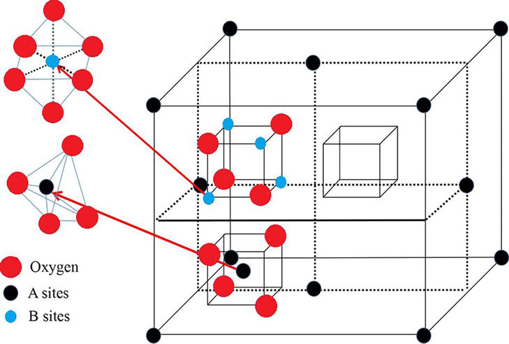
Figure 2.
Structure of spinel ferrite.
Ferrites may be treated as soft and hard ferrites, based on the magnetic property of coercivity (Hc), saturation magnetization (Ms), and retentivity (Mr). Soft ferrites can easily magnetize and demagnetize, the value of coercive field is around less than 40 A/m, which indicates lower value, but high saturation magnetization (Ms) and high permeability (μr), whereas for hard ferrites, it is opposite to that of soft ferrites in terms of magnetic properties.
1.3 Impact of Cerium in different ferrites
1.3.1 Cerium-doped cobalt ferrite
Elaya kumar
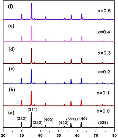
Figure 3.
X-ray diffraction (XRD) pattern of CoCexFe2-xO4(a = x = 0.0, b = x = 0.1, c = x = 0.2, d = x = 0.3, e = x = 0.4, and f = x = 0.5).
| Sample | x | Synthesized method | Structure | Crystallite size (nm) | Magnetic parameters | Reference | ||
|---|---|---|---|---|---|---|---|---|
| HC (Oe) | Mr (emu/g) | Ms (emu/g) | ||||||
| CoCexFe2-xO4 | 0 | Sol-gel | Single-phase cubic structure | 25.36 | 106.52 | 11.22 | 55.64 | [17] |
| 0.1 | 22.51 | 99.48 | 10.55 | 52.85 | ||||
| 0.2 | 20.98 | 95.47 | 9.94 | 47.87 | ||||
| 0.3 | 19.85 | 93.92 | 8.91 | 38.35 | ||||
| 0.4 | 18.67 | 91.95 | 7.45 | 35.46 | ||||
| 0.5 | 16.53 | 89.48 | 5.98 | 29.32 | ||||
| CuCe1-xFe2-xO4 | 0 | Sol-gel | Single-phase cubic structure | 25.36 | 25.45 | 9.25 | 4.668 | [18] |
| 0.1 | 23.88 | 24.98 | 8.36 | 4.548 | ||||
| 0.2 | 21.98 | 23.86 | 7.63 | 3.856 | ||||
| 0.3 | 20.65 | 22.68 | 6.85 | 3.365 | ||||
| 0.4 | 19.39 | 21.83 | 5.43 | 2.964 | ||||
| 0.5 | 18.53 | 20.75 | 4.82 | 2.322 | ||||
| CuFe2-xCexO4 | Auto-combustion sol-gel | Tetragonal phase structure | 52 | 1200 | 9 | 15 | [19] | |
| NiCexFe2-xO4 | 0 | Sol-gel | Single-phase cubic structure | 23.5 | 0.0289 | 0.358 | 65.38 | [20] |
| 0.1 | 22.8 | 0.0274 | 0.312 | 47.53 | ||||
| 0.2 | 22.3 | 0.0192 | 0.296 | 37.34 | ||||
| 0.3 | 21.7 | 0.0189 | 0.281 | 24.32 | ||||
| 0.4 | 21.2 | 0.0176 | 0.277 | 21.22 | ||||
| 0.5 | 20.9 | 0.0169 | 0.265 | 8.685 | ||||
| NiCexFe2-xO4 | 0 | Chemical co-precipitation method | Pure spinel phase structure | 55.8 | 63.5 | 7.27 | 51.9 | [21] |
| 0.05 | 42.2 | 92.2 | 7.17 | 46.9 | ||||
| 0.10 | 39.2 | 118.6 | 5.05 | 36.5 | ||||
| 0.15 | 33.5 | 130.9 | 3.19 | 22.6 | ||||
| 0.20 | 41.8 | 50.2 | 2.40 | 25.8 | ||||
| NiCexFe2-xO4 | 0 | Chemical route method | Pure spinel phase structure | 57 | 41.84 | — | 61.6 | [22] |
| 0.01 | 28 | 42.90 | — | 67.7 | ||||
| 0.02 | — | 41.34 | — | 25.16 | ||||
| 0.03 | — | 38.67 | — | 28.4 | ||||
| 0.04 | 11 | 41.14 | — | 14.46 | ||||
| 0.06 | 19 | 40.78 | — | 5.9 | ||||
| 0.08 | — | 41.35 | — | 15.1 | ||||
| 0.10 | — | 31.64 | — | 26.83 | ||||
| MgCexFe2-xO4 | 0.01 | Sol-gel method | Face-centred cubic structure | 51.113 | 140.86 | 0.13650 | 0.51660 | [23] |
| 0.012 | 82.12 | 134.46 | 0.14146 | 0.64289 | ||||
| 0.014 | 46.61 | 94.586 | 0.0775 | 0.65246 | ||||
| 0.016 | 67.22 | 144.40 | 0.18184 | 0.83079 | ||||
| MgCexFe2-xO4 | 0 | Sol–gel auto-combustion method | Single-phase spinel structure | 45.26 | 120 | 2.08 | 13.59 | [24] |
| 0.02 | 42.42 | 66.97 | 0.39 | 7.40 | ||||
| 0.04 | 42.82 | 95.26 | 1.11 | 9.65 | ||||
| 0.06 | 46.47 | 112.33 | 2.61 | 17.42 | ||||
| SrFe12-xCexO19 | 0 | Sol-gel auto-combustion method | — | 77 | — | — | 68.56 | [25] |
| 0.1 | 67 | — | — | 58.45 | ||||
| 0.2 | 65.1 | — | — | 53.12 | ||||
| 0.3 | 47.7 | — | — | 51.24 | ||||
| 0.4 | 63.8 | — | — | 47.41 | ||||
| 0.5 | 51.1 | — | — | 46.69 | ||||
Table 3.
Synthesize method, structure, crystallite size and magnetic parameters of Cerium-doped different nanoferrites.
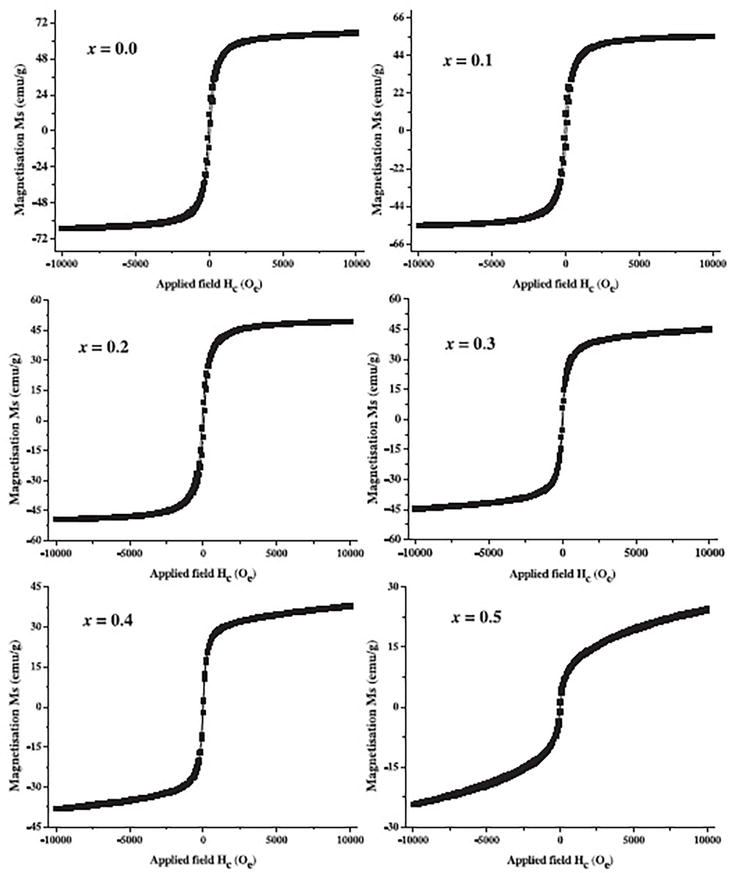
Figure 4.
Vibrating sample magnetometer (VSM) analysis of CoCexFe2-xO4(a = x = 0.0, b = x = 0.1, c = x = 0.2, d = x = 0.3, e = x = 0.4, and f = x = 0.5).
1.3.2 Cerium-doped copper ferrite
The powder XRD patterns of CuCe1-xFe2-xO4 samples, as depicted in Figure 5, align well with the standard values for copper ferrite (JCPDS file: 770010) reported by K. Elaya kumar
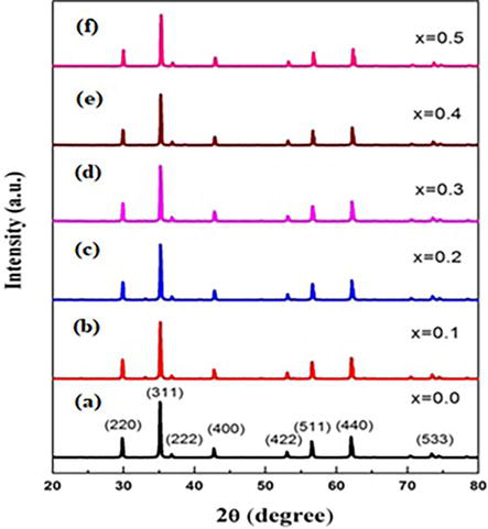
Figure 5.
X-ray diffraction (XRD) spectra of Ce3+ doped copper ferrite (a = x = 0.0, b = x = 0.1, c = x = 0.2, d = x = 0.3, e = x = 0.4, and f = x = 0.5).
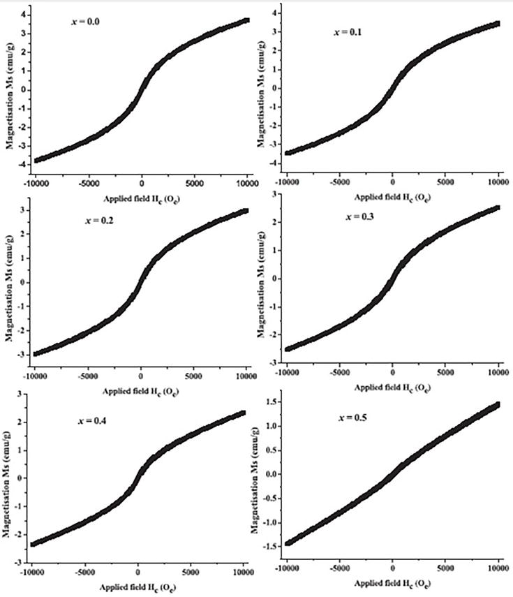
Figure 6.
Vibrating sample magnetometer (VSM) analysis of Ce3+ doped copper ferrite (a = x = 0.0, b = x = 0.1, c = x = 0.2, d = x = 0.3, e = x = 0.4, and f = x = 0.5).
Tetragonal phase of spinel copper ferrite nanoparticles is confirmed by the XRD pattern, which was reported by Mehdi Rahimi-Nasrabadi et al. [19]. The average crystalline size of CuFe2O4 nanoparticles is found to be 52 nm by the auto-combustion sol-gel method. Furthermore, CuFe2-xCexO4 nanoparticles exhibit the ferromagnetic characteristics at room temperature. From the hysteresis loop, the saturation magnetization (Ms) value is said to be 15 emu/g, the remnant magnetization (Mr) is noted as 9 emu/g, and the coercive field (Hc) value is 1200 Oe, respectively, and these values are listed in Table 3.
1.3.3 Cerium-doped nickel ferrite
The structural study of Cerium-doped Nickel ferrite nanoparticles, NiCexFe2-xO4, by the sol-gel method was successfully evaluated through the XRD technique, as reported by Elayakumar et al. [20]. The XRD patterns reveal that the pure-phase formation of spinel structural crystalline samples is shown in Figure 7. It shows that the Cerium ions (Ce3+) are entered into the Nickel ferrite lattice without any distortion. With the help of Scherrer formula, the crystalline sizes of the resultant samples are calculated and the values are listed in Table 3. The ionic radius of Ce3+ is 1.03 Å, which is greater than the ionic radius of Fe3+ that is 0.64 Å, which results in the impact of Cerium ions to penetrate into the spinel lattice. It was found that the crystalline size reduced from 23.5 to 20.9 nm, when the Cerium ions’ concentration was increased. The magnetic characterization of NiCexFe2-xO4 nanoparticles was analyzed by the vibrating sample magnetometer (VSM), which is shown in Figure 8. The magnetic parameters like saturation magnetization (Ms), coercivity (Hc), and remanence magnetization (Mr) were calculated. It was noted that the remarkable changes in the magnetic parameters of Nickel ferrite lattice took place due to the replacement of Fe3+ ions by Ce3+. Thus, it is weakening the sublattice interaction and creates reduction in the coercivity value of the products. The remanence magnetization (Mr) value was decreased to 0.265 from 0.365 emu/g and value are listed in Table 3, which indicates that the prepared samples exhibit superparamagnetic nature. Thus, it happens due to the Ce3+-Fe3+ interaction taking place on account of 3d-4f electron coupling.
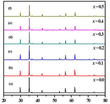
Figure 7.
Powder XRD patterns of NiCexFe2-xO4 (a = x = 0.0, b = x = 0.1, c = x = 0.2, d = x = 0.3, e = x = 0.4, and f = x = 0.5) samples.
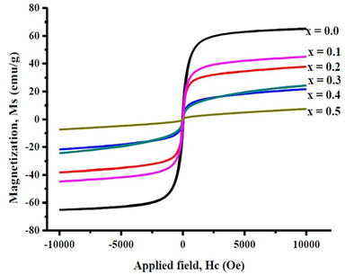
Figure 8.
Vibrating sample magnetometer (VSM) analysis of NiCexFe2-xO4 (x = 0.0, 0.1, 0.2, 0.3, 0.4, and 0.5) samples.
Priyadharshini et al. [21] synthesized Ce-doped NiFe2O4 nanoparticles by chemical co-precipitation method. It was noted that the lattice parameters are altered by the substitution of Cerium-doped Nickel ferrite lattice. By the substitution of Scherrer method, the crystalline size was gradually reduced from 55.8 to 41.8 mm with the substitution of Ce3+ ions and is listed in Table 3. When the amount of Cerium dopant increases, the NiFe2O4 lattice limits the crystalline growth. Well-deserved ‘S’-shaped curves are obtained from the VSM graph. The saturation magnetization (Ms), remanence value (Mr), and coercivity (Hc) values are altered with the dopant concentration of Ce3+ ions. The coercivity value (Hc) slowly increases from 63.05 Or to 130.9 Oe at the concentration of (x = 0.15 M) Cerium ions penetrating into the spinel lattice. The Ms. and Mr. values reduced to 22.6 and 3.19 emu/g when the Ce content is increased at (x = 0.15 M). Thus, it happens due to the weakening of the Fe3+-Fe3+ interactions, brought about by the replacement of Fe3+ ions by the Ce3+ ions.
Gagan Dixit et al. [22] synthesized the Ce-doped NiFe2O4 nanoparticles and characterized the structural, optical, and magnetic properties by the chemical route method. XRD reveals the pure-phase formation of spinel structure nanoparticles. The substitution of Ce3+ ions into the spinel lattice creates lattice strain and thus reduces the crystalline size from 57 to 17 nm, which is listed in Table 3. However, the other structural parameters remain constant. The magnetic parameters like saturation magnetization (Ms) and coercivity (Hc) are calculated from the VSM graph. The Ms. value is gradually decreased to 31.64 from 41.84 emu/g at the dopant concentration level of x = 0.10 M, and the Cerium dopant level is listed in Table 3. Thus, the Ce3+ ions penetrate the Nickel ferrite lattice.
1.3.4 Cerium-doped magnesium ferrite
Muthuraman et al. [23] reported the polycrystalline and single-phase formation of prepared samples, which was confirmed by the XRD techniques. The structural and magnetic properties of Cerium-doped Magnesium ferrite are studied by the XRD and VSM techniques. These Cerium-doped Magnesium ferrite nanoparticles are prepared by the sol-gel method. XRD graphs show the impurities-free Cerium-doped Magnesium ferrite nanoparticles. The structural parameters like lattice parameter and crystallite size were calculated from Bragg’s law and Debye Scherrer’s formula. The average crystalline size found to be in the range of 46.61 to 82.12 nm values is listed in Table 3. The lattice parameter increases from 8.3732 to 8.4076 at the Cerium concentration range of x = 0.014 M. From the hysteresis loop, magnetic parameters like saturation magnetization (Ms), coercivity (Hc), and retentivity (Mr) are measured. The least saturation magnetization value of 0.51660 emu was noted at a lower concentration of Ce3+ dopant and gradually increases to 0.83079 emu at the higher concentration of Cerium ions. The lower coercivity value of 95.586 Oe was found at the concentration of x = 0.016 M. And it was found to have the least retentivity value of 0.0775 at the concentration of x = 0.016 M values that are listed in Table 3.
Syed Ismail Ahmad et al. [24] reported the Cerium-doped Magnesium ferrite nanoparticles by the sol-gel auto-combustion technique. X-ray diffraction analysis reveals the single-phase spinel structure without any impurities. The addition of Ce3+ ions in the parental lattice decreases the intensity of the peaks, which indicates the strain produced due to the creation of oxygen vacancies. Thus, the reflections are gradually shifted to lower angles because the radii of Ce3+ ions are larger than those of the Fe3+ ions. The average crystallite size in the range of 42–46 nm values is listed in Table 3. The magnetic parameters were calculated from the M-H curves of vibrating sample magnetometer (VSM) device for the Cerium-doped Magnesium ferrite. Due to the porosity of the samples, the saturation magnetization (Ms) value decreases from 13.59 to 9.65 emu/g at the concentration range of Cerium (x = 0.04). After this saturation, magnetization slowly increases to 17.42 emu/g at the dopant range of (x = 0.06) concentration. Thus, the increase in (Ms) value is attributed to the migration of Mg2+ ions from B site to A site and segregation of Ce3+ ions at the level of grain boundary. The large amount of Ce3+ ion dopant may cause redistribution of cations, which means the divalent magnesium ions are partially placed in A site and equal number of Fe3+ ions move to B site, which makes the increase in saturation magnetization (Ms). The coercivity value (Hc) was found to be 120 Oe at (x = 0.00) of Cerium content and decreases to 95.26 Oe at (x = 0.04) of Cerium content. Once again, the coercivity value (Hc) increases to 112.33 Oe at higher concentration of (x = 0.06) of Cerium dopant into the Magnesium Ferrite values and is listed in Table 3. Overall, the coercivity (Hc) value decreases when compared with undoped Magnesium ferrite, since the coercivity (Hc) purely depends on the crystal anisotropy. From the magnetic parameters, it was concluded that all the samples exhibit superparamagnetic nature.
1.3.5 Cerium-doped strontium hexaferrite
SrFe12-xCexO19 magnetic nanoparticles are fabricated by the sol-gel auto-combustion method. The structural parameters were evaluated by the XRD technique at room temperature. All the noted peaks are well matched with the JCPDS Card No. 96–100-8857, which indicates the impurities-free Cerium-doped Strontium Hexaferrite. The ionic radius of Ce3+ is 1.05, which is larger than that of Fe3+ which is 0.64, and thus the replacement of Fe3+ ion by the Ce3+ ion takes place. The concentration of crystal axis takes place due to the strain, defects, and chemical composition of Cerium-doped Strontium Hexaferrite. The magnetic measurements were noted from the vibrating sample magnetometer technique. The saturation magnetization (Ms) value is 68.56 emu/g at 300 k that was noted and is listed in Table 1. The coercive field (Hc) of the Strontium Hexaferrite was 4337–5703 Oe at room temperature and 2808–4514 Oe at 10 k. The remanence magnetization (Mr) is measured in the range of 35–23 emu/g at room temperature and 50–32 emu/g at 10 k. These overall magnetic parameters reveal that the formation of Cerium-doped Strontium Hexaferrite exhibits the ferromagnetic characteristics. The magnetization expression is given by Eq. (1)
where
The values of saturation magnetization (Ms) are higher at lower temperature, when compared with those at room temperature. This may happen due to the reduction in thermal fluctuations of magnetic moments and thus strengthening the superexchange interaction.
Advertisement
2. Conclusion
Researchers have been drawn to Cerium-doped magnetic ferrites, a rare earth element, because of its distinct characteristics and wide range of uses in several sectors. The chapter provides a summary of the structural characteristics, including crystallite size and structures. Saturation magnetization (M-S) and coercivity values for several ferrites are listed, and other magnetic properties are recorded from the VSM investigation. Sol-gel analysis reveals that the majority of Cerium-doped magnetic ferrites have a single-phase spinel structure. Among these several ferrites doped with Ce3+ ions, the nanoparticles of CuFe2O4 and MgFe2O4 exhibit the least saturation magnetization (Ms). The crystalline size of all ferrites synthesized using the sol-gel technique is around 50 nm on average. As a result, the Cerium ions are successfully revealed by the differences in crystallite size and magnetic properties in all magnetic ferrites.
References
- 1.
Lide DR, editor. CRC Handbook of Chemistry and Physics. Boca Raton, FL: CRC press; 2004 - 2.
Mogensen M, Sammes NM, Tompsett GA. Physical, chemical and electrochemical properties of pure and doped ceria. Solid State Ionics. 2000; 129 (1-4):63-94 - 3.
Kilbourn BT. Cerium and Cerium compounds. In: Kirk-Othmer Encyclopedia of Chemical Technology. Vol. 1-23. Wiley Online Library; 2000 - 4.
Haxel G. Rare Earth Elements: Critical Resources for High Technology. US: US Department of the Interior, US Geological Survey; 2002 - 5.
O'Neil MJ. The Merck index: And encyclopedia of chemicals, drugs, and biologicals. In: The Merck Index: And Encyclopedia of Chemicals, Drugs, and Biologicals. 2006, 1756-1756 - 6.
Greenwood NN, Earnshaw A. Chemistry of the Elements. Burlington: Elsevier; 2012 - 7.
Gleń M, Grzmil B, Sreńscek-Nazzal J, Kic B. Effect of CeO2 and Sb2O3 on the phase transformation and optical properties of photostable titanium dioxide. Chemical Papers. 2011; 65 (2):203-212 - 8.
DaNa - Information on the safety of new, innovative materials and nanomaterials. Cerium Dioxide, 2024. Available from: https://www.mcgill.ca/crcf/files/crcf/2005-Increasing_Caseloads_OIS93-98.pdf - 9.
Trovarelli A. Catalysis by Ceria and Related Materials. London: World Scientific; 2002 - 10.
Rashad MM, Rayan DA, Turky AO, Hessien MM. Effect of Co2+ and Y3+ ions insertion on the microstructure development and magnetic properties of Ni0. 5Zn0. 5Fe2O4 powders synthesized using co-precipitation method. Journal of Magnetism and Magnetic Materials. 2015; 374 :359-366 - 11.
Mund HS, Ahuja BL. Structural and magnetic properties of Mg doped cobalt ferrite nano particles prepared by sol-gel method. Materials Research Bulletin. 2017; 85 :228-233 - 12.
Moradmard H, Shayesteh SF, Tohidi P, Abbas Z, Khaleghi M. Structural, magnetic and dielectric properties of magnesium doped nickel ferrite nanoparticles. Journal of Alloys and Compounds. 2015; 650 :116-122 - 13.
Xu J, Yang H, Fu W, Du K, Sui Y, Chen J, et al. Preparation and magnetic properties of magnetite nanoparticles by sol–gel method. Journal of Magnetism and magnetic Materials. 2007; 309 (2):307-311 - 14.
Vinayak V, Khirade PP, Birajdar SD, Gaikwad PK, Shinde ND, Jadhav KM. Low temperature synthesis of magnesium doped cobalt ferrite nanoparticles and their structural properties. International Advanced Research Journal in Science Engineering and Technology. 2015; 2 (3):55-58 - 15.
Kumar R, Kumar H, Singh RR, Barman PB. Variation in magnetic and structural properties of Co-doped Ni–Zn ferrite nanoparticles: A different aspect. Journal of Sol-Gel Science and Technology. 2016; 78 :566-575 - 16.
Kharisov BI, Dias HR, Kharissova OV. Mini-review: Ferrite nanoparticles in the catalysis. Arabian Journal of Chemistry. 2019; 12 (7):1234-1246 - 17.
Elayakumar K, Dinesh A, Manikandan A, Palanivelu M, Kavitha G, Prakash S, et al. Structural, morphological, enhanced magnetic properties and antibacterial bio-medical activity of rare earth element (REE) Cerium (Ce3+) doped CoFe2O4 nanoparticles. Journal of Magnetism and Magnetic Materials. 2019; 476 :157-165 - 18.
Elayakumar K, Manikandan A, Dinesh A, Thanrasu K, Raja KK, Kumar RT, et al. Enhanced magnetic property and antibacterial biomedical activity of Ce3+ doped CuFe2O4 spinel nanoparticles synthesized by sol-gel method. Journal of Magnetism and Magnetic Materials. 2019; 478 :140-147 - 19.
Rahimi-Nasrabadi M, Behpour M, Sobhani-Nasab A, Jeddy MR. Nanocrystalline Ce-doped copper ferrite: Synthesis, characterization, and its photocatalyst application. Journal of Materials Science: Materials in Electronics. 2016; 27 :11691-11697 - 20.
Elayakumar K, Sathana V, Kumar RT. Structural and magnetic characterization of rare earth element Cerium-doped nickel ferrite nanoparticles (NiCe x Fe2-x O4) by sol-gel method with antibacterial activity. Journal of Superconductivity and Novel Magnetism. 2020; 33 (7):2171-2178 - 21.
Priyadharshini P, Pushpanathan K. Synthesis of Ce-doped NiFe2O4 nanoparticles and their structural, optical, and magnetic properties. Chemical Physics Impact. 2023; 6 :100201 - 22.
Dixit G, Singh JP, Srivastava RC, Agrawal HM. Structural, optical and magnetic studies of Ce doped NiFe2O4 nanoparticles. Journal of Magnetism and Magnetic Materials. 2013; 345 :65-71 - 23.
Muthuraman K, Naidu V, Ahmed SK, Vasudevan T. Study of electrical and magnetic properties of Cerium doped nano smart magnesium ferrite material. International Journal of Computer Applications. 2013; 65 (23):18-24 - 24.
Ahmad SI, Kumar DR, Syed IA, Satar R, Ansari SA. Structural, spectroscopic and magnetic study of nanocrystalline Cerium-substituted magnesium ferrites. Arabian Journal for Science and Engineering. 2017; 42 :389-398 - 25.
Almessiere MA, Slimani Y, Baykal AJ. Structural and magnetic properties of Ce-doped strontium hexaferrite. Ceramics International. 2018; 44 (8):9000-9008