
Abstract
Ethnobotanical study is an important activity related to the research and development of drugs. The growing need to find alternatives for the treatment of chronic degenerative diseases, such as diabetes, hypertension, and metabolic syndrome, among others, justifies the study of medicinal plants used in traditional medicine. The therapeutic effects of plants are due to the content of different secondary metabolites such as essential oils, tannins, phenolic acids, sesquiterpenes, and flavonoids—for example, several reports about the beneficial effects of a wide range of plants to treat diabetes. In Mexico, most of the traditional knowledge about medicinal plants comes from pre-Hispanic times, and different ethnic groups still retain it.
Keywords
- medicinal plants
- oxidative stress
- antioxidants
- Eryngium
- Justicia
- Potentilla
1. Introduction
The ethnobotanical study is an important activity in the research and development of drugs. The growing need to find alternatives for the treatment of chronic degenerative diseases, such as diabetes, hypertension, and metabolic syndrome, among others, justifies the study of medicinal plants used in traditional medicine. The therapeutic effects of plants are due to the content of different secondary metabolites such as essential oils, tannins, phenolic acids, sesquiterpenes, flavonoids, among others [1]. In the literature are several reports about the beneficial effects of various plants in treating diabetes. In Mexico, most of the traditional knowledge about medicinal plants comes from pre-Hispanic times, and different ethnic groups still retain it. Also, the country’s biodiversity regions range from semi-desert regions in Northern Mexico to tropical and rainy Southern regions with many plants that grow under different extreme climate conditions, producing many chemical compounds. Some plants, such as
2. Diabetes mellitus and oxidative stress
Diabetes mellitus (DM) is a metabolic disorder of multiple etiologies characterized by chronic hyperglycemia and alterations in the metabolism of carbohydrates, lipids, and proteins because of defects in the secretion or action of insulin or a combination of both [6]. The prevalence of DM is growing exponentially worldwide being one of the main challenges for health systems [7], in fact, according to IDF data [6], it is estimated that there are 537 million people with DM worldwide, and by 2045 the figure will increase to 783 million. There are different types of diabetes: type 1 DM (T1DM), type 2 DM (T2DM), and gestational diabetes. T1DM represents approximately 5–10% of all cases, the pathophysiology of this type of diabetes results from the destruction of pancreatic beta cells, resulting in decreased insulin levels [8]. T2DM is the most prevalent type of diabetes, accounting for 90% of all cases and is associated with both deficiency and tissue resistance to insulin [9]. Gestational diabetes is caused by hormonal variation during pregnancy, which results in the appearance of chronic hyperglycemia [10].
Chronic hyperglycemia in non-insulin-dependent tissues causes a higher glycolytic rate, which leads to a greater conversion of pyruvate to acetyl-CoA, which feeds the Krebs cycle, and this, in turn, produces a greater amount of NADH y FADH2 than they donate their electrons to mitochondrial electron transport chain (ETC), and this causes an increase in the leak of electrons resulting in greater generation of reactive oxygen species (ROS) [11]. In this sense, the main source of ROS in the cell is the mitochondria, specifically complex I and III of ETC [12]. Chronic hyperglycemia, in addition, increases the activity of other metabolic pathways that together lead to the increase in generation of ROS and consequently the development of oxidative stress [13]. Among these, pathways related to hyperglycemia is overactivation of protein kinase C (PKC) due to the increase in
2.1 Eryngium carlinae F. Delaroche
Currently, different drugs are available for the treatment of diabetes [22]. However, these drugs only act by reducing glucose levels and not oxidative stress, and furthermore, there are different drawbacks to their use such as severe hypoglycemia, weight gain, lower therapeutic efficacy, ineffective dosage, solubility and permeability problems, low potency, and altered side effects due to drug metabolism [23]; due to these effects, the use of medicinal plant extracts that can exert hypoglycemic and antioxidant effects is of importance in the research of effective therapies against DM and oxidative stress.
Among the compounds that have been isolated from
2.2 Antioxidant, hypoglycemic, and hypolipidemic effects of E. Carlinae extracts
Pérez-Ramírez et al. [25] obtaining an aqueous decoction from the flowers, performed the phytochemical identification using the HPLC-ESI/MSD technique, where the presence of phenolic acids, flavonoids, phytosterols, and saponins was found. The main components of this extract were ellagic acid (38.3 ± 1.8 mA), campesteryl β-D-glucopyranoside (28.9 ± 1.4 mA), and caffeic acid (20.3 ± 1.5 mA); administration of this decoction in obese male Wistar rats induced with a high fat and fructose diet (13% protein, 18% lipids [6% saturated fat] and 43% carbohydrates [14% fructose]) at a dose of 0.6 g/day; this treatment reduces oxidative stress in kidneys because lipid peroxidation and protein carbonylation decreased by 29 and 18%, respectively, in the treated obese group compared to the control obese group. Trejo-Hurtado et al. [26] from an ethyl acetate extract from the inflorescences, carried out the identification and quantification of secondary metabolites where the main compounds were rosmarinic acid (3473.79 ± 146.18 μg/g of dried extract), chlorogenic acid (64.92 ± 1.24 μg/g of dried extract), and kaempferol-3-O-glucoside (50.42 ± 1.72 μg/g of dried extract); oral administration of this extract in streptozotocin-induced (45 mg/kg of body weight) diabetic male Wistar rats at a dose of 30 mg/kg of body weight for 60 days; this treatment did not show hypoglycemic effect; however, antioxidant effects were observed in the liver since the treated group showed a two-fold decrease in ROS generation and lipid peroxidation compared to diabetic group. These results were related to the restoration of the activity of antioxidant enzyme catalase in the treated group. Noriega-Cisneros et al. [27] identified the compounds present in the ethanolic extract of aerial part by gas chromatography/mass spectrometry (GC/MS), which were β-selinene (26.04% of abundance), α-selinene (17.54% of abundance), and stearic acid (14.54% of abundance); oral administration of this extract in streptozotocin-induced (45 mg/kg of body weight) diabetic male Wistar rats at a dose of 30 mg/kg of body weight for 40 days; this treatment showed hypolipidemic activity since it significantly decreased the levels of total cholesterol from 74 ± 7 to 55 ± 4 mg/dl, triglycerides from 224 ± 40 mg/dl, and non-HDL cholesterol from 61 ± 7 to 35 ± 4 mg/dl in the serum of the diabetic group with respect to the group administered respectively. Peña-Montes et al. [4] obtained a hexanic extract of inflorescences where identified the main compounds by GC/MS that were (Z)β-Farnesene (38.79% of abundance), β-pinene (17.53% of abundance), and calamenene (13.3% of abundance); oral administration of this extract in streptozotocin-induced (45 mg/kg of body weight) diabetic male Wistar rats at two doses of 3 and 30 mg/kg of body weight for 7 weeks; only the group treated with the dose of 30 mg/kg body weight showed antioxidant and protective activity against oxidative damage in brain, liver, and kidney because it significantly decreased lipid peroxidation, protein carbonylation, and ROS generation with respect to diabetic group, and blood glucose levels also decreased from 503.3 mg/dl in the diabetic group to 410 mg/dl in the group treated with the 30 mg dose. Furthermore, García-Cerrillo et al. [3] obtained a hexanic extract of inflorescences oral administration of this extract in streptozotocin-induced (45 mg/kg of body weight) diabetic male Wistar rats at a dose of 30 mg/kg of body weight for 7 weeks; this treatment showed hypoglycemic and hypolipidemic effects in serum because it significantly decreased glucose levels from 355.2 to 120 mg/dl and triacylglycerides from 274.8 to 111.8 mg/dl in the diabetic group compared with respect to administered group, respectively.
On the other hand, in recent years, nanoformulations have presented a new therapeutic alternative in DM treatment. Nanoparticles increase several key pharmacological characteristics of drugs such as solubility, rapid onset of action, controlled release, increased half-life, and optimized bioavailability [28]. Among all nanoparticle synthesis techniques is the green synthesis technique. This technique is based on the reduction of mono- or divalent metal ions, nucleation, and stabilization using biological species, such as fungi and bacteria, especially plant extracts [29]. The advantage of this method is that the compounds of the extract used first carry out the synthesis of nanoparticles, and subsequently these metabolites remain attached to the surface of these nanoparticles; this characteristic allows nanoparticles to increase the pharmacokinetic characteristics of secondary metabolites when they are administered in a biological system [30]. Lemus de la Cruz et al. [31], using an aqueous extract of aerial part of
3. Potentilla indica (Andrews) Th.Wolf
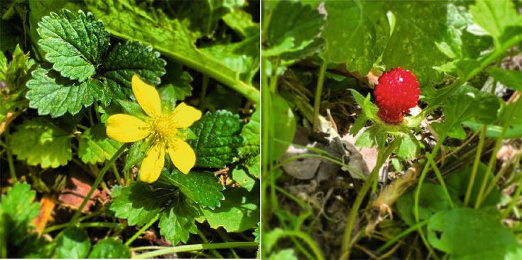
Figure 1.
This species is native to East Asia; however, it is widely distributed in the Asian, European, and American continents, which converts it into a plant with a favorable environmental adaptability.
The use of plants as a complementary therapy is an ancestral practice that nowadays continues to spread all over the world. Traditionally,
Medicinal plants have been of great interest to different research groups as they may represent an important natural source of bioactive compounds for the development of new and promising drugs in the search for an effective treatment with a lower degree of side effects for various diseases. These plant-derived bioactive compounds can exert their therapeutic effects through different mechanisms that may include, in general terms, their interaction with cytoplasmic membrane receptors, resulting in the modulation of distinct signaling cascades with the activation/inhibition of different regulatory kinases. Also, through their ability to regulate gene expression in target cells or by interacting directly with the aggressor agent.
It has been documented that the different extracts of
3.1 Antioxidant effect of Potentilla indica
It is well documented that oxidative stress represents a determinant factor in the development and evolution of multiple chronic degenerative diseases such as diabetes, metabolic syndrome, cardiovascular disease, cancer, Alzheimer’s, and Parkinson’s disease, and its incidence has been increasing exponentially in recent years [37]. Therefore, it is necessary to search for and implement drugs with antioxidant properties as a therapeutic strategy. In this sense, numerous studies have reported a potent antioxidant activity exhibited by different extracts of certain parts of
In addition to the fruit, other parts of the plant have been subjected to research and have been shown to possess antioxidant activity, both
In 2019, ferulic acid was demonstrated to activate Nrf2 protein, a factor involved in the positive transcriptional regulation of cellular antioxidant response,
3.2 Anti-inflammatory effect of Potentilla indica
There is accumulating evidence that dysregulation of inflammatory pathways is closely linked to the development and progression of various chronic diseases [49]. Several investigations have been reported an anti-inflammatory effect of
3.3 Anticancer effect of Potentilla indica
Cancer is one of the leading causes of mortality worldwide [52]. Cancer cells frequently experience resistance to chemotherapeutic drugs; however, it has been reported in different cellular models that several phytochemicals are able to increase the sensitivity and efficacy to chemotherapy through different mechanisms; therefore, they may contribute considerably to decrease the mortality of this disease [53]. In this context, it has been reported in different studies that
3.4 Antimicrobial effect of Potentilla indica
Currently, infectious diseases caused by different pathogenic microorganisms represent one of the main global threats to public health. Additionally, in recent decades, drug resistance to commercially available synthetic compounds has increased considerably [59], and natural products could represent a promising source to satisfy this demand. In this context,
On the other hand, 25 compounds have been identified by GC–MS in the essential oil of
3.5 Perspectives and limitations of using of Potentilla indica
The administration of the plant for specific therapeutic purposes may present some limitations in medical practice since years of clinical research are required for the establishment of a therapeutic dose with controlled studies to validate its efficacy and safety. However, no toxic side effects of
4. Justicia spicigera Schltdl
It is a plant that has been used in Mexico since pre-Hispanic times as a source of natural pigment to dye fabrics and crafts [68]. Furthermore, in traditional Mexican medicine,
Currently, various studies have been carried out aimed at the use of
4.1 Anticancer activity
Recent research has shown that extracts of dried plant material of
On the other hand, it was found that the ethanolic extract of
4.2 Hypoglycemic activity
In previous reports, the potential hypoglycemic effect present in the ethanolic extract of dried plant material of
4.3 Antioxidant activity
Various
4.4 Lipid-lowering and anti-inflammatory activity
Obesity is a proinflammatory disease that is related to certain chronic degenerative diseases such as type 2 diabetes, nonalcoholic fatty liver disease (NAFLD), and cancer [79]. Real-Sandoval [80], administered the ethanolic extract of
4.5 Antihypertensive activity
The effect of extracts of dry plant material of
4.6 Antimicrobial activity
Recent studies have shown that
In another sense,
In summary, Mexico is a country with a great diversity of medicinal plants, among which
5. Conclusions
According to the research on these medicinal plants, Eryngium native plant of Mexico, Potentilla widely in Asian traditional medicine, and Justicia distributed in Latin American, all represent an important natural source of bioactive compounds. Flavonoids it is the major compound in all plants and help improve lipid profiles, blood pressure, insulin resistance, and systemic inflammation and have important effects on various chronic degenerative diseases. However, seasonality, water, light supply, temperature, herbivory and microbes, and soil factors can be influenced by climate and can result in up to 50% increased or decreased content of secondary plant metabolites. It is for that reason that further studies need to be done to characterize new active compounds with the objective of making science and evidence-based hard claims for the functionality and efficacy of the phytonutrient blend. Synergistic activity in the compounds is what makes them have a beneficial effect because they contain each of the compounds in specific quantities demonstrating the claimed biological functionality and health benefit as specifically to the mixture of active compounds. However, such molecular blends are difficult to reproducibly generate and formulate it is because regulatory authorities are more in favor of approving single or few active principles rather than complex blends.
Acknowledgments
The authors appreciate the partial support from Scientific Research Coordination (18070, to ASM), Michoacana University of San Nicholas of Hidalgo.
References
- 1.
Noriega-Cisneros R, Ortiz- Avila O, Esquivel-Gutiérrez E, Clemente-Guerrero M, Manzo-Avalos S, Salgado-Garciglia R, et al. Hypolipidemic activity of Eryngium carlinae on streptozotocin-induced diabetic rats. Biochemistry Research International. 2012;2012 :603501. DOI: 10.1155/2012/603501 - 2.
Landa-Moreno CI, Trejo- Hurtado CM, Lemus-de la Cruz J, Peña-Montes D, Murillo-Villicaña M, Huerta-Cervantes M, et al. Antioxidant effect of the ethyl acetate extract of Potentilla indica on kidney mitochondria of streptozotocin-induced diabetic rats. Plants. 2023;12 :3196. DOI: 10.3390/plants12183196 - 3.
García-Cerrillo D, Noriega-Cisneros R, Peña-Montes D, Huerta-Cervantes M, Ríos-Silva M, Salgado-Garciglia R, et al. Asian Journal of Applied Sciences. 2018; 6 :308-314 - 4.
Peña-Montes DJ, Huerta- Cervantes M, Ríos-Silva M, Trujillo X, Huerta M, Noriega-Cisneros R, et al. Protective effect of the Hexanic extract of Eryngium carlinae inflorescencesin vitro , in yeast, and in Streptozotocin-induced diabetic male rats. Antioxidants (Basel). 2019;8 (3):73. DOI: 10.3390/antiox8030073 - 5.
Awad NE, Abdelkawy MA, Rahman EHA, Hamed MA, Ramadan NS. Phytochemical and in vitro screening ofJusticia spicigera ethanol extract for antioxidant activity andin vivo assessment againstSchistosoma mansoni infection in mice. Anti-Infective Agents. 2018;16 :49-56 - 6.
International Diabetes Federation, editor. IDF Diabetes Atlas. 10th ed. International Diabetes Federation; 2021 - 7.
Mayer-Davis EJ, Lawrence JM, Dabelea D, Divers J, Isom S, Dolan L, et al. Incidence trends of type 1 and type 2 diabetes among youths, 2002-2012. The New England Journal of Medicine. 2017; 376 (15):1419-1429. DOI: 10.1056/NEJMoa1610187 - 8.
American Diabetes Association. Diagnosis and classification of diabetes mellitus. Diabetes Care. 2014; 37 (Supplement 1):S81-S90 - 9.
Galicia-Garcia U, Benito-Vicente A, Jebari S, Larrea-Sebal A, Siddiqi H, Uribe KB, et al. Pathophysiology of type 2 diabetes mellitus. International Journal of Molecular Sciences. 2020; 21 (17):6275. DOI: 10.3390/ijms21176275 - 10.
Maraschin JF. “Classification of diabetes,” in diabetes. Advances in Experimental Medicine and Biology. 2013; 771 :12-19 - 11.
Misrani A, Tabassum S, Yang L. Mitochondrial dysfunction and oxidative stress in Alzheimer's disease. Frontiers in Aging Neuroscience. 2021; 18 (13):617588. DOI: 10.3389/fnagi.2021.617588 - 12.
Sivitz WI, Yorek MA. Mitochondrial dysfunction in diabetes: From molecular mechanisms to functional significance and therapeutic opportunities. Antioxidants & Redox Signaling. 2010; 12 (4):537-577. DOI: 10.1089/ars.2009.2531 - 13.
González P, Lozano P, Ros G, Solano F. Hyperglycemia and oxidative stress: An integral, updated and critical overview of their metabolic interconnections. International Journal of Molecular Sciences. 2023; 24 (11):9352. DOI: 10.3390/ijms24119352 - 14.
Kay AM, Simpson CL, Stewart JA Jr. The role of AGE/RAGE signaling in diabetes-mediated vascular calcification. Journal Diabetes Research. 2016; 2016 :6809703. DOI: 10.1155/2016/6809703 - 15.
Dong K, Ni H, Wu M, Tang Z, Halim M, Shi D. ROS-mediated glucose metabolic reprogram induces insulin resistance in type 2 diabetes. Biochemical and Biophysical Research Communications. 2016; 476 (4):204-211. DOI: 10.1016/j.bbrc.2016.05.087 - 16.
Mizukami H, Osonoi S. Pathogenesis and molecular treatment strategies of diabetic neuropathy collateral glucose-utilizing pathways in diabetic polyneuropathy. International Journal of Molecular Sciences. 2020; 22 (1):94. DOI: 10.3390/ijms22010094 - 17.
Roy Chowdhury S, Djordjevic J, Thomson E, Smith DR, Albensi BC, Fernyhough P. Depressed mitochondrial function and electron transport complex II-mediated H2O2 production in the cortex of type 1 diabetic rodents. Molecular and Cellular Neurosciences. 2018; 23 (90):49-59. DOI: 10.1016/j.mcn.2018.05.006 - 18.
Ortiz-Avila O, Sámano-García CA, Calderón-Cortés E, Pérez-Hernández IH, Mejía-Zepeda R, Rodríguez- Orozco AR, et al. Dietary avocado oil supplementation attenuates the alterations induced by type I diabetes and oxidative stress in electron transfer at the complex II-complex III segment of the electron transport chain in rat kidney mitochondria. Journal of Bioenergetics and Biomembranes. 2013; 45 (3):271-287. DOI: 10.1007/s10863-013-9502-3 - 19.
Raza H, John A, Howarth FC. Increased oxidative stress and mitochondrial dysfunction in zucker diabetic rat liver and brain. Cellular Physiology and Biochemistry. 2015; 35 (3):1241-1251. DOI: 10.1159/000373947 - 20.
Maiese K. New insights for oxidative stress and diabetes mellitus. Oxidative Medicine and Cellular Longevity. 2015; 2015 :875961. DOI: 10.1155/2015/875961 - 21.
Niki E. Oxidative stress and antioxidants: Distress or eustress? Archives of Biochemistry and Biophysics. 2016; 595 :19-24. DOI: 10.1016/j.abb.2015.11.017 - 22.
Chaudhury A, Duvoor C, Reddy Dendi VS, Kraleti S, Chada A, Ravilla R, et al. Clinical review of antidiabetic drugs: Implications for type 2 diabetes mellitus management. Frontiers in Endocrinology (Lausanne). 2017; 4 (8):6. DOI: 10.3389/fendo.2017.00006 - 23.
Padhi S, Nayak AK, Behera A. Type II diabetes mellitus: A review on recent drug-based therapeutics. Biomedicine & Pharmacotherapy. 2020; 131 :110708. DOI: 10.1016/j.biopha.2020.110708 - 24.
Cárdenas-Valdovinos JG, García-Ruiz I, Angoa-Pérez MV, Mena-Violante HG. Ethnobotany, biological activities and phytochemical compounds of some species of the genus Eryngium (Apiaceae), from the Central-Western region of Mexico. Molecules. 2023; 28 (10):4094. DOI: 10.3390/molecules28104094 - 25.
Pérez-Ramírez IF, Enciso- Moreno JA, Guevara-González RG, Gallegos-Corona MA, Loarca-Piña G, Reynoso-Camacho R. Modulation of renal dysfunction by Smilax cordifolia andEryngium carlinae , and their effect on kidney proteome in obese rats. Journal of Functional Foods. 2016;20 :545-555. DOI: 10.1016/j.jff.2015.11.024 - 26.
Trejo-Hurtado CM, Landa- Moreno CI, la Cruz JL, Peña-Montes DJ, Montoya-Pérez R, Salgado-Garciglia R, et al. An ethyl acetate extract of Eryngium carlinae inflorescences attenuates oxidative stress and inflammation in the liver of Streptozotocin-induced diabetic rats. Antioxidants (Basel). 2023;12 (6):1235. DOI: 10.3390/antiox12061235 - 27.
Noriega-Cisneros R, Peña-Montes DJ, Huerta-Cervantes M, Torres-Martínez R, Huerta M, Manzo-Avalos S, et al. Eryngium carlinae ethanol extract corrects lipid abnormalities in Wistar rats with experimental diabetes. Journal of Medicinal Food. 2020;23 (8):827-833. DOI: 10.1089/jmf.2019.0189 - 28.
Uppal S, Italiya KS, Chitkara D, Mittal A. Nanoparticulate-based drug delivery systems for small molecule anti-diabetic drugs: An emerging paradigm for effective therapy. Acta Biomaterialia. 2018; 81 :20-42. DOI: 10.1016/j.actbio.2018.09.049 - 29.
Shah M, Fawcett D, Sharma S, Tripathy SK, Poinern GEJ. Green synthesis of metallic nanoparticles via biological entities. Materials (Basel). 2015; 8 (11):7278-7308. DOI: 10.3390/ma8115377 - 30.
Sahu T, Ratre YK, Chauhan S, Bhaskar L, Nair MP, Verma HK. Nanotechnology based drug delivery system: Current strategies and emerging therapeutic potential for medical science. The Journal of Drug Delivery Science and Technology. 2021; 63 :102487. DOI: 10.1016/j.jddst.2021.102487 - 31.
Lemus-de la Cruz J, Trejo-Hurtado M, Landa-Moreno C, Peña-Montes D, Landeros-Páramo JL, Cortés-Rojo C, et al. Antioxidant effects of silver nanoparticles obtained by green synthesis from the aqueous extract of Eryngium carlinae on the brain mitochondria of streptozotocin-induced diabetic rats. Journal of Bioenergetics and Biomembranes. 2023;55 (2):123-135. DOI: 10.1007/s10863-023-09963-w - 32.
Xiang B, Yu X, Li B, Xiong Y, Long M, He Q. Caracterización, actividades antioxidantes y anticancerígenas de un polisacárido neutro de Duchesnea indica (Andr.) Focke. Revista de bioquímica de alimentos. 2019; 43 (7):e12899 - 33.
Zhu M, Dong X, Guo M. Phenolic profiling of Duchesnea indica combining macroporous resin chromatography (MRC) with HPLC-ESI-MS/MS and ESI-IT-MS. Molecules. 2015; 20 (12):22463-22475 - 34.
Zhao L, Zhang SL, Tao JY, Jin F, Pang R, Guo YJ, et al. Anti-inflammatory mechanism of a folk herbal medicine, Duchesnea indica (Andr) Focke at RAW264. 7 cell line. Immunological Investigations. 2008; 37 (4):339-357 - 35.
Hu W, Han W, Huang C, Wang MH. Protective effect of the methanolic extract from Duchesnea indica against oxidative stress in vitro andin vivo . Environmental Toxicology and Pharmacology. 2011;31 (1):42-50 - 36.
Yang WE, Ho YC, Tang CM, Hsieh YS, Chen PN, Lai CT, et al. Duchesnea indica extract attenuates oral cancer cells metastatic potential through the inhibition of the matrix metalloproteinase-2 activity by down-regulating the MEK/ERK pathway. Phytomedicine. 2019; 63 :152960 - 37.
Sharifi-Rad J, Quispe C, Castillo CMS, Caroca R, Lazo-Vélez MA, Antonyak H, et al. Ellagic acid: A review on its natural sources, chemical stability, and therapeutic potential. Oxidative Medicine and Cellular Longevity. 2022; 2022 :3848084 - 38.
Qin C, Li Y, Zhang R, Niu W, Ding Y. Separation and elucidation of anthocyanins in the fruit of mock strawberry (Duchesnea indica Focke). Natural Product Research. 2009; 23 (17):1589-1598 - 39.
Alappat B, Alappat J. Anthocyanin pigments: Beyond aesthetics. Molecules. 2020; 25 (23):5500 - 40.
Shan S, Huang S, Shah MH, Abbasi AM. Evaluation of polyphenolics content and antioxidant activity in edible wild fruits. BioMed Research International. 2019; 2019 :1381989. DOI: 10.1155/2019/1381989 - 41.
Huneif MA, Alqahtani SM, Abdulwahab A, Almedhesh SA, Mahnashi MH, Riaz M, et al. α-Glucosidase, α-amylase and antioxidant evaluations of isolated bioactives from wild strawberry. Molecules. 2022; 27 (11):3444 - 42.
Azeem M, Hanif M, Mahmood K, Ameer N, Chughtai FRS, Abid U. An insight into anticancer, antioxidant, antimicrobial, antidiabetic and anti-inflammatory effects of quercetin: A review. Polymer Bulletin. 2023; 80 (1):241-262 - 43.
Liaqat MUHA, Kakar MMAD, Akram IU, Muhammad SHAHZAD, Kakar MU, Ahmad NADEEM, et al. Antimicrobial and phytochemical exploration of Duchesnea indica plant. Plant Cell Biotechnology and Molecular Biology. 2021; 22 :74-85 - 44.
Li D, Rui YX, Guo SD, Luan F, Liu R, Zeng N. Ferulic acid: A review of its pharmacology, pharmacokinetics and derivatives. Life Sciences. 2021; 284 :119921 - 45.
Mancuso A, Cristiano MC, Pandolfo R, Greco M, Fresta M, Paolino D. Improvement of ferulic acid antioxidant activity by multiple emulsions: In vitro andin vivo evaluation. Nanomaterials. 2021;11 (2):425 - 46.
Choe E. Roles and action mechanisms of herbs added to the emulsion on its lipid oxidation. Food Science and Biotechnology. 2020; 29 (9):1165-1179 - 47.
Mahmoud AM, Hussein OE, Abd El-Twab SM, Hozayen WG. Ferulic acid protects against methotrexate nephrotoxicity via activation of Nrf2/ARE/HO-1 signaling and PPARγ, and suppression of NF-κB/NLRP3 inflammasome axis. Food & Function. 2019; 10 (8):4593-4607 - 48.
Roghani M, Kalantari H, Khodayar MJ, Khorsandi L, Kalantar M, Goudarzi M, et al. Alleviation of liver dysfunction, oxidative stress and inflammation underlies the protective effect of ferulic acid in methotrexate-induced hepatotoxicity. Drug Design, Development and Therapy. 2020:1933-1941 - 49.
Kunnumakkara AB, Shabnam B, Girisa S, Harsha C, Banik K, Devi B, et al. Inflammation, NF-κB, and chronic diseases: How are they linked? Critical Reviews™ in Immunology. 2020; 40 (1):1-39 - 50.
Ullah HA, Lee YY, Kim SD, Rhee MH. Duchesnea indica extract attenuates coal fly ash-induced inflammation in murine alveolar macrophages through the NF-kappa B pathway. Evidence-Based Complementary and Alternative Medicine. 2021; 2021 :1-11 - 51.
Lee YY, Yuk HJ, Saba E, Kim SD, Kim DS, Kopalli SR, et al. Duchesnea indica extract ameliorates LPS-induced septic shock in mice. Evidence-Based Complementary and Alternative Medicine. 2022; 2022 (14):5783867 - 52.
Deo SVS, Sharma J, Kumar S. GLOBOCAN 2020 report on global cancer burden: Challenges and opportunities for surgical oncologists. Annals of Surgical Oncology. 2022; 29 (11):6497-6500 - 53.
Khatoon E, Banik K, Harsha C, Sailo B, Thakur KK, Khwairakpam AD, et al. Phytochemicals in cancer cell chemosensitization: Current knowledge and future perspectives. In: Seminars in Cancer Biology. Vol. 80. Academic Press; 2022. pp. 306-339 - 54.
Kürbitz Heise D, Redmer T, Goumas F, Arlt A, Lemke J, et al. Epicatechin gallate and catechin gallate are superior to epigallocatechin gallate in growth suppression and anti-inflammatory activities in pancreatic tumor cells. Cancer Science. 2011; 102 (4):728-734 - 55.
Xu YQ , Gao Y, Granato D. Effects of epigallocatechin gallate, epigallocatechin and epicatechin gallate on the chemical and cell-based antioxidant activity, sensory properties, and cytotoxicity of a catechin-free model beverage. Food Chemistry. 2021; 339 :128060 - 56.
Chen PN, Yang SF, Yu CC, Lin CY, Huang SH, Chu SC, et al. Duchesnea indica extract suppresses the migration of human lung adenocarcinoma cells by inhibiting epithelial–mesenchymal transition. Environmental Toxicology. 2017; 32 (8):2053-2063 - 57.
Peng B, Chang Q , Wang HQ , Wang Y, Tang J, Liu X. Suppression of human ovarian SKOV-3 cancer cell growth by Duchesnea phenolic fraction is associated with cell cycle arrest and apoptosis. Gynecologic Oncology. 2008; 108 (1):173-181 - 58.
Peng B, Hu Q , Liu X. Duchesnea phenolic fraction inhibits in vitro and in vivo growth of cervical cancer through induction of apoptosis and cell cycle arrest. Experimental Biology and Medicine. 2009; 234 (1):74-83 - 59.
Dadgostar P. Antimicrobial resistance: Implications and costs. Infection and Drug Resistance. 2019; 12 :3903-3910 - 60.
Liaqat M, Kakar IU, Akram M, Hussain SHAHZAD, Kakar MU, Ahmad Nade EM, et al. Antimicrobial and phytochemical exploration of Duchesnea indica plant. Plant Cell Biotechnology and Molecular Biology. 2021;2019 (22):74-85 - 61.
Biharee A, Sharma A, Kumar A, Jaitak V. Antimicrobial flavonoids as a potential substitute for overcoming antimicrobial resistance. Fitoterapia. 2020; 146 :104720 - 62.
Umesh BT, Thoppil JE. Comparison of chemical constituents of tissue cultured and field grown plants of Duchesnea indica, (Andr.) Focke. Journal of Pharmacognosy and Phytochemistry. 2014; 3 (3):68-70 - 63.
Justicia spicigera Schltdl. in GBIF Secretariat. GBIF Backbone Taxonomy, GBIF Secretariat. Available from: GBIF.org [Accessed: October 2, 2023]. doi: 10.15468/39omei - 64.
Fonseca-Chávez RE, Rivera- Levario LA, Vázquez-García L. Guía ilustrada de plantas medicinales en el Valle de México. Meixico: Instituto Nacional de los Pueblos Indígenas; 2020 - 65.
Castro-Muñoz R, León-Becerril E, García-Depraect O. Beyond the exploration of Muicle ( Justicia spicigera ): Reviewing its biological properties, bioactive molecules and materials chemistry. Processes. 2022;10 (5):1035 - 66.
Baqueiro-Peña I, Guerrero- Beltrán JA. Uses of Justicia spicigera in medicine and as a source of pigments. Functional Foods in Health and Disease. 2014;4 (9):401-414 - 67.
Ortiz-Andrade R, Cabañas-Wuan A, Arana-Argáez VE, Alonso-Castro AJ, Zapata-Bustos R, Salazar-Olivo LA, et al. Antidiabetic effects of Justicia spicigera schltdl (acanthaceae). Journal of Ethnopharmacology. 2012;143 (2):455-462 - 68.
Baqueiro-Peña I, Guerrero-Beltrán JÁ. Physicochemical and antioxidant characterization of Justicia spicigera . Food Chemistry. 2017;218 :305-312 - 69.
Anaya-Esparza LM, Ramos-Aguirre D, Zamora-Gasga VM, Yahia E, Montalvo-González E. Optimization of ultrasonic-assisted extraction of phenolic compounds from Justicia spicigera leaves. Food Science and Biotechnology. 2018;27 :1093-1102 - 70.
Rodríguez-Garza NE, Quintanilla-Licea R, Romo-Sáenz CI, Elizondo-Luevano JH, Tamez-Guerra P, Rodríguez-Padilla C, et al. In vitro biological activity and lymphoma cell growth inhibition by selected Mexican medicinal plants. Life. 2023;13 (4):958 - 71.
Jacobo-Salcedo MDR, Alonso-Castro AJ, Salazar-Olivo LA, Carranza-Alvarez C, González-Espíndola LÁ, Domínguez F, et al. Antimicrobial and cytotoxic effects of Mexican medicinal plants. Natural Product Communications. 2011; 6 (12):1925-1928 - 72.
Fernández-Pomares C, Juárez-Aguilar E, Domínguez-Ortiz MÁ, Gallegos-Estudillo J, Herrera-Covarrubias D, Sánchez-Medina A, et al. Hydroalcoholic extract of the widely used Mexican plant Justicia spicigera Schltdl. Exert- s a cytostatic effect on LNCaP prostate cancer cells. Journal of Herbal Medicine. 2018;12 :66-72 - 73.
Ramírez G, Zavala M, Pérez J, Zamilpa A. In vitro screening of medicinal plants used in Mexico as antidiabetics with glucosidase and lipase inhibitory activities. Evidence-Based Complementary and Alternative Medicine. 2012;2012 :701261 - 74.
Murillo-Villicaña M, Noriega-Cisneros R, Peña-Montes DJ, Huerta-Cervantes M, Aguilera-Méndez A, Cortés-Rojo C, et al. Antilipidemic and hepatoprotective effects of ethanol extract of Justicia spicigera in streptozotocin diabetic rats. Nutrients. 2022;14 (9):1946 - 75.
Gulcin İ. Antioxidants and antioxidant methods: An updated overview. Archives of Toxicology. 2020; 94 (3):651-715 - 76.
De La Cruz-Jiménez L, Hernández-Torres MA, Monroy-García IN, Rivas-Morales C, Verde-Star MJ, Gonzalez-Villasana V, et al. Biological activities of seven medicinal plants used in Chiapas, Mexico. Plants. 2022; 11 (14):1790 - 77.
García-Márquez E, Román- Guerrero A, Pérez-Alonso C, Cruz-Sosa F, Jiménez-Alvarado R, Vernon-Carter EJ. Effect of solvent-temperature extraction conditions on the initial antioxidant activity and total phenolic content of muitle extracts and their decay upon storage at different pH. Revista mexicana de ingeniería química. 2012; 11 (1):1-10 - 78.
Awad NE, Abdelkawy MA, Hamed MA, Souleman AM, Abdelrahman EH, Ramadan NS. Antioxidant and hepatoprotective effects of Justicia spicigera ethyl acetate fraction and characterization of its anthocyanin content. International Journal of Pharmacy and Pharmaceutical Sciences. 2015;7 (8):91-96 - 79.
Cordero P, Li J, Oben JA. Obesity and NAFLD. In: Obesity: Pathogenesis, Diagnosis, and Treatment. Cham, Switzerland: Springer International Publishing; 2019. pp. 179-194 - 80.
Real-Sandoval SA, Gutiérrez- López GF, Domínguez-López A, Paniagua-Castro N, Michicotl-Meneses MM, Jaramillo-Flores ME. Downregulation of proinflammatory liver gene expression by Justicia spicigera and kaempferitrin in a murine model of obesity-induced by a high-fat diet. Journal of Functional Foods. 2020;65 :103781 - 81.
Esquivel-Gutiérrez E, Noriega-Cisneros R, Arellano-Plaza M, Ibarra-Barajas M, Salgado-Garciglia R, Saavedra-Molina A. Antihypertensive effect of Justicia spicigera in L-NAME-induced hypertensive rats. Pharmacology Online. 2013;2 :120-127 - 82.
Caesar LK, Cech NB. Synergy and antagonism in natural product extracts: When 1+ 1 does not equal 2. Natural Product Reports. 2019; 36 (6):869-888