
Abstract
The zebrafish (Danio rerio) is a freshwater species native to South Asia belonging to the Cyprinidae family. Due to its easy housing and breeding, high fecundity, and rapid development, it has become a popular laboratory animal. Furthermore, zebrafish is an increasingly successful and widely used vertebrate model organism in scientific research, e.g., in drug discovery, particularly preclinical development, mainly because of its immune response and well-known genome. Nevertheless, zebrafish health in laboratory facilities is crucial. Both the water quality and pathogens control are directly related to the zebrafish welfare, which, under inappropriate conditions, may confound experimental findings, leading to irreproducible outcomes and invalid or misinterpreted results. Infections by Aeromonas and mycobacterium, for example, interfere with the results of experiments by altering physiological parameters. Likewise, infection of the nervous system by Pseudoloma neurophilia causes behavioral changes in zebrafish, leading to misinterpretation in behavioral studies. In this chapter, we seek to provide valuable contributions about zebrafish housing and husbandry conditions known to influence animal health, drawing attention to the most common diseases and pathogens that eventually may affect zebrafish in the laboratory.
Keywords
- immune system
- laboratory animal
- opportunistic pathogens
- aquatic system
- behavior
- freshwater fish
- infection
- Mycobacteriosis
- Pseudoloma neurophilia
1. Introduction
Research in several areas using the vertebrate zebrafish (
Maintaining zebrafish health in experimental environments is vital, and providing adequate water and food following ideal standards will ensure stability, immunity, and well-being. In these conditions of well-being, the probability of infections being manifested will be low [2]. On the contrary, a low sanitary aspect in zebrafish housing can induce changes in physiology, behavior, and immune response, detectable through altered characteristics and clinical signs, such as lethargy, opercular enlargement, swallowing air at the water surface, loss of floatability control, eating less or not eating, and signs in their appearance like ulcer, injuries, hemorrhage, changes in color or hydropsy [3].
Given the increasing growth of zebrafish research facilities worldwide, these organisms are housed within a spectrum of settings ranging from modest setups involving a few tanks to large collaborative core facilities boasting thousands of aquaria. In these scenarios, the typical configuration consists of numerous compact aquaria integrated into a recirculation framework using commercially designed “racks”, encompassing approximately 30 tanks to large systems supporting hundreds of tanks. Irrespective of scale, the fundamental layout involves the recirculation of effluent water through both mechanical and biological filtration stages, succeeded by ultraviolet sterilization procedures. Supplementary water is commonly sourced from reverse osmosis outlets to which salts are introduced to control conductivity level.
Moreover, the technologies applied to create mutants or transgenic lines have boosted the use of zebrafish in research. More often than not, these lines might suffer from the same diseases that wild-type animals, although depending on the type of gene edition, it could impair their health vulnerability and needs.
Understanding the specific requirements of zebrafish and comprehending the nuances of their immune system is paramount for maintaining this species in laboratory environments that ensure their optimal well-being and health. Zebrafish, as a model organism, offer valuable insights into various biological processes, but their intricate needs, both in terms of habitat and immune responses, must be met to uphold their viability as research subjects.
Adequate knowledge of their demands, such as optimal water quality, appropriate diet, and suitable housing conditions, directly influences their behavior, growth rates, immunity, reproductive success, survival, and life expectancy. Moreover, delving into their immune system intricacies equips researchers with the tools to address potential disease outbreaks anticipatively, promote disease resistance, and reduce mortality. By refining our understanding of zebrafish requirements and immune dynamics, we fortify the foundation for productive research while upholding the ethical responsibility to ensure their comfort and health within laboratory settings.
Concisely, during early-life stages, zebrafish rely on their innate immune response, which involves innate molecules, barriers (skin, gills, gastrointestinal - GI tract), phagocytic cells, and soluble mediators, such as cytokines, pore-forming toxins, and lectins, which regulate immune cell interactions and activities [4, 5, 6]. On the other hand, juvenile and adult zebrafish possess a well-developed complement system and an adaptive immune response similar to other vertebrates. Surface barriers like skin, gills, and GI tract are coated by mucus, offering both physical and chemical protection against pathogen microorganisms or aggressors, besides promoting osmotic balance. Further, zebrafish surfaces are provided with phagocytic macrophages and neutrophils. Also, GI tract secretions produce adverse environments for exogenous microorganisms [7].
This book chapter intends to provide valuable contributions about zebrafish housing and husbandry conditions known to influence animal welfare, drawing attention to the most common diseases and pathogens that might affect zebrafish in laboratories.
2. Biotic and abiotic factors and the zebrafish health
Zebrafish are subject to water quality concerns similar to those of other fish reared in intensive recirculating systems. The zebrafish housing facility acts as a life support system, similar to natural habitats, and alterations or poor water quality compromise several biological processes in zebrafish, such as growth, metabolism, immune function, behavior, and stress [8]. Thus, although zebrafish are quite tolerant to water quality parameter fluctuations, the standardization of husbandry procedures must be pursued to optimize experimental techniques and reach reliable results [9].
Therefore, physical, chemical, and biological parameters must be fulfilled to achieve conditions, where the fish perform their best once inadequate water quality produces important subclinical and clinical manifestations with severe morbidity and mortality [10].
Zebrafish are naturally found in South Asia, inhabiting slow-flowing water bodies in different ranges of water parameters, temperatures varying from 10 to 40°C, pH between 6 and 10, and up to 60 cm of water column [9]. According to Schaefer & Ryan (2006), thermal tolerance varies between individuals, and for zebrafish, the optimum range is between 24 and 28°C [11]. Temperature also interferes with other phenomena like sex determination, where masculinization is related to high rearing temperatures (reviewed in [8]). Higher temperatures also decrease the saturation of dissolved oxygen (DO), which leads to an imbalance of total ammonia nitrogen (TAN) and enhances the prevalence of unionized ammonia (NH3), increasing the NH3 toxicity [12].
The TAN balance is also influenced by pH concentration. Unionized ammonia and ammonium ions (NH3 and NH4+, respectively) rely on pH and must be regulated in order to prevent disbalance. Ammonia is oxidized to nitrite (NO2−), which is also toxic by oxidizing hemoglobin and converting it to meta-hemoglobin. Thus, the respiratory process is affected, leading to potential asphyxiation and death. The conversion of nitrite to nitrate (NO3−) is essential once this molecule presents minor toxicity, and levels up to 200 mg.L−1 are considered safe for zebrafish [13].
Water quality diseases caused by ammonia toxicity generally lead to behavioral alterations, like hyperexcitability and anorexia, which can be observed in the aquarium, and lesions observed in additional exams, such as gill hyperplasia and hypertrophy (reviewed in [2]). Sporadically, mats of bacteria are associated with hyperplastic gill lesions, although this is also most often secondary to suboptimal water quality and not a primary bacterial problem [14].
pH values reflect the concentration of hydrogen ions present in the solution and characterize the water as acidic, alkaline, or neutral when around pH 7. The production of TAN in the creation system acidifies the water (pH <7); thus, neutralization with buffer solutions is necessary [12]. The tolerable pH range for freshwater fish is between 6.5 and 9, and for zebrafish, it stands around 6.2 and 8.5, with no behavioral or physiological alterations (reviewed in [8]). Acidification or alkalinization of water has effects on fish, resulting in several physiological alterations in gills that are in direct contact with water, but also in gas exchange and electrolyte balance [15].
Moreover, pH alterations cause massive mucus production, besides behavioral changes [16], glycemia, a decrease in red blood cells (RBC), and an increase in white blood cells (WBC) [17]. Additionally, any disturbance in the osmotic state leads to physiological stress and disease (reviewed in [8]).
Biological processes like neuronal signaling, muscle contraction, and osmoregulation depend on essential ions—calcium, magnesium, and ferrous iron, among others—that are absorbed from rearing water; these ions’ concentration is represented by water hardness [18]. According to Chen
The concentration of DO is affected by external factors like water temperature, feeding rates, and fish density in an inversely proportional manner. Oxygen concentration between 6 and 8 ppm is suitable for zebrafish culture [12]. Meanwhile, high concentrations of carbon dioxide affect fish by diminishing hemoglobin affinity to oxygen and acidifying blood. Thus, it is imperative to keep lower concentrations of carbon dioxide. Besides DO and carbon dioxide, water nitrogen levels interfere with fish physiology. When water nitrogen supersaturation occurs, fish develop a syndrome known as gas bubble disease (Figure 1).
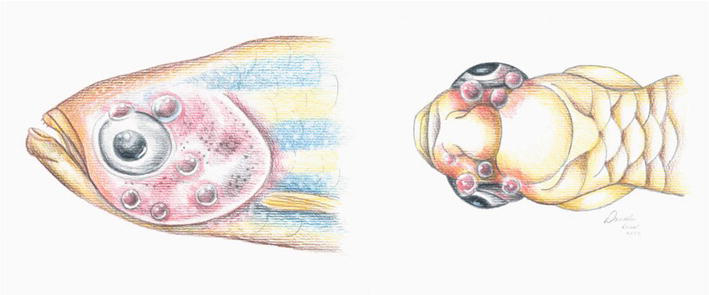
Figure 1.
Illustration of gas bubble disease; lateral view (
In housing systems, leaks on suction water pumps or intensive air injection on water can cause water gas supersaturation. Also, abrupt temperature alters gas concentration stability. Supersaturated water produces emboli (free gas bubbles) in the vascular system that spread through other tissues, mainly subcutaneous tissue [8], which is particularly problematic. Local inflammation and behavioral abnormalities can occur in affected zebrafish. Also, air emboli in the vascular system may cause obstruction of blood vessels, leading to local ischemia and tissue damage, and the severity of the disease depends on the intensity of the formed emboli. Necrosis areas derived from disease can suffer from secondary bacterial infections. Eyeballs are frequently injured by tissue gas bubbles, and clinical signs, such as exophthalmos (Figure 2), eye inflammation, cataracts, and blindness may occur [14]. Chronically affected fish can present emaciation, buoyancy defects, and reduced activity.
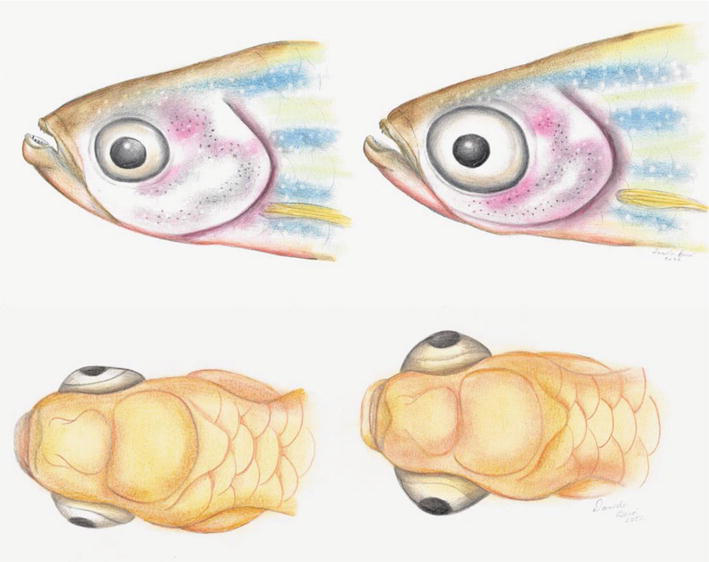
Figure 2.
Illustration exophthalmia; healthy (
Likewise, nephrocalcinosis is also triggered by rough water quality. A range of nephrocalcinosis lesions in zebrafish can be assessed by histology, most of which are diagnosed in clinically normal fish. Calcium deposits in renal tubules and ducts may occur as a consequence of high dissolved CO2 [14].
Abnormal behaviors, such as frequent aggression, lethargy, and erratic swimming, are reliable indicators of welfare disruption, as well as specific clinical signs indicating punctual situations, such as opercular flaring (Figure 3), reflecting respiratory distress.
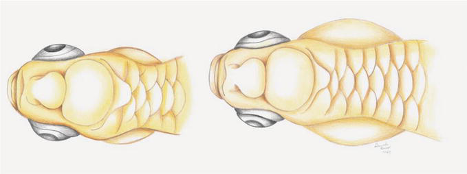
Figure 3.
Illustration of opercular flaring.
3. Stress and zebrafish
As highly evidenced, among all the critical modulating factors on health and equilibrium, stress has a lot to do with many biological responses as a state of threatened homeostasis. Stress is the consequence of external or internal factors (stressors) implicated in changes in biological equilibrium. It might initiate an adaptive response to restore baseline physiology, seen as a change in physiology, psychological state, or behavior.
In laboratory zebrafish, stress can induce alterations notably in four broad physiologic categories: behavior, the autonomic nervous system (ANS), the neuroendocrine system, and immune function [20]. Consequent modifications in these functions can be assessed by evaluating behavioral patterns,
Lastly, zebrafish nutrition is an additional important factor for their health and proper development. Some diseases that affect zebrafish are caused by a nutritional deficiency. In this regard, for example, vitamin E or selenium deficiency might cause muscle degeneration, vitamin C deficiency can lead to scoliosis, and pantothenic acid deficiency is likely to cause gill disease.
4. Bacterial infections
Clinical or subclinical infections may distract experimental findings, leading to irreproducible outcomes and invalid or misinterpreted results [22]. In general, local lesions are noticeable and may indicate bacterial, fungal, or protozoal infections located on the skin and fins, with hemorrhagic areas or ulcerative lesions. These clinical signs vary in severity according to the intensity of infection and are the base for correct diagnosis, along with microorganism culture [23].
In fish, skin infections are caused by bacteria such as
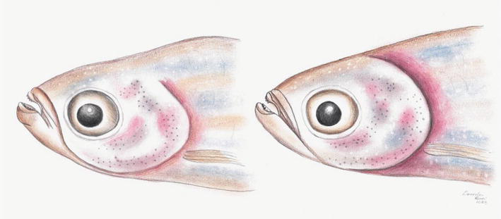
Figure 4.
Illustration of gill inflammation in zebrafish.
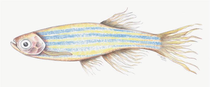
Figure 5.
Illustration of zebrafish fins and tail with frayed edges.
4.1 Columnariosis
The infection by the opportunistic bacteria
Zebrafish with compromised immune system are especially vulnerable to
As an infectious disease, the affected fish must be isolated from healthy individuals and treated with antibiotics—oxytetracycline or florfenicol. Improving water quality and general handling, as well as quarantine adoption of new fishes, tend to be effective against the
4.2 Edwardsiellosis
The gram-negative bacterium
Diffuse erythema on the skin and necrosis in intern organs are common clinical findings, and the olfactory neural pathway is also a target for the infection [29]. Quarantine is key to prevention, once infected fishes rapidly evolve the characteristic lesions, approximately two weeks, antibiotics are the usual treatment for infected fishes.
Besides local signs, infections in zebrafish also have a systemic character, with clinical manifestations such as peritonitis and generalized edema. The edematous condition of skin and abdominal distension is called dropsy (Figure 6) and is derived from fluid accumulation into the elomatic cavity caused by systemic infections [23, 30].
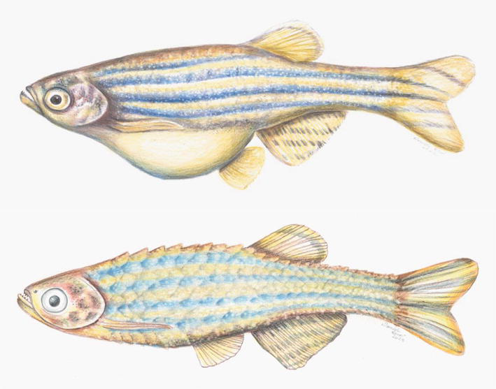
Figure 6.
Illustration of dropsy zebrafish with fluid accumulation in the coelomic cavity (
According to Pullium
4.3 Mycobacteriosis
Bacterial infections usually cause notable symptoms, provoking mortality or health decline in the zebrafish population. However, subclinical infections lead to uncontrollable biases in experimental protocols [34], so in laboratory conditions, the fish must be observed, and good practices should be taken to prevent these infections.
Mycobacteria are ubiquitous, naturally occurring gram-positive, acid-fast, and aerobic bacteria. They are found in both environmental and laboratory water besides soil, and several species have been described to infect zebrafish, like
Poor water quality and stressed animals are the pre-existing factors for this organism to establish itself. The clinical presentation varies greatly. Some lesions observed in the fish will be discoloration (Figure 7), skin ulcerations, or granulomas (nodules) in the abdominal cavity (Figure 8). However, lethargy may be another sign of infection. This is a chronic disease, and if not properly treated, it can lead to death. Mortality and morbidity of Mycobacteriosis vary according to species, where
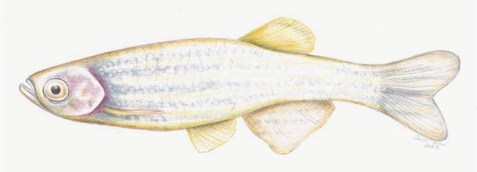
Figure 7.
Illustration of zebrafish showing epithelial discoloration.
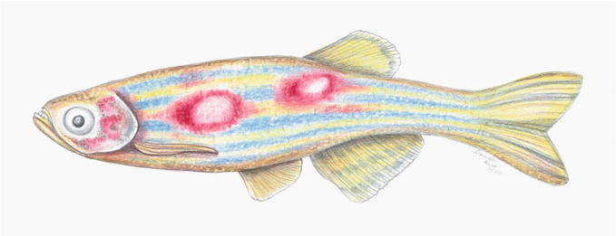
Figure 8.
Illustration of zebrafish with skin ulcerations.
Zebrafish facilities affected by Mycobacteriosis must adopt rigorous protocols to eliminate the contamination. Preventive measures, such as quarantine of newly introduced fish, embryo disinfection, effective sterilization of system water, and satisfactory sanitation of rearing tanks, must be taken in every zebrafish facility [36].
Mycobacteria are oligotrophic organisms and, thus, can survive and proliferate in a plethora of environments. In zebrafish aquaria, when sanitation is not efficient, biofilms are formed, which are adequate environments for the survival of mycobacteria despite the cleanness of water [37].
Chang, Lewis, and Whipps (2019) reported that
Research programs with zebrafish can be severely affected by bacterial outbreaks in the facilities. Generally, mycobacteria are opportunistic pathogens; however, two species are of special relevance:
The gastrointestinal system is also considered a route for mycobacteria infection. There have been reports of salmonids and medaka infected through feed, and mycobacteria were identified in the zebrafish intestinal tract (reviewed in [39]). Studies pointed out that transmission through other organisms could maximize contamination of Mycobacteriosis since Japanese medaka were more infected by mycobacteria-contaminated mosquitoes, and infected protozoan
Mycobacteriosis is considered a zoonotic disease, and non-tuberculous mycobacteria (NTM)-induced cutaneous infections have been described in dermatological care clinics. Most of the cases were caused by
5. Fungal infections
Fungal infections generally present a cotton or wool-like form that can derive from necrotic lesions, although species like
5.1 Saprolegnia
Saprolegniosis is an infectious disease caused by the fungi of the Saprolegnia genus, notably
The affected fish can present lesions on the skin, gills, and fins, showing a “fuzzy” appearance, and with the disease aggravation, a significant portion of the body is affected [42]. The affected surface area, which may be composed of large areas, predicts the severity of the disease. Thus, destruction of the epidermis and respiratory collapse due to infection of the gills cause behavioral changes, with lethargy leading to a high risk of predation. The impairment of physiological processes such as osmoregulation and the eventual entrance of fungal hyphae in internal organs in advanced stages can lead to organ failure.
Pre-existent skin lesions and infection by other pathogens could represent a serious risk for contracting Saprolegnia infections. Moreover, physiological stressors such as poor water quality, high density, and sudden temperature change have an important impact on zebrafish immune homeostasis, increasing the susceptibility to opportunistic infections, such as Saprolegniosis. Improving water quality, avoiding stressing factors, quarantining newly introduced fish, and regularly cleaning tanks and equipment are key actions to prevent fungal occurrence [43].
Infected fishes must be isolated from the experimental aquaria, along with antifungal treatments in order to preserve sanitary conditions of the experimental set and prevent further infections. Also, proper monitoring of fish is essential to recognize and take immediate action for treatment.
6. Parasitic infections
6.1 Ichthyophthiriasis
Ichthyophthiriasis is a parasitic infection caused by the protozoan
The Ichthyophthiriasis characteristic white spots consist in the trophont stage of the parasite, which is the stage when the parasite adheres to fish skin. The skin infection leads to behavior alteration, and the fish starts to rub against objects’ surfaces, increasing mucus production. This behavior represents a visible irritation and discomfort of infected fishes, and the stressful situation provokes immunodepression, making them more susceptible to
Containment measures like regular monitoring of fish behavior, avoiding high density, and keeping water quality seem to be effective in avoiding the parasite. Meanwhile, infected fish must be isolated for therapeutic intervention and to avoid the spread of the disease [46]. The treatment can be executed through two approaches: the interruption of the parasite’s life cycle, with a gradual elevation of salinity and temperature, or with chemical substances, such as formalin or copper-based medication.
6.2 Pseudoloma neurophilia
Microsporidia are obligate intracellular spore-forming parasites that can infect approximately 150 fish species, as well as terrestrial animals such as rodents, rabbits, and primates including humans [47]. Microsporidiosis is a common disease in laboratory fish that affects the central nervous system and can cause emaciation and spinal deformity [48, 49]. Also, infection by microsporidia is characterized by xenomas, a cellular growth formed by hypertrophied, unorganized, and unstructured polyploid cells due to parasite proliferation [50].
In zebrafish, microsporidia infection was described by de Kinkelin (1980) [51], and the most common parasite was identified as
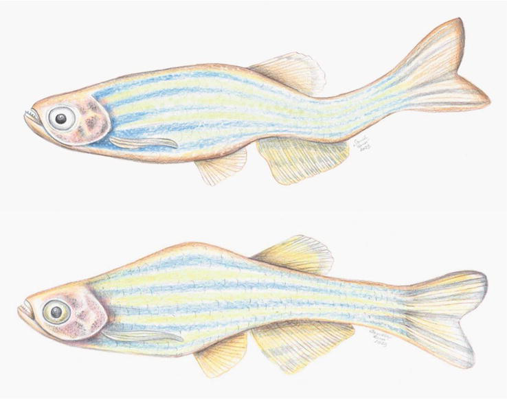
Figure 9.
Illustration of zebrafish with abnormal morphology, spinal deformity (
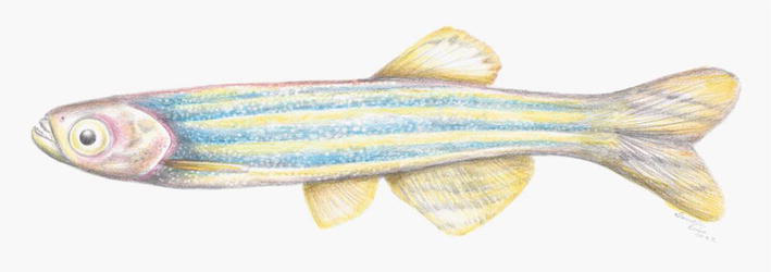
Figure 10.
Illustration of zebrafish with emaciation.
The spinal cord is associated with locomotor ability and motor function, and areas of the hindbrain control neural pathways associated with behaviors like anxiety, fear, and learning. Thus, it has been suggested that lesions caused by
According to Spagnoli, Sanders, & Kent [53], zebrafish infected with
7. Viral infections
Health management in zebrafish laboratories is mandatory since this model system becomes strikingly important to biomedical sciences in both larvae and adult stages [55]. Although several mammalian viral infections are modeled in zebrafish [56], naturally occurring viral infections are scarcely studied in zebrafish [57].
As the importance of zebrafish in science rises, aquatic laboratory facilities also start to emerge in research centers, and the utilization of shared systems exacerbates the possibilities for transmission of undiagnosed viral infections [58, 59]. The implementation of different mutant lines, such as immunocompromised fish, to studies in the immunology field contributes to the introduction of new diseases in the population, including viral infections (reviewed in [57]).
Bermúdez
8. Concluding remarks
Zebrafish have been considered an outstanding model for biomedical research. One of many advantages of the zebrafish is the possibility to screen small molecules to identify potential therapeutics with precision. Moreover, the conservation of immune mechanisms among vertebrates strengthens the translational character of zebrafish as a model system.
However, like other animal models, zebrafish are subject to suffering from disorders, including being contaminated and transmitting infectious diseases.
The study of natural diseases that can occur in zebrafish, mainly in fish kept in a scientific experimentation environment, may provide the opportunity to directly visualize the pathogen-host dynamics and to carry out chemical screenings, which facilitates the development and testing of new therapies, ensuring animal welfare.
Acknowledgments
We thank the São Paulo Research Foundation (FAPESP) for the support—notably through the Center for Toxins, Immune Response, and Cell Signaling (CeTICS). All illustrations presented here were made exclusively for this chapter by Daniela Bená.
This work was supported by the São Paulo Research Foundation—FAPESP (#2019/27677-7; #2021/08891-8 and #2013/07467-1). The funders had no role in the study design, data collection and analysis, decision to publish, or preparation of the manuscript.
References
- 1.
Masud S, Torraca V, Meijer AH. Modeling infectious diseases in the context of a developing immune system. Current Topics in Developmental Biology. 2017; 124 :277-329. DOI: 10.1016/bs.ctdb.2016.10.006 - 2.
Esmail MY, Astrofsky KM, Lawrence C, Serluca FC. Chapter 20 - The biology and management of the Zebrafish. In: Fox JG, Anderson LC, Otto GM, Pritchett-Corning KR, Whary MT, editors. American College of Laboratory Animal Medicine. 3rd ed. Academic Press; 2015. pp. 1015-1062. DOI: 10.1016/B978-0-12-409527-4.00020-1 - 3.
Baker DG. Natural pathogens of laboratory mice, rats, and rabbits and their effects on research. Clinical Microbiology Reviews. 1998; 11 :231-266. DOI: 10.1128/CMR.11.2.231 - 4.
de Abreu MS, Giacomini ACVV, Zanandrea R, Dos Santos BE, Genario R, de Oliveira GG, et al. Psychoneuroimmunology and Immunopsychiatry of Zebrafish. Psychoneuroendocrinology. 2018; 92 :1-12. DOI: 10.1016/j.psyneuen.2018.03.014 - 5.
Brinchmann M, Patel D, Pinto N, Iversen M. Functional aspects of fish mucosal lectins—Interaction with non-self. Molecules. 2018; 23 :1119. DOI: 10.3390/molecules23051119 - 6.
Lima C, Disner GR, Falcão MAP, Seni-Silva AC, Maleski ALA, Souza MM, et al. The Natterin proteins diversity: A review on phylogeny, structure, and immune function. Toxins (Basel). 2021; 13 :538. DOI: 10.3390/toxins13080538 - 7.
Sahoo S, Banu H, Prakash A, Tripathi G. Immune system of fish: An evolutionary perspective. In: Antimicrobial Immune Response. London, UK, London, UK: IntechOpen; 2021 - 8.
Hammer HS. Water quality for zebrafish culture. In: The Zebrafish in Biomedical Research. The Netherlands: Elsevier; 2020. pp. 321-335. DOI: 10.1016/B978-0-12-812431-4.00029-4 - 9.
Aleström P, D’Angelo L, Midtlyng PJ, Schorderet DF, Schulte-Merker S, Sohm F, et al. Zebrafish: Housing and husbandry recommendations. Laboratory Animals. 2020; 54 :213-224. DOI: 10.1177/0023677219869037 - 10.
Murray KN, Lains D, Spagnoli ST. Water quality and idiopathic diseases of laboratory zebrafish. In: Cartner S, Eisen J, Farmer S, Guillemin K, Kent ML, Sanders GS, editors. The Zebrafish in Biomedical Research: Biology, Husbandry, Diseases and Research Applications. London: Elsevier; 2020. pp. 463-478 - 11.
Schaefer J, Ryan A. Developmental plasticity in the thermal tolerance of zebrafish Danio rerio . Journal of Fish Biology. 2006;69 :722-734. DOI: 10.1111/j.1095-8649.2006.01145.x - 12.
Timmons MB, Ebeling JM. Recirculating Aquaculture. 3rd ed. Ithaca, NY: Ithaca Publishing Company LLC; 2013 - 13.
Learmonth C, Carvalho AP. Acute and chronic toxicity of nitrate to early life stages of zebrafish—Setting nitrate safety levels for zebrafish rearing. Zebrafish. 2015; 12 :305-311. DOI: 10.1089/zeb.2015.1098 - 14.
Kent ML, Sanders JL, Spagnoli S, Al-Samarrie CE, Murray KN. Review of diseases and health Management in Zebrafish Danio rerio (Hamilton 1822) in research facilities. Journal of Fish Diseases. 2020;43 :637-650. DOI: 10.1111/jfd.13165 - 15.
Kwong RWM, Kumai Y, Perry SF. The physiology of fish at low PH: The zebrafish as a model system. The Journal of Experimental Biology. 2014; 217 :651-662. DOI: 10.1242/jeb.091603 - 16.
Magalhães DP, Buss DF, da Cunha RA, Linde-Arias AR, Baptista DF. Analysis of individual versus group behavior of zebrafish: A model using PH sublethal effects. Bulletin of Environmental Contamination and Toxicology. 2012; 88 :1009-1013. DOI: 10.1007/s00128-012-0608-9 - 17.
Zahangir MM, Haque F, Mostakim GM, Islam MS. Secondary stress responses of zebrafish to different PH: Evaluation in a seasonal manner. Aquaculture Reports. 2015; 2 :91-96. DOI: 10.1016/j.aqrep.2015.08.008 - 18.
Boyd CE, Tucker CS, Somridhivej B. Alkalinity and hardness: Critical but elusive concepts in aquaculture. Journal of World Aquaculture Society. 2016; 47 :6-41. DOI: 10.1111/jwas.12241 - 19.
Chen Y, Lu F, Hwang P. Comparisons of calcium regulation in fish larvae. The Journal of Experimental Zoology. 2003; 295A :127-135. DOI: 10.1002/jez.a.10195 - 20.
Pekow C. Defining, measuring, and interpreting stress in laboratory animals. Contemporary Topics in Laboratory Animal Science. 2005; 44 :41-45 - 21.
Valcarce DG, Riesco MF, Cuesta-Martín L, Esteve-Codina A, Martínez-Vázquez JM, Robles V. Stress decreases spermatozoa quality and induces molecular alterations in zebrafish progeny. BMC Biology. 2023; 21 :70. DOI: 10.1186/s12915-023-01570-w - 22.
Estes JM, Altemara ML, Crim MJ, Fletcher CA, Whitaker JW. Behavioral and reproductive effects of environmental enrichment and Pseudoloma Neurophilia infection on adult zebrafish ( Danio rerio ). Journal of the American Association for Laboratory Animal Science. 2021;60 :249-258. DOI: 10.30802/AALAS-JAALAS-20-000113 - 23.
Matthews JL. Common diseases of laboratory zebrafish. Methods in Cell Biology. Vol. 77. Academic Press; 2004. pp. 617-643. DOI: 10.1016/S0091-679X(04)77033-8 - 24.
Roberts RJ. Fish Pathology. Wiley Online Library. Glasgow: Blackwell Publishing Ltd.; 2012. DOI: 10.1002/9781118222942 - 25.
Moyer TR, Hunnicutt DW. Susceptibility of Zebra fish Danio rerio to infection by Flavobacterium Columnare andF. Johnsoniae . Diseases of Aquatic Organ. 2007;76 :39-44. DOI: 10.3354/dao076039 - 26.
Hawke JP, Kent M, Rogge M, Baumgartner W, Wiles J, Shelley J, et al. Edwardsiellosis caused by Edwardsiella Ictaluri in laboratory populations of zebrafishDanio rerio . Journal of Aquatic Animal Health. 2013;25 :171-183. DOI: 10.1080/08997659.2013.782226 - 27.
Evans JJ, Klesius PH, Plumb JA, Shoemaker CA. Edwardsiella Septicemias. In: Fish Diseases and Disorders. Vol. 3. CABI Digital Library; 2011. pp. 512-569 - 28.
Petrie-Hanson L, Romano CL, Mackey RB, Khosravi P, Hohn CM, Boyle CR. Evaluation of zebrafish Danio rerio as a model for enteric Septicemia of catfish (ESC). Journal of Aquatic Animal Health. 2007;19 :151-158. DOI: 10.1577/H06-026.1 - 29.
Whipps CM, Kent ML. Bacterial and fungal diseases of zebrafish. In: The Zebrafish in Biomedical Research. The Netherlands: Elsevier; 2020. pp. 495-508. DOI: 10.1016/B978-0-12-812431-4.00041-5 - 30.
López Nadal A, Ikeda-Ohtsubo W, Sipkema D, Peggs D, McGurk C, Forlenza M, et al. Microbiota, and gut immunity: Using the zebrafish model to understand fish health. Frontiers in Immunology. 2020; 11 :114. DOI: 10.3389/fimmu.2020.00114 - 31.
Pullium JK, Dillehay DL, Webb S. High mortality in zebrafish ( Danio rerio ). Contemporary Topics in Laboratory Animal Science. 1999;38 :80-83 - 32.
Harikrishnan R, Balasundaram C. Modern trends in Aeromonas Hydrophila disease management with fish. Reviews in Fisheries Science. 2005; 13 :281-320. DOI: 10.1080/10641260500320845 - 33.
Shotts EB Jr, Kleckner AL, Gratzek JB, Blue JL. Bacterial Flora of aquarium fishes and their shipping waters imported from Southeast Asia. Journal of Fisheries and Research Board Canada. 1976; 33 :732-735. DOI: 10.1139/f76-089 - 34.
Kent ML, Spitsbergen JM, Matthews JM, Fournie JW, Murray KN, Westerfield M. Diseases of Zebrafish in Research Facilities. Zebrafish International Resource Center; 2012. Available from: https://zebrafish.org/wiki/health/disease_manual/start#dokuwiki__top - 35.
Zebrafish International Resource Center’s (ZIRC) Diagnostic Services [ZIRC Public Wiki]. Available from: https://zebrafish.org/wiki/health/submission/report [Accessed: February 08, 2023] - 36.
Whipps CM, Lieggi C, Wagner R. Mycobacteriosis in zebrafish colonies. ILAR Journal. 2012; 53 :95-105. DOI: 10.1093/ilar.53.2.95 - 37.
Chang CT, Lewis J, Whipps CM. Source or sink: Examining the role of biofilms in transmission of Mycobacterium Spp. in laboratory zebrafish. Zebrafish. 2019; 16 :197-206. DOI: 10.1089/zeb.2018.1689 - 38.
Mason T, Snell K, Mittge E, Melancon E, Montgomery R, McFadden M, et al. Strategies to mitigate a Mycobacterium Marinum outbreak in a zebrafish research facility. Zebrafish. 2016; 13 :S-77-S-87. DOI: 10.1089/zeb.2015.1218 - 39.
Chang CT, Benedict S, Whipps CM. Transmission of Mycobacterium Chelonae and Mycobacterium Marinum in laboratory zebrafish through live feeds. Journal of Fish Diseases. 2019; 42 :1425-1431. DOI: 10.1111/jfd.13071 - 40.
Peterson T, Ferguson J, Watral V, Mutoji K, Ennis D, Kent M. Paramecium Caudatum enhances transmission and infectivity of Mycobacterium Marinum and M. Chelonae in zebrafish Danio rerio . Diseases of Aquatic Organisms. 2013;106 :229-239. DOI: 10.3354/dao02649 - 41.
Astrofsky KM, Bullis RA, C.G.S. Chapter 19 - biology and Management of the Zebrafish. In: American College of Laboratory Animal Medicine. 2nd ed. The Netherlands: Elsevier; 2002. pp. 861-883 - 42.
Stoskopf MK. Fish Medicine. Philadelphia: W.B. Sannders; 1993 - 43.
Noga EJ. Fish Disease. United States: Wiley; 2010 - 44.
von Jørgensen L. The fish parasite Ichthyophthirius Multifiliis – Host immunology, vaccines and novel treatments. Fish & Shellfish Immunology. 2017; 67 :586-595. DOI: 10.1016/j.fsi.2017.06.044 - 45.
Mathiessen H, Kjeldgaard-Nintemann S, Gonzalez CMF, Henard C, Reimer JA, Gelskov SV, et al. Acute immune responses in zebrafish and evasive behavior of a parasite - who is winning? Frontier in Cellular Infectious Microbiology. 2023; 13 :1190931. DOI: 10.3389/fcimb.2023.1190931 - 46.
Christoffersen TB, Kania PW, von Gersdorff Jørgensen L, Buchmann K. Zebrafish Danio rerio as a model to study the immune response against infection with Ichthyophthirius Multifiliis. Journal of Fish Diseases. 2017;40 :847-852. DOI: 10.1111/jfd.12543 - 47.
Lom J, Nilsen F. Fish microsporidia: Fine structural diversity and phylogeny. International Journal for Parasitology. 2003; 33 :107-127. DOI: 10.1016/S0020-7519(02)00252-7 - 48.
Matthews JL, Brown AMV, Larison K, Bishop-Stewart JK, Rogers P, Kent ML. Pseudoloma Neurophilia n. g., n. Sp., a new microsporidium from the central nervous system of the zebrafish ( Danio rerio ). The Journal of Eukaryotic Microbiology. 2001;48 :227-233. DOI: 10.1111/j.1550-7408.2001.tb00307.x - 49.
Kent ML, Bishop-Stewart JK. Transmission and tissue distribution of Pseudoloma Neurophilia (microsporidia) of zebrafish, Danio rerio (Hamilton). Journal of Fish Diseases. 2003;26 :423-426. DOI: 10.1046/j.1365-2761.2003.00467.x - 50.
Ramsay JM, Watral V, Schreck CB, Kent ML. Pseudoloma Neurophilia infections in zebrafish Danio rerio : Effects of stress on survival, growth, and reproduction. Diseases of Aquatic Organisms. 2009;88 :69-84. DOI: 10.3354/dao02145 - 51.
Kinkelin P. Occurrence of a microsporidian infection in Zebra Danio Brachy Danio rerio (Hamilton-Buchanan). Journal of Fish Diseases. 1980;3 :71-73. DOI: 10.1111/j.1365-2761.1980.tb00187.x - 52.
Midttun HLE, Vindas MA, Whatmore PJ, Øverli Ø, Johansen IB. Effects of Pseudoloma Neurophilia infection on the brain transcriptome in zebrafish ( Danio rerio ). Journal of Fish Diseases. 2020;43 :863-875. DOI: 10.1111/jfd.13198 - 53.
Spagnoli S, Sanders J, Kent ML. The common neural parasite Pseudoloma Neurophilia causes altered shoaling behaviour in adult laboratory zebrafish ( Danio rerio ) and its implications for Neurobehavioural research. Journal of Fish Diseases. 2017;40 :443-446. DOI: 10.1111/jfd.12512 - 54.
Spagnoli ST, Xue L, Murray KN, Chow F, Kent ML. Pseudoloma Neurophilia: A retrospective and descriptive study of nervous system and muscle infections, with new implications for pathogenesis and Behavioral phenotypes. Zebrafish. 2015; 12 :189-201. DOI: 10.1089/zeb.2014.1055 - 55.
Rosa JGS, Lima C, Lopes-Ferreira M. Zebrafish larvae behavior models as a tool for drug screenings and pre-clinical trials: A review. International Journal of Molecular Sciences. 2022; 23 :6647. DOI: 10.3390/ijms23126647 - 56.
Maleski ALA, Rosa JGS, Bernardo JTG, Astray RM, Walker CIB, Lopes-Ferreira M, et al. Recapitulation of retinal damage in zebrafish larvae infected with Zika virus. Cell. 2022; 11 :1457. DOI: 10.3390/cells11091457 - 57.
Crim MJ. Viral diseases. In: The Zebrafish in Biomedical Research. The Netherlands: Elsevier; 2020. pp. 509-526 - 58.
Crim MJ, Riley LK. Viral diseases in zebrafish: What is known and unknown. ILAR Journal. 2012; 53 :135-143. DOI: 10.1093/ilar.53.2.135 - 59.
Kent ML, Feist SW, Harper C, Hoogstraten-Miller S, Law J, et al. Recommendations for control of pathogens and infectious diseases in fish research facilities. Comparative Biochemistry and Physiology. 2009; 149 :240-248. DOI: 10.1016/j.cbpc.2008.08.001 - 60.
Bermúdez R, Losada AP, de Azevedo AM, Guerra-Varela J, Pérez-Fernández D, Sánchez L, et al. First description of a natural infection with spleen and kidney necrosis virus in zebrafish. Journal of Fish Diseases. 2018; 41 :1283-1294. DOI: 10.1111/jfd.12822 - 61.
Altan E, Kubiski SV, Boros Á, Reuter G, Sadeghi M, Deng X, et al. A highly divergent picornavirus infecting the gut epithelia of zebrafish ( Danio rerio ) in research institutions worldwide. Zebrafish. 2019;16 :291-299. DOI: 10.1089/zeb.2018.1710 - 62.
Kent ML, Harper C, Wolf JC. Documented and potential research impacts of subclinical diseases in zebrafish. ILAR Journal. 2012; 53 :126-134. DOI: 10.1093/ilar.53.2.126