 Open access peer-reviewed chapter
Abstract
To describe ophthalmological fundoscopic findings in patients with COVID-19 admitted to the intensive care unit (ICU) of the largest third-level referral center for COVID-19 in Mexico City. In this cross-sectional single-center study, consecutive patients admitted to the ICU with a diagnosis of COVID-19 underwent fundus examination with an indirect ophthalmoscope. Clinical photographs were taken using a posterior-pole camera. We explored the association between ocular manifestations and demographic characteristics, inflammatory markers, hemodynamic factors, and comorbidities. Of 117 patients examined, 74 were male; the median age was 54 years (range: 45–63 years). Forty-two patients had ophthalmological manifestations (unilateral in 23 and bilateral in 19), and 10 of these patients had more than one ophthalmological manifestation. Ocular findings were papillitis (n = 13), cotton-wool spots (n = 12), retinal hemorrhages (n = 5), retinal nerve fiber layer edema (n = 8), macular whitening (n = 5), retinal vascular tortuosity (n = 4), papillophlebitis (n = 3), central retinal vein occlusion (n = 1), and branch retinal vein occlusion (n = 1). Ocular fundus manifestations were not associated with demographic characteristics, inflammatory markers, hemodynamic factors, or comorbidities. Over one-third of patients with severe COVID-19 had ophthalmological manifestations. The most frequent fundoscopic findings were optic nerve inflammation, microvasculature occlusion, and major vascular occlusions. We recommend long-term follow-up to prevent permanent ocular sequelae.
Keywords
- chorioretinal vasculopathy
- uveitis
- retinal vein occlusion
- optic neuritis
- acute retinal ischemia
- SARS-CoV-2
- Covid-19
- PAMM
1. Introduction
The COVID-19 pandemic was declared on March 11, 2020, by the World Health Organization (WHO). As is already known, the coronavirus belongs to the genus
The coronavirus causes a severe acute respiratory syndrome (SARS), which was responsible for the death of approximately 6,881,955 people worldwide, according to the Johns Hopkins University as of March 10, 2023 [3]. For the virus to enter human cells, it uses the angiotensin-converting enzyme 2 (ACE2), which is expressed in the lungs, heart, central nervous system (CNS), adrenal glands, and in the ocular globe. The structures that have this enzyme include the cornea, conjunctiva, sclera, ciliary body, retina, aqueous humor, vitreous humor, and iris [4, 5]. This explains the wide range of clinical manifestations that can occur in COVID-19 infection.
It has been found that the cluster of differentiation 147 (CD147) is expressed in the corneal and conjunctival epithelia and plays an important role in severe acute respiratory syndrome coronavirus 2 (SARS-CoV-2) infection [6], along with ACE2, which has been identified in the Müller cells of the retina, mainly in the inner nuclear layer [7], as well as in endothelial cells. This is where the theory of SARS-CoV-2 entry into the retina arises, which begins on the corneal and conjunctival surface and then spreads hematogenously to reach the choroid plexus, leading to the breakdown of the blood-retinal barrier mediated by CD147, facilitated by an inflammatory microenvironment [8, 9, 10].
The clinical findings of the anterior segment commonly described in COVID-19 infections occur in a range of 2–32% [11, 12, 13]. There are very few reports of posterior segment manifestations in hospitalized patients. A review of the literature showed a mean age of 47.4 ± 14.8 years [14], and ocular manifestations have been reported in 36% [15] of patients in the Intensive Care Unit (ICU) within a range of 4–55 days after the onset of the initial symptoms.
The exact role that SARS-CoV-2 plays in the retina during the infection is still not known for certain. Studies have been conducted using immunohistochemical analysis, where no RNA or spike protein was found in biopsies from deceased COVID-19 patients [16, 17]. On the other hand, Menuchin demonstrated in three-dimensional (3D) retinal tissue cultures, referred to as retinal organoids, evidence that SARS-CoV-2 can actively replicate in the retina [18].
2. Posterior segment ocular findings
2.1 Optic neuritis
The SARS-CoV induces neuronal death in the medullary respiratory center through the upregulation of inflammatory markers or through direct autophagy. The same hypothesis has been proposed for SARS-CoV-2, in which the virus infects a peripheral neuron and travels retrograde to the brain via synaptic transport [19].
The most common ocular finding reported by Romero [15] was inflammation of the optic nerve, including papillitis and papillophlebitis, in hospitalized patients. The reason why ganglion cells and the optic nerve are affected by COVID infection is explained by Menuchin’s study [18] with retinal organoids, which suggests that the ganglion cell layer had a higher probability of being infected by SARS-CoV-2, up to 40%. It concluded that progenitor cells are less susceptible to infection than neurons. However, the pathophysiology is still not clear and may be related to vascular dysfunction caused by hypercoagulability or a process similar to viral infection-induced vasculitis in severely ill patients [20]. Neuro-ophthalmic manifestations in COVID-19 infection include optic neuritis, acute transverse myelitis, viral encephalitis, toxic encephalopathy, leukoencephalopathy, and acute disseminated encephalomyelitis [21].
Optic neuritis is defined as an inflammatory demyelinating condition that causes acute visual loss, generally monocular, and rarely binocular, very few cases of optic neuritis have been reported days after the onset of symptoms in patients with mild disease. Mechanisms responsible for these neuro-ophthalmic manifestations have been postulated, such as neurotropism and molecular mimicry, where viral antigens trigger a host immune response directed toward central nervous system myelin proteins [22, 23]. It can be associated with the spectrum of neuromyelitis optica, a disease associated with myelin oligodendrocyte glycoprotein [23], or infectious disease. The latter is more common in children, with common organisms being
Oculomotor paralysis of cranial nerves III, IV, or VI and VII [24, 25, 26] can result from direct viral infection or neurological complications, such as Guillain-Barré syndrome, Miller Fisher syndrome [27], or acute disseminated encephalomyelitis, and can occur in isolation or as polyneuropathies.
The prone position can cause anterior or posterior ischemic neuropathy, since this position has been shown to increase intraocular pressure to more than 40 mmHg [28]. On the other hand, this position has shown improved survival in patients with acute respiratory distress syndrome (ARDS) [29].
2.2 Retinal vascular occlusions
2.2.1 Central retinal vein occlusion (CRVO)
Central retinal vein occlusion (CRVO) is a common manifestation of the disease when it comes to posterior segment abnormalities. This was reported by Fonollosa, who found that 25 out of 35 patients presented with central retinal vein occlusion, and in some cases, it occurred bilaterally. CRVO occurs more frequently in the acute stages of COVID-19 infection, and it has been shown that it does not necessarily involve thrombotic and occlusive lesions in other organs but can be limited to the retina [30].
The involvement of retinal vasculature in COVID-19 infection can be explained by the state of hypercoagulability during the acute phase, leading to endothelial damage [31].
CRVO is characterized by painless visual loss accompanied by intraretinal hemorrhages in a flame-shaped pattern across the retinal surface, unlike branch retinal vein occlusion where hemorrhages are localized to the affected vessel’s course. Other features may include cotton-wool spots and, in some cases, macular edema.
Risk factors associated with the development of CRVO include age over 50 years (in 90% of cases), systemic arterial hypertension, diabetes mellitus, and hyperlipidemia [32, 33]. These risk factors also predispose individuals to severe COVID-19 infection. In a large cohort of patients with a history of COVID-19 and CRVO, 68% of patients were under 40 years of age and had mild disease compared to the typical age of presentation. However, 36% of them had cardiovascular risk factors, and the procoagulant state during the acute phase of the disease increased the risk of ischemic events in this cohort [32, 33].
2.2.2 Central retinal artery occlusion (CRAO)
Thrombotic events in COVID-19 infection have been reported in 39% of patients [34]. Central retinal artery occlusion (CRAO) tends to develop frequently in patients during the acute phase of COVID-19 infection. For example, Acharya [35] reported the first case in a 60-year-old patient with acute respiratory distress syndrome, who had comorbidities, such as hypertension, dyslipidemia, stable coronary artery disease, and chronic obstructive pulmonary disease (COPD). After being placed on mechanical ventilation, the patient experienced sudden and painless vision loss, and ophthalmological examination revealed a cherry-red spot with retinal whitening.
While hypercoagulability is a leading cause of ischemic alterations, it’s essential to recognize that this patient had comorbidities that increased the risk of such an event.
Macular whitening has also been reported as a finding in the posterior segment of hospitalized COVID-19 patients [15], likely due to cilioretinal artery hypoperfusion, believed to be secondary to vascular dysfunction caused by elevated levels of D-dimer, prothrombin, fibrinogen, partial thromboplastin time, and patient comorbidities such as hypertension and obesity.
2.3 Acute paracentral middle maculopathy (PAMM) and acute macular neuroretinopathy (NMA)
Acute paracentral middle maculopathy (PAMM) and acute macular neuroretinopathy (NMA) are caused by microvascular alterations in the deep retinal vessels [36, 37, 38].
Cases of concurrent NMA and PAMM with a COVID-19 diagnosis have been reported. Castro [37] reviewed the literature on reported cases and noted that all patients were under 55 years of age, predominantly female, without prior comorbidities, and that these conditions occurred 1 month after the diagnosis of SARS-CoV-2 infection as the sole systemic complication. These findings are significant because PAMM was previously known to predominantly occur in patients with cardiovascular risk factors [39].
A decrease in macular capillary density by 8% has been demonstrated in severe COVID-19 patients, as well as a decrease in the density of intermediate and deep retinal capillary plexuses in patients without previous comorbidities. There appears to be a correlation between the severity of the disease and the decrease in retinal vessel density [40].
2.4 Vascular alterations
The mechanism by which COVID-19 can affect retinal vasculature is still unknown, but several theories have been proposed. The angiotensin-converting enzyme 2 (ACE2) is believed to play a significant role in producing vasoconstriction, triggering thrombogenicity by increasing platelet and leukocyte adhesion [41]. It has been reported that COVID-19 infection can cause alterations in retinal microvasculature up to 8.86 times more [42].
Ivernizzi [43] evaluated the diameter of retinal veins 30 days after the onset of systemic symptoms and found that the diameter of both retinal veins and arteries was increased compared to the control group. Furthermore, the diameter was even larger in the more severe cases, leading to the conclusion that the dilation of retinal and pulmonary vessels could be secondary to increased blood supply due to the inflammatory response and endothelial damage. This theory about endothelial damage is supported by a study that observed decreased vessel density in the superficial capillary plexus, deep capillary plexus, and choriocapillaris, along with an increase in the foveal avascular zone, through optical coherence tomography (OCT) in a six-month follow-up after COVID-19 infection [44].
The use of retinal lesions as a biomarker for assessing the extent of COVID-19’s impact on the brain has been proposed by de Figuereido [9]. This is based on theories about the mechanism of SARS-CoV-2 entry into the eye and its proximity to the central nervous system (Figure 1).
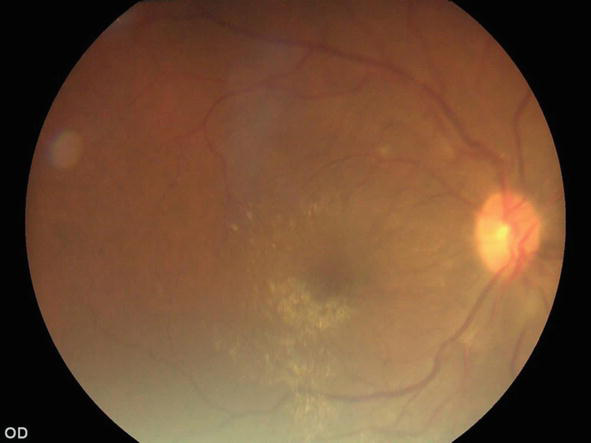
Figure 1.
Color fundus photograph showing tortuosity of vessels, hemorrhages in the temporal vascular arches and cottony exudates in the inferior temporal vascular arch with macular edema.
2.5 Vasculitis
Kawasaki-like [45] manifestations have been reported in association with COVID-19. The exact cause of this condition is still uncertain, but the most accepted theory is an aberrant immune response to a pathogen in genetically predisposed patients [46]. Endothelial cells can also be affected by COVID-19, leading to inflammation, apoptosis, and dysfunction [47].
There have been investigations regarding coronaviruses within the coronavirus family, such as the New Heaven virus identified in respiratory secretions of children with Kawasaki disease [48]. However, this finding has been highly debated, because the series of cases reported as Kawasaki-like has occurred in patients older than the usual age, with respiratory and gastrointestinal involvement, as well as meningeal and cardiovascular signs [46]. These clinical manifestations have also been reported in COVID patients, making it difficult to determine the exact etiology of these alterations.
Another form of vasculitis reported in the literature is seen in pediatric patients with chilblains and areas of vasculitis. Although no other abnormalities were found in these patients, vasculitis presentation is believed to be an autoimmune response in COVID-19 patients, possibly triggered by interferon type 1 (IFN), leading to acral skin lesions and vasculitis [49].
2.6 Retinal abnormalities
The exact route of SARS-CoV-2 infection to the retina is not known with certainty. It could occur through hematogenous dissemination or via nerve fibers through the optic nerve, potentially causing changes in the optic nerve and retina [21].
Cotton-wool spots have been reported in various series [15, 21, 50, 51] and have been referred to as retinal microangiopathy associated with COVID-19 [52]. These cotton-wool spots are not exclusive to COVID-19, as they have been observed in conditions like hypertension, human immunodeficiency virus (HIV) retinopathy [53], diabetic retinopathy [54], and giant cell arteritis [55]. Cotton-wool spots have been proposed as an indicator of some acute vascular event [56] or as a biomarker of systemic vascular disease [52]; however, imaging studies have not conclusively identified whether these cotton-wool spots are secondary to patients’ comorbidities or simply the result of the treatments used [51].
However, cotton-wool spots have not only been reported in patients with severe illness. Landecho [52] and Pereira [56] observed these findings in asymptomatic patients 43 days after the onset of the first symptoms. The location can vary, either appearing on the surface of the retina or following the course of venous or arterial vessels.
In a study of hospitalized patients, cotton-wool spots, retinal nerve fiber layer edema, and retinal hemorrhages were the second most common ocular findings [15]. These findings are consistent with previous reports where flame hemorrhages were seen in 22.2% and peripheral retinal hemorrhages in 11.1% of severe patients [56] and support the hypothesis that the retinal tropism of SARS-CoV-2 is associated with the disruption of the blood-retinal barrier [9].
Zago [57] and Costa [36] reported the presence of localized white-yellowish points in the outer retina that were self-limiting and did not result in visual sequelae. Optical coherence tomography (OCT) revealed hyporeflectivity in the pigment epithelium of the retina and the ellipsoid layer, as well as a discontinuation of the outer segments of photoreceptors. Notably, these findings were observed in patients who had recovered from COVID-19 within an average time of 82 ± 36.4 days after the onset of their initial symptoms. Of this group, 51.5% had a severe disease course and 37.5% had a critical illness.
2.7 Choroidal abnormalities
It has been suggested that the retina and the choroid could be key sites for ocular infection due to the presence of angiotensin-converting enzyme, which is expressed in both the choroid and the retina. Among the observed changes, a decreased choroidal thickness has been reported in the subfoveal, temporal, and nasal quadrants in recovered patients without anticoagulant or antiplatelet therapy [58]. This is speculated to be linked to an exaggerated immune response during the early stages of the disease, leading to an increase in choroidal thickness [59], followed by a subsequent decrease due to endothelial damage, resulting in reduced choroidal thickness [60].
Irregular, wall-less dark holes among choroidal vessels have been reported in 21% of severe COVID-19 patients. These findings are similar to those seen in conditions like geographic atrophy and pachychoroid. These holes may represent a new drainage pathway for choroidal vessels through the lymphatic system or spaces filled with lipids that regulate the inflammatory process [61].
References
- 1.
Chilamakuri R, Agarwal S. COVID-19: Characteristics and therapeutics. Cells [Internet]. 2021; 10 (2):206. DOI: 10.3390/cells10020206 - 2.
Sharma A, Ahmad Farouk I, Lal SK. COVID-19: A review on the novel coronavirus disease evolution, transmission, detection, control and prevention. Viruses [Internet]. 2021; 13 (2):202. DOI: 10.3390/v13020202 - 3.
COVID-19 map [Internet]. Johns Hopkins Coronavirus Resource Center. Available from: https://coronavirus.jhu.edu/map.html [Accessed: October 3, 2023] - 4.
Yaguchi S, Ogawa Y, Shimmura S, Hatou S, Nakamura S, Inaba T, et al. Presence and physiologic function of the renin–angiotensin system in mouse lacrimal gland. Investigative Ophthalmology & Visual Science [Internet]. 2012; 53 (9):5416. DOI: 10.1167/iovs.12-9891 - 5.
Senanayake PD, Drazba J, Shadrach K, Milsted A, Rungger-Brandle E, Nishiyama K, et al. Angiotensin II and its receptor subtypes in the human retina. Investigative Ophthalmology & Visual Science [Internet]. 2007; 48 (7):3301-3311. DOI: 10.1167/iovs.06-1024 - 6.
Hamashima K, Gautam P, Lau KA, Khiong CW, Blenkinsop TA, Li H, et al. Potential modes of COVID-19 transmission from human eye revealed by single-cell atlas [internet]. bioRxiv. 2020. DOI: 10.1101/2020.05.09.085613 - 7.
Tikellis C, Johnston CI, Forbes JM, Burns WC, Thomas MC, Lew RA, et al. Identification of angiotensin converting enzyme 2 in the rodent retina. Current Eye Research [Internet]. 2004; 29 (6):419-427. DOI: 10.1080/02713680490517944 - 8.
Zhang Y, Stewart JM. Retinal and choroidal manifestations of COVID-19. Current Opinion in Ophthalmology [Internet]. 2021; 32 (6):536-540. DOI: 10.1097/icu.0000000000000801 - 9.
de Figueiredo CS, Raony Í, Giestal-de-Araujo E. SARS-CoV-2 targeting the retina: Host–virus interaction and possible mechanisms of viral tropism. Ocular Immunology and Inflammation [Internet]. 2020; 28 (8):1301-1304. DOI: 10.1080/09273948.2020.1799037 - 10.
Varga Z, Flammer AJ, Steiger P, Haberecker M, Andermatt R, Zinkernagel AS, et al. Endothelial cell infection and endotheliitis in COVID-19. Lancet [Internet]. 2020; 395 (10234):1417-1418. DOI: 10.1016/S0140-6736(20)30937-5 - 11.
Ulhaq ZS, Soraya GV. The prevalence of ophthalmic manifestations in COVID-19 and the diagnostic value of ocular tissue/fluid. Arbeitsphysiologie [Internet]. 2020; 258 (6):1351-1352. DOI: 10.1007/s00417-020-04695-8 - 12.
Zhou Y, Duan C, Zeng Y, Tong Y, Nie Y, Yang Y, et al. Ocular findings and proportion with conjunctival SARS-COV-2 in COVID-19 patients. Ophthalmology [Internet]. 2020; 127 (7):982-983. DOI: 10.1016/j.ophtha.2020.04.028 - 13.
Inomata T, Kitazawa K, Kuno T, Sung J, Nakamura M, Iwagami M, et al. Clinical and prodromal ocular symptoms in coronavirus disease: A systematic review and meta-analysis. Investigative Ophthalmology & Visual Science [Internet]. 2020; 61 (10):29. DOI: 10.1167/iovs.61.10.29 - 14.
Sen M, Honavar SG, Sharma N, Sachdev MS. COVID-19 and eye: A review of ophthalmic manifestations of COVID-19. Indian Journal of Ophthalmology [Internet]. 2021; 69 (3):488-509. DOI: 10.4103/ijo.IJO_297_21 - 15.
Romero-Castro RM, Ruiz-Cruz M, Alvarado-de la Barrera C, González-Cannata MG, Luna-Villalobos YA, García-Morales AK, et al. Posterior segment ocular findings in critically ill patients with covid-19. Retina [Internet]. 2022; 42 (4):628-633. DOI: 10.1097/iae.0000000000003457 - 16.
Casagrande M, Fitzek A, Spitzer M, Püschel K, Glatzel M, Krasemann S, et al. Detection of SARS-CoV-2 genomic and subgenomic RNA in retina and optic nerve of patients with COVID-19. British Journal of Ophthalmology [Internet]. 2022; 106 (9):1313-1317. DOI: 10.1136/bjophthalmol-2020-318618 - 17.
Casagrande M, Fitzek A, Püschel K, Aleshcheva G, Schultheiss H-P, Berneking L, et al. Detection of SARS-CoV-2 in human retinal biopsies of deceased COVID-19 patients. Ocular Immunology and Inflammation [Internet]. 2020; 28 (5):721-725. DOI: 10.1080/09273948.2020.1770301 - 18.
Menuchin-Lasowski Y, Schreiber A, Lecanda A, Mecate-Zambrano A, Brunotte L, Psathaki OE, et al. SARS-CoV-2 infects and replicates in photoreceptor and retinal ganglion cells of human retinal organoids. Stem Cell Reports [Internet]. 2022; 17 (4):789-803. DOI: 10.1016/j.stemcr.2022.02.015 - 19.
Baig AM, Khaleeq A, Ali U, Syeda H. Evidence of the COVID-19 virus targeting the CNS: Tissue distribution, host–virus interaction, and proposed neurotropic mechanisms. ACS Chemical Neuroscience [Internet]. 2020; 11 (7):995-998. DOI: 10.1021/acschemneuro.0c00122 - 20.
Tang N, Li D, Wang X, Sun Z. Abnormal coagulation parameters are associated with poor prognosis in patients with novel coronavirus pneumonia. Journal of Thrombosis and Haemostasis [Internet]. 2020; 18 (4):844-847. DOI: 10.1111/jth.14768 - 21.
Alomari SO, Abou-Mrad Z, Bydon A. COVID-19 and the central nervous system. Clinical Neurology and Neurosurgery [Internet]. 2020; 198 (106116):106116. DOI: 10.1016/j.clineuro.2020.106116 - 22.
Sawalha K, Adeodokun S, Kamoga G-R. COVID-19-induced acute bilateral optic neuritis. Journal of Investigative Medicine High Impact Case Reports [Internet]. 2020; 8 . DOI: 10.1177/2324709620976018 - 23.
Zhou S, Jones-Lopez EC, Soneji DJ, Azevedo CJ, Patel VR. Myelin oligodendrocyte glycoprotein antibody–associated optic neuritis and myelitis in COVID-19. Journal of Neuro-Ophthalmology [Internet]. 2020; 40 (3):398-402. DOI: 10.1097/wno.0000000000001049 - 24.
Behera G, Gera P, Stephen M, Jose A, Thabah MM, Wadwekar V. Bilateral optic neuritis and facial palsy following COVID-19 infection. Cureus [Internet]. 2022; 14 (9):e28735. DOI: 10.7759/cureus.28735 - 25.
Dinkin M, Gao V, Kahan J, Bobker S, Simonetto M, Wechsler P, et al. COVID-19 presenting with ophthalmoparesis from cranial nerve palsy. Neurology [Internet]. 2020; 95 (5):221-223. DOI: 10.1212/wnl.0000000000009700 - 26.
Tonkal A, Alamri AA, AlMaghrabi SJ, Mozahim NF, Mozahim SF, Alsubaie SA, et al. Cranial nerve impairment associated with COVID-19 infections: A systematic review. Cureus [Internet]. 2022; 14 (11):e31997. DOI: 10.7759/cureus.31997 - 27.
Gutiérrez-Ortiz C, Méndez-Guerrero A, Rodrigo-Rey S, San Pedro-Murillo E, Bermejo-Guerrero L, Gordo-Mañas R, et al. Miller fisher syndrome and polyneuritis cranialis in COVID-19. Neurology [Internet]. 2020; 95 (5):e601-e605. DOI: 10.1212/wnl.0000000000009619 - 28.
Mao L, Jin H, Wang M, Hu Y, Chen S, He Q , et al. Neurologic manifestations of hospitalized patients with coronavirus disease 2019 in Wuhan, China. JAMA Neurology [Internet]. 2020; 77 (6):683. DOI: 10.1001/jamaneurol.2020.1127 - 29.
Kharat A, Simon M, Guérin C. Prone position in COVID 19-associated acute respiratory failure. Current Opinion in Critical Care [Internet]. 2022; 28 (1):57-65. DOI: 10.1097/mcc.0000000000000900 - 30.
Fonollosa A, Hernández-Rodríguez J, Cuadros C, Giralt L, Sacristán C, Artaraz J, et al. Characterizing covid-19–related retinal vascular occlusions: A case series and review of the literature. Retina [Internet]. 2022; 42 (3):465-475. DOI: 10.1097/iae.0000000000003327 - 31.
Voicu S, Ketfi C, Stépanian A, Chousterman BG, Mohamedi N, Siguret V, et al. Pathophysiological processes underlying the high prevalence of deep vein thrombosis in critically ill COVID-19 patients. Frontiers in Physiology [Internet]. 2020; 11 :608788. DOI: 10.3389/fphys.2020.608788 - 32.
Klein R. The 15-year cumulative incidence of retinal vein occlusion: The beaver dam eye study. Archives of Ophthalmology [Internet]. 2008; 126 (4):513. DOI: 10.1001/archopht.126.4.513 - 33.
O’Mahoney PRA, Wong DT, Ray JG. Retinal vein occlusion and traditional risk factors for atherosclerosis. Archives of Ophthalmology [Internet]. 2008; 126 (5):692-699. DOI: 10.1001/archopht.126.5.692 - 34.
Sanjay S, Srinivasan P, Jayadev C, Mahendradas P, Gupta A, Kawali A, et al. Post COVID-19 ophthalmic manifestations in an Asian Indian male. Ocular Immunology and Inflammation [Internet]. 2021; 29 (4):656-661. DOI: 10.1080/09273948.2020.1870147 - 35.
Acharya S, Diamond M, Anwar S, Glaser A, Tyagi P. Unique case of central retinal artery occlusion secondary to COVID-19 disease. IDCases [Internet]. 2020; 21 (e00867):e00867. DOI: 10.1016/j.idcr.2020.e00867 - 36.
Costa ÍF, Bonifácio LP, Bellissimo-Rodrigues F, Rocha EM, Jorge R, Bollela VR, et al. Ocular findings among patients surviving COVID-19. Scientific Reports [Internet]. 2021; 11 (1):11085. DOI: 10.1038/s41598-021-90482-2 - 37.
Castro CS, Ferreira AS, Silva NP, Lume MR, Furtado MJ. Paracentral acute middle maculopathy after COVID-19 disease: Multimodal evaluation. Retinal Cases and Brief Reports [Internet]. 2022; 17 (6):791-796. DOI: 10.1097/icb.0000000000001301 - 38.
Virgo J, Mohamed M. Paracentral acute middle maculopathy and acute macular neuroretinopathy following SARS-CoV-2 infection. Eye [Internet]. 2020; 34 (12):2352-2353. DOI: 10.1038/s41433-020-1069-8 - 39.
Rahimy E, Kuehlewein L, Sadda SR, Sarraf D. Paracentral acute middle maculopathy: What we knew then and what we know now. Retina [Internet]. 2015; 35 (10):1921-1930. DOI: 10.1097/IAE.0000000000000785 - 40.
Kalaw FGP, Warter A, Cavichini M, Knight D, Li A, Deussen D, et al. Retinal tissue and microvasculature loss in COVID-19 infection. Scientific Reports [Internet]. 2023; 13 (1):5100. DOI: 10.1038/s41598-023-31835-x - 41.
Verdecchia P, Cavallini C, Spanevello A, Angeli F. COVID-19: ACE2centric infective disease? Hypertension [Internet]. 2020; 76 (2):294-299. DOI: 10.1161/HYPERTENSIONAHA.120.15353 - 42.
Teo KYC, Invernizzi A, Staurenghi G, Cheung CMG. COVID-19-related retinal micro-vasculopathy – A review of current evidence. American Journal of Ophthalmology [Internet]. 2022; 235 :98-110. DOI: 10.1016/j.ajo.2021.09.019 - 43.
Invernizzi A, Torre A, Parrulli S, Zicarelli F, Schiuma M, Colombo V, et al. Retinal findings in patients with COVID-19: Results from the SERPICO-19 study. EClinicalMedicine [Internet]. 2020; 27 (100550):100550. DOI: 10.1016/j.eclinm.2020.100550 - 44.
Kal M, Winiarczyk M, Zarębska-Michaluk D, Odrobina D, Cieśla E, Płatkowska-Adamska B, et al. Long-term effect of SARS-CoV-2 infection on the retinal and choroidal microvasculature. Journal of Clinical Medicine [Internet]. 2023; 12 (7):2528. DOI: 10.3390/jcm12072528 - 45.
Verdoni L, Mazza A, Gervasoni A, Martelli L, Ruggeri M, Ciuffreda M, et al. An outbreak of severe Kawasaki-like disease at the Italian epicentre of the SARS-CoV-2 epidemic: An observational cohort study. Lancet [Internet]. 2020; 395 (10239):1771-1778. DOI: 10.1016/s0140-6736(20)31103-x - 46.
Becker RC. COVID-19 update: Covid-19-associated coagulopathy. Journal of Thrombosis and Thrombolysis [Internet]. 2020; 50 (1):54-67. DOI: 10.1007/s11239-020-02134-3 - 47.
Shulman ST, Rowley AH. Kawasaki disease: Insights into pathogenesis and approaches to treatment. Nature Reviews Rheumatology [Internet]. 2015; 11 (8):475-482. DOI: 10.1038/nrrheum.2015.54 - 48.
Esper F, Shapiro ED, Weibel C, Ferguson D, Landry ML, Kahn JS. Association between a novel human coronavirus and Kawasaki disease. The Journal of Infectious Diseases [Internet]. 2005; 191 (4):499-502. DOI: 10.1086/428291 - 49.
Quintana-Castanedo L, Feito-Rodríguez M, Fernández-Alcalde C, Granados-Fernández M, Montero-Vega D, Mayor-Ibarguren A, et al. Concurrent chilblains and retinal vasculitis in a child with COVID-19. Journal of the European Academy of Dermatology and Venereology [Internet]. 2020; 34 (12):e764-e766. DOI: 10.1111/jdv.16801 - 50.
Sanjay S, Agrawal S, Jayadev C, Kawali A, Gowda PB, Shetty R, et al. Posterior segment manifestations and imaging features post-COVID-19. Medical Hypothesis, Discovery & Innovation in Ophthalmology [Internet]. 2021; 10 (3):95-106. DOI: 10.51329/mehdiophthal1427 - 51.
Chan AX, Ritter M, Bakhoum MF. Bilateral cotton wool spots after ambulatory COVID-19. International Journal of Infectious Diseases [Internet]. 2021; 105 :414-415. DOI: 10.1016/j.ijid.2021.02.119 - 52.
Landecho MF, Yuste JR, Gándara E, Sunsundegui P, Quiroga J, Alcaide AB, et al. COVID-19 retinal microangiopathy as an in vivo biomarker of systemic vascular disease? Journal of Internal Medicine [Internet]. 2021; 289 (1):116-120. DOI: 10.1111/joim.13156 - 53.
Jaworski C. Morphology of the HIV versus the diabetic cotton wool spot. Optometry and Vision Science [Internet]. 2000; 77 (11):600-604. DOI: 10.1097/00006324-200011000-00010 - 54.
Akram MU, Akbar S, Hassan T, Khawaja SG, Yasin U, Basit I. Data on fundus images for vessels segmentation, detection of hypertensive retinopathy, diabetic retinopathy and papilledema. Data Brief [Internet]. 2020; 29 (105282):105282. DOI: 10.1016/j.dib.2020.105282 - 55.
Rai AS, Freund P, Margolin EA, Micieli JA. Numerous cotton wool spots from giant cell arteritis. Journal of Clinical Rheumatology [Internet]. 2020; 26 (5):e124. DOI: 10.1097/RHU.0000000000000995 - 56.
Pereira LA, Soares LCM, Nascimento PA, Cirillo LRN, Sakuma HT, Veiga GL da, et al. Retinal findings in hospitalised patients with severe COVID-19. British Journal of Ophthalmology [Internet]. 2022;106(1):102-105. DOI: 10.1136/bjophthalmol-2020-317576 - 57.
Zago Filho LA, Lima LH, Melo GB, Zett C, Farah ME. Vitritis and outer retinal abnormalities in a patient with COVID-19. Ocular Immunology and Inflammation [Internet]. 2020; 28 (8):1298-1300. DOI: 10.1080/09273948.2020.1821898 - 58.
Hepokur M, Gunes M, Durmus E, Aykut V, Esen F, Oguz H. Long-term follow-up of choroidal changes following COVID-19 infection: Analysis of choroidal thickness and choroidal vascularity index. Canadian Journal of Ophthalmology [Internet]. 2023; 58 (1):59-65. DOI: 10.1016/j.jcjo.2021.06.020 - 59.
Bayram N, Gundogan M, Ozsaygılı C, Adelman RA. Posterior ocular structural and vascular alterations in severe COVID-19 patients. Arbeitsphysiologie [Internet]. 2022; 260 (3):993-1004. DOI: 10.1007/s00417-021-05420-9 - 60.
Bariş Üçer M, Cevher S. How does Covid-19 affect the choroidal structures at the early post-infectious period? Journal Francais D Ophtalmologie [Internet]. 2023; 46 (2):106-113. DOI: 10.1016/j.jfo.2022.08.003 - 61.
Abdelmassih Y, Azar G, Bonnin S, Scemama Timsit C, Vasseur V, Spaide RF, et al. COVID-19 associated choroidopathy. Journal of Clinical Medicine [Internet]. 2021; 10 (20):4686. DOI: 10.3390/jcm10204686