
Abstract
Diabetic foot ulcers (DFUs) are a debilitating complication frequently observed in long-term diabetes patients. These ulcers are categorized into neuropathic, ischemic, and neuroischemic, with neuroischemia being the most prevalent. Subclinical inflammation plays a vital role in the development of diabetes complications, contributing to the severity of foot ulcers. Peripheral vascular disease and neuropathy are significant predisposing factors for DFUs. This chapter delves into the pathogenesis of DFUs, focusing on three key elements: neuropathy, impaired vasculature, and immune responses. Neuropathy in diabetes is induced by metabolic disruptions, such as hyperglycemia and advanced glycation end products (AGEs), resulting in structural and functional nerve impairments. It diminishes pain perception, increasing the risk of unnoticed injuries. Impaired vasculature, particularly atherosclerosis, plays a pivotal role in diabetic vascular complications. PKC, hyperactive metabolic pathways, and oxidative stress disrupt vascular function and contribute to atherosclerosis development, directly impacting the risk of DFUs. Immune responses within DFUs involve impaired macrophages, neutrophils, keratinocytes, and fibroblasts, which collectively hinder the healing process. Additionally, elevated glucose levels negatively affect endothelial cells, angiogenesis, and stem cells, further delaying wound repair. Understanding these intricate mechanisms is essential in developing effective interventions for preventing and treating DFUs in diabetes.
Keywords
- diabetes
- ulcer
- neuropathy
- ischemia
- peripheral arterial disease
- inflammation
1. Introduction
Diabetic foot ulcer is a disabling complication, and it is common in patients with long-term diabetes [1]. Etiology-wise, diabetic foot ulcers can be categorized as neuropathic, ischemic, or neuroischemic [1]. The current prevalence of each category is 35%, 15%, and 50%, respectively [1]. Therefore, the most common etiology of diabetic foot ulcers is neuroischemia. Furthermore, subclinical inflammation is a vital risk factor for the development of diabetes complications. Many studies have shown that foot ulcers and their severity are associated with a significant increase in acute-phase proteins, chemokines, and cytokines regardless of infections [2]. In addition, the presence of peripheral vascular disease or neuropathy predispose patients with diabetes to the development of foot ulcers [3, 4]. The potential mechanisms responsible for DFUs have been the subject of growing research interest. Previous studies have specifically examined three key elements associated with DFUs: Neuropathy, impaired vasculature, and immune responses. This chapter will discuss the pathogenesis of diabetic foot ulcers in terms of neuropathy, vascular impairment, and immune responses.
2. Neuropathy
Diabetes goes beyond its well-known impact on blood sugar levels. Disturbed metabolic conditions in diabetes unleash a cascade of pathological alterations that extend their malevolent influence deep into the nervous system. This profound connection between diabetes and nerve health is a critical, yet often underappreciated aspect of this disease.
2.1 Pathological alteration in the nervous system in diabetes
Hyperglycemia and elevated levels of advanced glycation end products (AGEs) disrupt the integrity and functionality of nerve cells, leading to motor, sensory, and other functional impairments. Due to reduced pain perception, individuals with diabetes face a significantly higher risk of sustaining injuries, resulting in skin damage and ulcers that can go unnoticed for extended periods, both by the patients themselves and medical professionals [5]. AGEs, on one hand, induce alterations or loss of protein functions. On the other hand, when they bind to specific receptors known as RAGEs, they initiate changes in gene expression and facilitate intracellular signal transduction. This, in turn, leads to an increased production of inflammatory mediators and free radicals, which significantly disrupt the release and transportation of neuronal transmitters. Additionally, the excessive availability of substrates and a saturated delivery system cause the conversion of acetyl-CoA molecules into acylcarnitine. This conversion triggers a heightened stress response and mitochondrial dysfunction in Schwann cells and dorsal root ganglion (DRG) neurons, ultimately resulting in axonal degeneration. Consequently, irreversible damage is inflicted upon the nervous system of individuals with diabetes [6]. The primary signs of motor neuropathy in a clinical context include muscle wasting in the leg and foot, alongside the potential occurrence of motor weakness and a decrease in muscle reflexes. An early indicator of motor neuropathy is the impairment of the Achilles tendon reflex [7]. Autonomic neuropathy frequently leads to impaired regulation of blood vessels in the lower limbs, causing the formation of arteriovenous shunts within the cutaneous vascular network of the lower extremities [8]. Moreover, this type of neuropathy can disrupt sweat gland activity and raise blood circulation to the deeper layers of the skin, leading to elevated skin temperatures [9]. Irregularities in the sweat gland secretion within the skin can result in excessive evaporation of sweat, causing the foot skin to become dry. This further compromises the skin’s natural protective ability, elevating the susceptibility to foot ulcers [10]. The simultaneous occurrence of sensory and motor peripheral neuropathies contributes to imbalanced pressure distribution on the foot and impaired walking patterns. Over a duration, the buildup of excess skin (hyperkeratosis) in pressure-prone areas can lead to the formation of a hematoma, which, due to neuropathy and increased pressure on the sole, can rupture and eventually develop into a challenging-to-heal ulcer [11].
2.2 Impact of elevated glucose levels on nervous system molecules
Research conducted on animal models indicates that elevated blood sugar levels disrupt the functioning of crucial adaptive molecules within the nervous system. This disruption extends to the way neuromodulin, β-tubulin, heat-shock protein, and poly-ADP-ribose polymerase are expressed within the dorsal root ganglia (DRG) [12, 13]. Altered functionality within the dorsal root ganglia (DRG), such as modifications in spliceosome activity, shifts in the expression of motor neuron proteins, and the heightened presence of GW-bodies (sites for mRNA processing), play crucial roles in the context of diabetic neuropathy [14].
2.3 (ROS) and other metabolic pathways
Furthermore, within individuals with diabetes, reactive oxygen species (ROS) bring about the oxidation of plasma low density lipoproteins (LDLs). These oxidized LDLs subsequently attach to receptors such as oxidized LDL receptors 1 and 4, as well as RAGE. This sequence of events triggers a cascade of signaling pathways, encompassing caspase 3 and ribonucleic acid pathways, consequently fostering further inflammatory reactions. Ultimately, this process results in the buildup of reactive oxygen free radicals, culminating in potentially irreversible harm to nerve tissues [15, 16]. Certain investigations have identified irregularities in polyol and inositol metabolic pathways in diabetic individuals. These anomalies encompass the degradation of the Na/K-adenosine triphosphate (ATP) enzyme, neurovascular abnormalities, disturbances in neurotrophic functions, issues with axonal transport, and the occurrence of nonenzymatic glycosylation affecting neurons and transport proteins within the nerves of individuals with diabetes [17, 18]. Disturbances in these pivotal pathways can result in anomalous handling of proteins, oxidative harm, and impaired functioning of mitochondria within neurons. These factors collectively contribute to the decline in peripheral nerve function [19].
3. Impaired vasculature
The most severe manifestations of diabetes are related to its vascular complications. These include diabetic retinopathy, diabetic nephropathy, and diabetic foot ulcers. One of the mechanisms of vascular impairment in diabetes is atherosclerosis.
3.1 Atherosclerosis and diabetes
Atherosclerosis leads to peripheral arterial disease, and it is the primary cause of reduced lifespan in individuals with diabetes [3]. Atherosclerosis is also associated with the development of diabetic foot ulcers [1]. Multiple microvascular mechanisms of injury induced by hyperglycemia lead to atherosclerosis and impaired vasculature and, subsequently diabetic foot ulcers. Progress in comprehending the vascular disease linked to diabetes has revealed that the development of diabetic vascular problems is influenced by an equilibrium between mechanisms causing injury at the molecular level and endogenous protective elements within the body [3]. In normal state, the protective factors prevent the development of vascular diseases. These include anti-inflammatory factors, insulin, antioxidant enzymes, platelet-derived growth factor (PDGF), vascular endothelial growth factor (VEGF), and activated protein C. In diabetes, however, these factors are overwhelmed by the mechanisms of injury induced by hyperglycemia. Many abnormalities in cellular function have been described in diabetes, including gene expression, cell signaling, and cell biology. These abnormalities happen simultaneously during the development of vascular impairment [3]. These cellular mechanisms that lead to vascular impairment in diabetes involve protein kinase C (PKC), hyperactive metabolic pathways, and oxidative stress.
3.2 Protein kinase C (PKC)
PKC is an enzyme found throughout the body and is involved in various intracellular processes. In diabetes, its function becomes more active in vascular tissue, as seen in major arteries, the renal glomeruli, and the retina. Among the 10 PKC isoforms present in mammals, the α, β, and δ forms are the most regularly associated with vascular issues arising from diabetes. For example, in apoE null mice with PKCβ knockout, atherosclerosis was reduced significantly [3]. PKC isoforms are categorized into three groups—classic, novel, and atypical—according to their structural characteristics and methods of activation. Among these, PKCβ falls into the classic group, while PKCδ is part of the novel group. Both of these groups can be activated by diacylglycerol (DAG) [3]. In the context of diabetes, there is an elevated level of intracellular DAG present in vascular tissue. This increase can occur either by generating new DAG molecules using glyceraldehyde 3-phosphate and phosphatidic acid, or by utilizing non-esterified fatty acids. Elevated levels of glucose can also lead to increased PKC activity by triggering transcriptional upregulation. This phenomenon is demonstrated by the increased expression of PKCδ in vascular cells [20, 21].
3.3 Hyperactive metabolic pathways
When there is hyperglycemia, there is increased cellular absorption of glucose, this leads to the enhancement of the polyol pathway, alternatively named sorbitol pathway [22]. Figure 1 illustrates that polyol pathway utilizes NADPH during the aldose reductase reaction and diminishes NAD+ in sorbitol reductase reaction.
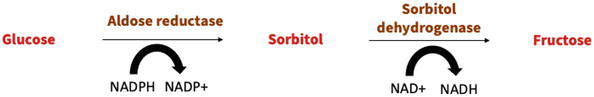
Figure 1.
Polyol pathway.
An overactive polyol pathway can have a negative impact on cellular balance by reducing the levels of cytosolic NADPH. This NADPH is crucial for keeping the main internal antioxidant, glutathione, in its active form. Furthermore, higher glucose levels can also deplete cellular NADPH through another process: the increased glucose concentrations hinder the activity of glucose 6-phosphate dehydrogenase. This enzyme is responsible for initiating the first step in the pentose phosphate pathway, which is the main supplier of NADPH within the cell [23]. Initial investigations on blocking aldose reductase in animals displayed potential to influence vascular impairment. However, these effects have not been proven in diabetic patients [3]. Widespread elevation of aldose reductase levels in mice led to heightened atherosclerosis [24]. Interestingly, contrary outcomes were noted in mice where the aldose reductase gene was deactivated or when they were administered an inhibitor for aldose reductase; both instances resulted in increased atherosclerosis [25]. Hence, additional investigation is required to ascertain the precise role of the aldose reductase pathway in the progression of atherosclerosis in the context of diabetes.
3.4 Oxidative stress
The generation of superoxide and other reactive oxygen species (ROS) within the blood vessel lining significantly contributes to the development of vascular disorders due to vascular endothelial damage, especially within the context of diabetes [3]. A specific oxidative enzyme that favors NADH contributes significantly to the increase in the amount of superoxidase in the vascular wall [26]. This enzyme is present in the endothelial cells and smooth muscle cells [26]. The levels and function of NADH oxidase in blood vessels are heightened in rat models of both type 1 [27] and type 2 [28] diabetes. This particular enzyme could be triggered by an elevation in the ratio of NADH to NAD+, which might result from increased activity of the polyol pathway in diabetes [29].
All these mechanisms of injury favor the development of atherosclerosis in diabetic individuals. Peripheral angiopathy stands as one of the cardinal triggers for diabetic foot ulcers (DFUs), as well as the foremost contributor to both amputation and mortality [30]. Atherosclerosis constitutes the principal pathological mechanism within peripheral vascular disease. The rupture of atherosclerotic plaques, particularly in the context of diabetes, can directly result in peripheral arterial thrombosis, subsequently leading to arterial blockage and lower limb ischemia. This sequence of events ultimately culminates in the emergence of DFUs [30]. Furthermore, the impact of atherosclerosis on the lower extremities differs between diabetic and non-diabetic patients [30]. In individuals with diabetes, the inferior genicular artery (posterior tibial artery and anterior tibial artery) is predominantly affected, while there is comparatively less involvement of the femoral and popliteal artery segments (superficial femoral artery and popliteal artery). Generally, the main iliac artery remains unaffected in diabetic patients. In cases where arterial perfusion to the foot is insufficient to uphold skin integrity, the development of tibial artery occlusion or proximal artery occlusion can lead to ischemic ulcers or gangrene [30].
4. Immune responses
The healing process of diabetic foot ulcers (DFUs) is a complex interplay of diverse cellular components and unique aspects, setting it apart from the healing of normal tissue.
4.1 Components of healing in DFU
The healing process of DFUs involves various components, including immune cells, keratinocytes, fibroblasts, endothelial cells, and a range of cytokines. During the inflammatory phase, the presence of infiltrating monocytes/macrophages within the wound plays a crucial role in transitioning the wound environment from being pro-inflammatory to an anti-inflammatory state [31]. During the advanced phase of typical wound inflammation, macrophages undergo a shift from a pro-inflammatory phenotype to an anti-inflammatory one. However, in individuals with DFUs, there is an impairment in the function and phenotypic transition of macrophages, resulting in the maintenance of a pro-inflammatory state by these cells [32].
4.2 Unique aspects of DFU healing
Clinical and experimental findings indicate that the healing process of DFUs differs from that of normal tissue [33]. Macrophages in DFU wounds have a diminished ability to effectively remove necrotic tissue, as their phagocytic function is significantly impaired. Alongside macrophage dysfunction, neutrophils contribute to an intensified inflammatory response, further impeding the healing of diabetic wounds. In diabetic wounds, the disruption of phagocytosis, neutrophil degranulation, and the anti-infective effects of reactive oxygen species (ROS) exacerbate the inflammatory condition [34]. Furthermore, elevated blood sugar levels lead to the increased expression of neutrophil protein arginine deiminase (PAD)-4. This heightened expression affects neutrophils’ capacity to release NETs (neutrophil extracellular traps) upon entering the wound, causing a delay in the healing process [35]. Macrophages and neutrophils release proteases in an inactive form known as zymogen, which becomes activated outside the cells and breaks down extracellular matrix (ECM) proteins like elastin and interstitial collagen. An instance is matrix metalloproteinases (MMP), which breaks down fibronectin into fragments and additionally activates other MMPs. These fibronectin fragments lead to the infiltration of leukocytes, tissue injury, and continuous inflammation. Hence, diverse cells will contribute to the healing of diabetic wounds with distinct functions in a therapeutic capacity [36].
4.3 Re-epithelialization and dermal repair in DFU healing
In the healing phase of diabetic foot ulcers (DFUs), the regeneration of skin tissue relies heavily on re-epithelialization and dermal repair [37]. Research indicates that during the process of wound re-epithelialization, keratinocytes move to the wound site and undergo proliferation and differentiation to reconstruct the epidermis’s structural and functional integrity [38]. This progression involves all of the skin layers. In the initial stages of wound healing, keratinocytes have the capability to directly combat invading pathogens by releasing cytokines, chemokines, antimicrobial peptides, and extracellular vesicles. These substances contribute to facilitating interactions between keratinocytes and circulating immune cells, promoting wound healing. However, the elevated glucose levels present in diabetic wound environments disrupt the normal functioning of keratinocytes, leading to delayed wound re-epithelialization. Dermal repair primarily depends on fibroblasts’ actions, encompassing their proliferation, differentiation, and secretion of extracellular matrix (ECM) components. In the upper papillary layer, specific fibroblasts can form hair papillae and regulate hair follicle growth and regeneration. Conversely, fibroblasts in the lower reticular layer mainly maintain the structural integrity of the dermis, creating a stable environment to support activities such as angiogenesis, nerve regeneration, and immune clearance [39]. Furthermore, fibroblasts have the ability to transform into myofibroblasts. These myofibroblasts play a role in contracting wounds, releasing enzymes and MMPs to break down the inflammatory matrix, and secreting collagen and other components of the extracellular matrix to aid in the creation of granulation tissue. Collagen III within the extracellular matrix is swapped out with collagen I, which boasts greater tensile strength. However, elevated blood sugar levels and the buildup of AGEs result in compromised fibroblast function. This translates to reduced cell growth, faster-programmed cell death, and hindered movement toward the wound site. Consequently, these factors collectively contribute to impaired skin restoration and the delayed healing observed in diabetic wounds [40].
4.4 Endothelial cells and neovascularization
The importance of wound healing is closely linked to the condition of endothelial cells and the process of neovascularization. Typically situated along the interior lining of blood vessels, endothelial cells are responsible for controlling the constriction and expansion of blood vessels by adjusting the concentrations of vasoactive substances like eNOS [41]. During the process of wound healing, endothelial cells undergoing various stages of angiogenesis are primarily under the control of VEGF. In the initial inflammatory phase, VEGF heightens the permeability of blood vessels, impacts the presence of selectin and intercellular adhesion molecules within endothelial cells, and fosters the attraction of white blood cells to the site of injury. As the proliferation stage ensues, VEGF significantly triggers both the replication and movement of endothelial cells. Subsequently, during the remodeling stage, VEGF prompts the arrangement of endothelial cells, facilitating the creation of the vascular lumen [42]. Research conducted within living organisms has demonstrated that arterial endothelial cells, when exposed to elevated glucose levels, experience a deterioration in their structural integrity. This renders them more susceptible to programmed cell death and detachment, allowing them to enter the bloodstream. Consequently, this situation disrupts the process of angiogenesis [43]. This impairment can be primarily attributed to the following five mechanisms: (a) the polyol pathway, (b) an elevation in intracellular AGEs, (c) an increase in the expression of RAGE, (d) the activation of multiple forms of protein kinase C, and (e) an excessive activation of the hexosamine pathway [44]. In diabetic conditions, reduced levels of nitric oxide synthase (NOS) due to peripheral neuropathy and peripheral arterial disease result in decreased peripheral blood flow due to blood vessel constriction. Furthermore, the absence of endothelial progenitor cells (EPCs) at the wound site hampers the creation of new blood vessels, consequently leading to a delay in the healing of wounds [45].
4.5 Stem cells and their role in DFU healing
Stem cells play a pivotal role in the recovery of diabetic foot ulcers (DFU) by overseeing the process of skin restoration after injuries and during regular maintenance. These stem cells possess distinctive attributes such as uneven replication, robust self-regeneration capabilities, and the ability to transform into various cell types [46]. Specifically, the operational condition of endothelial progenitor cells (EPCs) and epidermal stem cells (ESCs) significantly impacts the progression of wound healing. The abilities of EPCs, including migration, differentiation, adhesion, and the formation of tubes, become compromised under the hyperglycemic conditions associated with diabetes [47]. This impairment leads to persistent wound nonhealing over extended periods, especially in cases of chronic wounds like those seen in individuals with diabetes [48]. Acting as precursors to endothelial cells, endothelial progenitor cells (EPCs) move from the bone marrow into the peripheral bloodstream in response to factors like hypoxia-inducible factor-1, stromal cell-derived factor-1α, and VEGF. These cells are subsequently directed to ischemic sites where they contribute to the formation of fresh blood vessels, utilizing processes such as adhesion, proliferation, differentiation, and the creation of tubular structures, all of which aid in wound repair [49]. Furthermore, epidermal stem cells (ESCs) also hold a vital role in the wound-healing process. Laboratory tests conducted outside the body indicate that ESCs enhance the growth and movement of diabetic fibroblasts and macrophages (Mφ), while also promoting a shift toward an alternative or M2 Mφ polarization state [50]. In the case of wounds in db/db mice, the administration of ESCs accelerates wound healing by reducing inflammation, boosting cell growth at the wound site, fostering the development of new blood vessels, and encouraging the polarization of M2 macrophages [50].
5. Conclusion
Diabetic foot ulcers (DFUs) are a significant and debilitating complication of long-term diabetes, with neuropathy, impaired vasculature, and immune responses being the key contributors to their pathogenesis. Neuropathy, driven by hyperglycemia and advanced glycation end products (AGEs), disrupts nerve cell structure and function, leading to sensory, motor, and autonomic impairments. The combination of these factors reduces pain perception, increasing the risk of unnoticed injuries and subsequent ulceration. Impaired vasculature, marked by atherosclerosis and microvascular dysfunction, leads to reduced protective factors and increased susceptibility to vascular diseases, including DFUs. Key factors in this process include PKC activation, hyperactive metabolic pathways, and oxidative stress, all of which contribute to vascular damage.
Immune responses also play a significant role in the development and progression of DFUs. Chronic inflammation driven by dysfunctional immune cells, impaired macrophage function, neutrophil dysfunction, and compromised keratinocyte, fibroblast, and endothelial cell activities hinder the normal wound healing process. Additionally, stem cell dysfunction, especially in endothelial progenitor cells (EPCs) and epidermal stem cells (ESCs), further delays wound healing in diabetic individuals.
Understanding the multifaceted pathogenesis of DFUs is crucial for developing effective prevention and treatment strategies. Addressing neuropathy through glycemic control, AGE reduction, and targeted therapies could mitigate nerve damage and reduce the risk of injury. Targeting vascular impairment with interventions focused on PKC inhibition, metabolic pathway modulation, and oxidative stress reduction could improve blood vessel integrity. Furthermore, interventions that modulate immune responses and restore normal function in immune cells, keratinocytes, fibroblasts, endothelial cells, and stem cells could enhance the healing process.
In conclusion, diabetic foot ulcers result from the intricate interplay of neuropathy, impaired vasculature, and immune responses. A comprehensive approach that targets each of these components holds promise for the effective prevention and management of diabetic foot ulcers, thereby improving the quality of life for individuals with diabetes.
References
- 1.
Armstrong DG, Cohen K, Courric S, Bharara M, Marston W. Diabetic foot ulcers and vascular insufficiency: Our population has changed, but our methods have not. Journal of Diabetes Science and Technology. 2011; 5 (6):1591-1595 - 2.
Weigelt C, Rose B, Poschen U, Ziegler DAN, Friese G, Kempf K, et al. Immune mediators in patients with acute diabetic foot syndrome. Diabetes Care. 2009; 32 (8):1491-1496 - 3.
Rask-Madsen C, King GL. Vascular complications of diabetes: Mechanisms of injury and protective factors. Cell Metabolism. 2013; 17 (1):20-33 - 4.
Tecilazich F, Veves A. Chapter 7 - Role of peripheral neuropathy in the development of foot ulceration and impaired wound healing in diabetes mellitus. In: Nutritional and Therapeutic Interventions for Diabetes and Metabolic Syndrome. 2nd ed. Academic Press; 2018. pp. 95-104 - 5.
Peltier A, Goutman SA, Callaghan BC. Painful diabetic neuropathy [published correction]. BMJ. 2014; 348 :g1799 - 6.
Viader A, Sasaki Y, Kim S, et al. Aberrant Schwann cell lipidmetabolism linked to mitochondrial deficits leads to axondegeneration and neuropathy. Neuron. 2013; 77 (5):886-898 - 7.
Andersen H. Motor dysfunction in diabetes. Diabetes/Metabolism Research and Reviews. 2012; 28 (Suppl 1):89-92 - 8.
Kamenov ZA, Traykov LD. Diabetic autonomic neuropathy. Advances in Experimental Medicine and Biology. 2012; 771 :176-193 - 9.
Molines L, Darmon P, Raccah D. Charcot’s foot: Newest findings on its pathophysiology, diagnosis and treatment. Diabetes & Metabolism. 2010; 36 (4):251-255 - 10.
Vinik AI, Maser RE, Mitchell BD, Freeman R. Diabetic autonomic neuropathy. Diabetes Care. 2003; 26 (5):1553-1579 - 11.
Volmer-Thole M, Lobmann R. Neuropathy and diabetic foot syndrome. International Journal of Molecular Sciences. 2016; 17 (6):917 - 12.
Ma J, Pan P, Anyika M, Blagg BSJ, Dobrowsky RT. Modulating molecular chaperones improves mitochondrial bioenergetics and decreases the inflammatory transcriptome in diabetic sensory neurons. ACS Chemical Neuroscience. 2015; 6 (9):1637-1648 - 13.
Lupachyk S, Shevalye H, Maksimchyk Y, Drel VR, Obrosova IG. PARP inhibition alleviates diabetes-induced systemic oxidative stress and neural tissue 4-hydroxynonenal adduct accumulation: Correlation with peripheral nerve function. Free Radical Biology & Medicine. 2011; 50 (10):1400-1409 - 14.
Kobayashi M, Chandrasekhar A, Cheng C, et al. Diabetic polyneuropathy, sensory neurons, nuclear structure and spliceosome alterations: A role for CWC22. Disease Models & Mechanisms. 2017; 10 (3):215-224 - 15.
Vincent AM, Hayes JM, McLean LL, Vivekanandan-Giri A, Pennathur S, Feldman EL. Dyslipidemia-induced neuropathy in mice: The role of oxLDL/LOX-1. Diabetes. 2009; 58 (10):2376-2385 - 16.
Keller JN, Hanni KB, Markesbery WR. Oxidized low-density lipoprotein induces neuronal death: Implications for calcium, reactive oxygen species, and caspases. Journal of Neurochemistry. 1999; 72 (6):2601-2609 - 17.
Sandireddy R, Yerra VG, Areti A, Komirishetty P, Kumar A. Neuroinflammation and oxidative stress in diabetic neuropathy: Futuristic strategies based on these targets. International Journal of Endocrinology. 2014; 2014 :674987 - 18.
Singh VP, Bali A, Singh N, Jaggi AS. Advanced glycation end products and diabetic complications. Korean Journal of Physiology & Pharmacology. 2014; 18 (1):1-14 - 19.
Court FA, Hendriks WT, MacGillavry HD, Alvarez J, van Minnen J. Schwann cell toaxon transfer of ribosomes: Toward a novel understanding of the role of glia in the nervous system. The Journal of Neuroscience. 2008; 28 (43):11024-11029 - 20.
Suzuma K, Takahara N, Suzuma I, Isshiki K, Ueki K, Leitges M, et al. Characterization of protein kinase C β isoform's action on retinoblastoma protein phosphorylation, vascular endothelial growth factor-induced endothelial cell proliferation, and retinal neovascularization. Proceedings of the National Academy of Sciences. 2002; 99 (2):721-726 - 21.
Ohshiro Y, Ma RC, Yasuda Y, Hiraoka-Yamamoto J, Clermont AC, Isshiki K, et al. Reduction of diabetes-induced oxidative stress, fibrotic cytokine expression, and renal dysfunction in protein kinase Cβ–null mice. Diabetes. 2006; 55 (11):3112-3120 - 22.
Brownlee M. Biochemistry and molecular cell biology of diabetic complications. Nature. 2001; 414 (6865):813-820 - 23.
Xu Y, Zhang Z, Hu J, Stillman IE, Leopold JA, Handy DE, et al. Glucose-6-phosphate dehydrogenase-deficient mice have increased renal oxidative stress and increased albuminuria. The FASEB Journal. 2010; 24 (2):609 - 24.
Vikramadithyan RK, Hu Y, Noh HL, Liang CP, Hallam K, Tall AR, et al. Human aldose reductase expression accelerates diabetic atherosclerosis in transgenic mice. The Journal of Clinical Investigation. 2005; 115 (9):2434-2443 - 25.
Srivastava S, Vladykovskaya E, Barski OA, Spite M, Kaiserova K, Petrash JM, et al. Aldose reductase protects against early atherosclerotic lesion formation in apolipoprotein E-null mice. Circulation Research. 2009; 105 (8):793-802 - 26.
Lassègue B, San Martín A, Griendling KK. Biochemistry, physiology, and pathophysiology of NADPH oxidases in the cardiovascular system. Circulation Research. 2012; 110 (10):1364-1390 - 27.
Hink U, Li H, Mollnau H, Oelze M, Matheis E, Hartmann M, et al. Mechanisms underlying endothelial dysfunction in diabetes mellitus. Circulation Research. 2001; 88 (2):e14-e22 - 28.
Kim YK, Lee MS, Son SM, Kim IJ, Lee WS, Rhim BY, et al. Vascular NADH oxidase is involved in impaired endothelium-dependent vasodilation in OLETF rats, a model of type 2 diabetes. Diabetes. 2002; 51 (2):522-527 - 29.
Garcia Soriano F, Virag L, Jagtap P, Szabo E, Mabley JG, Liaudet L, et al. Diabetic endothelial dysfunction: The role of poly (ADP-ribose) polymerase activation. Nature Medicine. 2001; 7 (1):108-113 - 30.
Deng H, Li B, Shen Q , Zhang C, Kuang L, Chen R, et al. Mechanisms of diabetic foot ulceration: A review. Journal of Diabetes. 2023; 15 (4):299-312 - 31.
Wynn TA, Vannella KM. Macrophages in tissue repair, regeneration, and fibrosis. Immunity. 2016; 44 (3):450-462 - 32.
Baltzis D, Eleftheriadou I, Veves A. Pathogenesis and treatment of impaired wound healing in diabetes mellitus: Newinsights. Advances in Therapy. 2014; 31 (8):817-836 - 33.
Dinh T, Tecilazich F, Kafanas A, Doupis J, Gnardellis C, Leal E, et al. Mechanisms involved in the development and healing of diabetic foot ulceration. Diabetes. 2012; 61 (11):2937-2947. DOI: 10.2337/db12-0227 - 34.
Kolaczkowska E, Kubes P. Neutrophil recruitment and function in health and inflammation. Nature Reviews. Immunology. 2013; 13 (3):159-175 - 35.
Wang Y, Xiao Y, Zhong L, Ye D, Zhang J, Tu Y, et al. Increased neutrophil elastase and proteinase 3 and augmented NETosis are closely associated with β-cell autoimmunity in patients with type 1 diabetes. Diabetes. 2014; 63 (12):4239-4248 - 36.
Widgerow AD. Chronic wound fluid–thinking outside the box. Wound Repair and Regeneration. 2011; 19 (3):287-291 - 37.
Singer AJ, Clark RA. Cutaneous wound healing. New England Journal of Medicine. 1999; 341 (10):738-746 - 38.
Raja SK, Garcia MS, Isseroff RR. Wound re-epithelialization: Modulating keratinocyte migration in wound healing. Frontiers in Bioscience. 2007; 12 :2849-2868 - 39.
Dekoninck S, Blanpain C. Stem cell dynamics, migration and plasticity during wound healing. Nature Cell Biology. 2019; 21 (1):18-24 - 40.
Desta T, Li J, Chino T, Graves DT. Altered fibroblast proliferation and apoptosis in diabetic gingival wounds. Journal of Dental Research. 2010; 89 (6):609-614 - 41.
Ignarro LJ, Buga GM, Wood KS, Byrns RE, Chaudhuri G. Endothelium-derived relaxing factor produced and released from artery and vein is nitric oxide. Proceedings of the National Academy of Sciences of the United States of America. 1987; 84 (24):9265-9269 - 42.
Huang X, Liang P, Jiang B, et al. Hyperbaric oxygen potentiates diabetic wound healing by promoting fibroblast cell proliferation and endothelial cell angiogenesis. Life Sciences. 2020; 259 :118246 - 43.
Yu JQ , Liu XF, Chin LK, Liu AQ , Luo KQ. Study of endothelial cell apoptosis using fluorescence resonance energy transfer (FRET) biosensor cell line with hemodynamic microfluidic chip system. Lab on a Chip. 2013; 13 (14):2693-2700 - 44.
Giacco F, Brownlee M. Oxidative stress and diabetic complications. Circulation Research. 2010; 107 (9):1058-1070 - 45.
Thum T, Fraccarollo D, Schultheiss M, et al. Endothelial nitric oxide synthase uncoupling impairs endothelial progenitor cell mobilization and function in diabetes. Diabetes. 2007; 56 (3):666-674 - 46.
Ding DC, Shyu WC, Lin SZ. Mesenchymal stem cells. Cell Transplantation. 2011; 20 (1):5-14 - 47.
Wang K, Dai X, He J, et al. Endothelial overexpression of metallothionein prevents diabetes-induced impairment in ischemia angiogenesis through preservation of HIF-1α/SDF1/VEGF signaling in endothelial progenitor cells. Diabetes. 2020; 69 :1779-1792 - 48.
Kaushik K, Das A. Endothelial progenitor cell therapy for chronic wound tissue regeneration. Cytotherapy. 2019; 21 :1137-1150 - 49.
Wan G, Chen Y, Chen J, et al. Regulation of endothelial progenitor cell functions during hyperglycemia: New therapeutic targets in diabetic wound healing. Journal of Molecular Medicine (Berlin). 2022; 100 (4):485-498 - 50.
Wang P, Theocharidis G, Vlachos IS, et al. Exosomes derived from epidermal stem cells improve diabetic wound healing. Journal of Investigative Dermatology. 2022; 142 (9):2508-2517