Open Access is an initiative that aims to make scientific research freely available to all. To date our community has made over 100 million downloads. It’s based on principles of collaboration, unobstructed discovery, and, most importantly, scientific progression. As PhD students, we found it difficult to access the research we needed, so we decided to create a new Open Access publisher that levels the playing field for scientists across the world. How? By making research easy to access, and puts the academic needs of the researchers before the business interests of publishers.
We are a community of more than 103,000 authors and editors from 3,291 institutions spanning 160 countries, including Nobel Prize winners and some of the world’s most-cited researchers. Publishing on IntechOpen allows authors to earn citations and find new collaborators, meaning more people see your work not only from your own field of study, but from other related fields too.
Background: Determining injuries in relation to death have been highly imperative to forensic anthropologist since they provide clues on the norms, cultures and pattern of death of an individual. Aim: The study aimed at determining injuries among skeletal elements in the Anatomy Musuem of Delta State University, Abraka, Nigeria. Methodology: The study was crosssectional and a total of 150 bones which included the skulls, calvarium and pelvis were examined for the presence of ante, peri and postmortem injuries. Other injuries such as blunt force, sharp force and ballistic injuries were also studied. Chisquare test was used to evaluate an association between the bones and the time of injuries. Statistical assessment was done using SPSS 21 Software Version. Significance was accepted at p < 0.05. Result: The study showed that postmortem injuries was the most predominant (98%) among the bones. This was followed by perimortem and antemortem injuries (11.3, 10%). The percentages of blunt force, ballistic and sharp force were 98, 1.3 and 0.7% respectively. Further findings showed that there was no association between the bones and the nature of injuries that were observed (p = 0.837; 0.713). Conclusion: The study had shown that there are so many hidden facts from skeletal remains which can be of vital importance to forensic science.
Faculty of Basic Medical Sciences, Department of Human Anatomy, Delta State University, Abraka, Nigeria
Efe Jennifer Jaiyeoba-Ojigho*
Faculty of Basic Medical Sciences, Department of Human Anatomy, Delta State University, Abraka, Nigeria
*Address all correspondence to: efemenadelsu@gmail.com
1. Introduction
Investigating bones for injuries have become a vital tool in forensic studies [1]. They can establish an individual’s identity when compared with past medical records or ascertain the circumstances of death [1]. These injuries can be classified into ante-, peri- and postmortem depending on the time of occurrence [2, 3, 4]. Injuries are termed antemortem when they occur before death and are evidenced with partial or complete healing, visible at the fracture edges of bones as compared to peri-mortem injuries that show no signs of healing [1, 3, 5, 6]. Peri-mortem injuries occur at the time of death and they are distinguished in bones by the presence of fractures that interpret the cause death [1]. According to Elena [1], they are expressed by plastic deformation seen at the site of injury. Peri-mortem injuries also present a soft preponderant texture and regular outline as compared to postmortem injuries that are characterized with a rough preponderant texture and outline [1, 7, 8, 9]. It has been documented that postmortem bones have fracture patterns that are squared with sharp edges that are likely to cause enormous disintegration on dry bones [1, 7, 8, 9]. Post damages on bones classically reveal right-angled fractured margins, while those of peri-damages show obtuse or acute fracture angles [1, 7, 8, 9]. This have also been confirmed from computed tomographic scans [10]. It has also been acknowledged that injuries can also be blunt force, sharp force, ballistic and thermal [4]. This depends on the instruments that cause death at different time frame [1]. The blunt force injuries have been described as the most frequent type of injuries and they are associated with blunt objects or surfaces [1]. It must be frazzled that forensic assessment of dead bodies for injuries have now become a regular task for forensic pathologists, medical examiners and forensic anthropologists in various countries. Currently, examination of injuries includes the evaluation of ante-, peri-, or postmortem injuries, identification of injury patterns as well as possible association with certain objects. They have been limiting information on assessing injuries from remains of corpses in Nigeria, hence this study investigated them on dry bones in the Anatomy Museum of Delta State University, Abraka, Delta State Nigeria.
The study was crosssectional and a total of 150 bones which included the skulls, calvarium and pelvis were investigated for the presence of ante-, peri- and postmortem injuries. They were also examined for other injuries such as the blunt force, sharp force and ballistic injuries. Data were represented in frequencies and percentages while a Chisquare test showed an association between bones and the time of injuries. Ethical approval for this work was obtained from the Department of Human Anatomy, Faculty of Basic Medical Sciences, Delta State University, Abraka, Delta State.
Table 1 showed that 78.7, 11 and 10% of the bones had post-, peri- and antemortem injuries. Findings also showed that blunt force (98%), ballistic injuries (13%) and sharp force damages were observed among the investigated bones (Table 2). Table 3 presented 45.3% calvariums, 10% pelvis, 44.7% skulls while Table 4 illustrated the injuries that were present among these bones. It can be depicted fromTable 4 that 8.8, 20, 9%; 10.3, 13.3, 11.9%; 80, 66.7, 79.1% of the calvarium, pelvis and skull bones had ante-, peri- and postmortem injuries. Other injuries that were observed were blunt force, ballistic and sharp force (Table 5). These injuries were specific to the bones that were studied. According to Table 5, ballistic injuries was explicit on the skull and calvaria bones (98.5, 1.5%) while sharp force was specific to the calvarium (1.5%). Blunt force injury was noted on the calvarium (97%), pelvis and skull (100%) each respectively. Further findings showed that there was no association between injuries and the skeletal elements invested (p = 0.837; 0.713) (Tables 6 and 7).
Time of injury
Frequency (%)
Antemortem
15 (10.0)
Perimortem
17 (11.3)
Postmortem
118 (78.7)
Total
150 (100.0)
Table 1.
Distribution of time of injury.
Nature of injury
Frequency (%)
Sharp force
1 (0.7)
Blunt force
147 (98.0)
Ballistic force
2 (1.3)
Total
150 (100.0)
Table 2.
Distribution of nature of injury.
Skeletal elements
Frequency (%)
Calvarium
68 (45.3)
Pelvis
15 (10.0)
Skull
67 (44.7)
Total
150 (100.0)
Table 3.
Distribution of examined skeletal elements.
Skeletal elements
Frequency
(%)
Calvarium
Antermorterm
6
8.8
Perimorterm
7
10.3
Postmorterm
55
80.9
Total
68
100.0
Pelvis
Antermorterm
3
20.0
Perimorterm
2
13.3
Postmorterm
10
66.7
Total
15
100.0
Skull
Antermorterm
6
9.0
Perimorterm
8
11.9
Postmorterm
53
79.1
Total
67
100.0
Total
200
(100.0)
Table 4.
Distribution of examined skeletal elements based on time of injury.
Skeletal elements
Frequency
(%)
Calvarium
Ballistic force
1
1.5
Blunt force
66
97.1
Sharp force
1
1.5
Total
68
100.0
Pelvis
Blunt force
15
100.0
Total
1
1.5
Skull
Ballistic force
66
98.5
Blunt force
67
100.0
Total
1
1.5
Total
200
(100.0)
Table 5.
Distribution of examined skeletal elements based on nature of injuries.
Skeletal elements
Nature of injury
Chi-square
Df
P-value
Ballistic force
Blunt force
Sharp force
Calvarium
1 (0.7)
66 (44.0)
1 (0.7)
1.442
4
0.837
Pelvis
—
15 (10.0)
—
Skull
1 (0.7)
66 (42.0)
—
Total
2 (1.3)
147 (98.0)
1 (0.7)
Table 6.
Chi-square test of association between time of injury and nature of injury.
Skeletal elements
Time of injury
Chi-square
Df
P-value
Antemortem
Perimortem
Postmortem
Calvarium
6 (4.0)
7 (4.7)
55 (36.7)
Pelvis
3 (2.0)
2 (1.3)
10 (6.7)
2.125
4
0.713
Skull
6 (4.0)
8 (5.3)
53 (35.3)
Total
15 (10.0)
17 (11.3)
118 (78.7)
Table 7.
Chi-square test of association between injury time and examined skeletal elements.
The study had shown that all bones that were studied had more postmortem damages. Features that were observed for this injury were desiccation and fragility. This could be as a result of exposure of these bones to the environment. Several authors are of the opinion that changes that occur to corpses after death are due to complex results from physicochemical and environmental processes [1, 4]. They are affected by factors surrounded by the cadaver and the environment that they are kept [1, 4]. These factors influence the onset, increases the pace of post-mortem changes or impede it. The bones we observed having blunt force had an impact region, cranial and facial fractures. These evidences were highlighted by Casali et al. in her study on blunt force trauma carried out in Milan Italy [11]. According to these scholars, of the 307 victims studied, 40 and 30% had cranial and facial fractures [11]. Blunt force injury which was predominant from our study could be as a result of suicides, accidents, falls, described as factors associated with it [1]. This injury has been defined as one caused by a low-velocity contact resulting from a blunt surface or item [1]. According to Galloway [4], they occur as a consequence of accidents, sticks, and falls, with a diversity of broken patterns reliant on both internal and extrinsic circumstances. Features of antemortem injuries observed from this study were the presence of a poros, rounded and broken edges. This was consistent with the findings of Byers, who stated that the first signal of antemortem damage was the porosity around the fracture areas, indicative of bone activity, reabsorption, and fracture healing [12]. According to some scholars, the minor rounding or remodeling of the shattered edges, was the second trait of an antemortem harm, signifying that the damage occurred at least 7 days prior to death [2, 12, 13, 14]. Ballistic and sharp force injuries were also identified among the bones. The existence of an entry wound was seen in most of the ballistic bones from our study. This is the most prominent feature of a ballistic injury [15, 16]. The ballistic bones from this investigation also had a circumferential and radially branching fracture. A number of authors have associated ballistic injuries to the occurrence of projectiles, fractures connected with high impacts, wrecked materials found in either bone or the environment. In most instances, the presence of an entrance wound that is lesser than an exit wound also characterizes a ballistic injury [15, 16]. Sharp force injuries from our study were evidence with sharp lines around and within the bones suggestive of incised wounds. Our findings were not different from those of Thompson and Inglis who were of the opinion that this injury was marked by incisions that are gotten from tools pointed or those with marks in form of slashes [17].
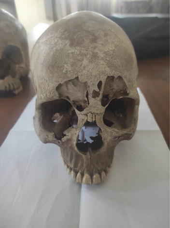
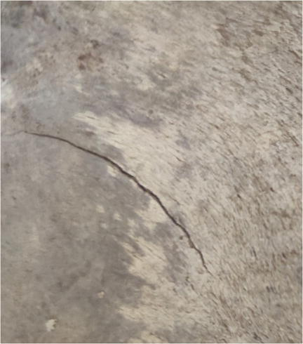
It has been established that several injuries discovered on bones can be of immense value to forensic anthropologist in investigating crimes, nature of death of an individual as well as tell a people way of life because of the instruments associated with the death of an individual (Figures 1 and 2).
Figure 1.
A skull illustrating blunt force injury.
Figure 2.
A calvarium illustrating sharp force injury.
References
1.Elena FK. Forensic investigation of cranial injuries due to blunt force trauma: Current best practice. Journal of Research and Reports in Forensic Medical Science. 2015;5:25-37
2.Sauer N. The timing of injuries and mannerof death: Distinguishing among antemortem, perimortem, and post-mortem trauma. In: Reichs KJ, editor. Forensic Osteology: Advances in the Identification of Human Remains. 2nd ed. Springfield, IL: Charles C. Thomas; 1998. pp. 321-332
3.Rodriguez-Martin C. Identification and differential diagnosis of traumatic lesions of the skeleton. In: Schmitt A, Cunha E, Pinheiro J, editors. Forensic Anthropology and Medicine: Complementary Sciences from Recovery to Cause of Death. New Jersey: Humana Press; 2006. pp. 197-221
4.Galloway A. The biomechanics of fracture production. In: Galloway A, editor. Broken Bones: An Anthropological Analysis of Blunt Force Trauma. Springfield, IL: Charles C. Thomas; 1999. pp. 35-62
5.Iscan MY, Steyn M, editors. The Human Skeleton in Forensic Medicine. 3rd ed. Springfield, IL: Charles C. Thomas; 2013
6.Lovell NC. Trauma analysis in paleopathology. Yearbook Physical Anthropology. 1997;40:139-170
7.Dirkmaat DC, Cabo LL, Ousley SD, Symes SA. New perspectives in forensic anthropology. Yearbook Physical Anthropology. 2008;51:33-52
8.Morlan RE. Toward the definition of criteria for the recognition of artificial bone alterations. Quaternary Research. 1984;22:160-171
9.Fleming-Farrell D, Michailidis K, Karantanas A, Roberts N, Kranioti EF. Virtual assessment of perimortem and postmortem blunt force cranial trauma. Forensic Science International. 2013;229(1-3):162.e1-162.e6
10.Bonnichsen R. Pleistocene Bone Technology in the Beringian Refugium. National Museum of Man Mercury Series, Archaeological Survey of Canada Paper No 89. Ottawa, Canada; 1979
11.Casali MB, Battistini A, Blandino A, Cattaneo C. The injury pattern in fatal suicidal falls from a height: An examination of 307 cases. Forensic Science International. 2014;244:57
12.Byers SN. Introduction to Forensic Anthropology: A Textbook. 2nd ed. Pearson Education: Boston, MA; 2005
13.Aufderheide AC, Rodriguez-Martin C. The Cambridge Encyclopedia of Human Paleopathology. Cambridge: Cambridge University Press; 1998
14.Konstantinos M, Chara S. Identification and differentiation diagnosis of perimortem blunt force trauma in tubular long bones. Forensic Science Medical Pathology. 2006;2(4):221-230
15.Smith OC, Pope EJ, Symes SA. Look until you see: Identification of trauma in skeletal material. In: Steadman DW, editor. Hard Evidence: Case Studies in Forensic Anthropology. Upper Saddle River, NJ: Prentice Hall; 2003. pp. 138-154
16.Berryman HE, Symes SA. Recognizing gunshot and blunt cranial trauma through fracture interpretation. In: Reichs KJ, editor. Forensic Osteology: Advances in the Identification of Human Remains. Springfield, IL: Charles C. Thomas; 1998. pp. 333-352
17.Thompson TJU, Inglis J. Differentiation of serrated and non-serrated blades from stab marks in bone. International Journal of Legal Medicine. 2009;123(2):129-135
Written By
Lilan Ebele Chris-Ozoko and Efe Jennifer Jaiyeoba-Ojigho
Submitted: 29 June 2023Reviewed: 30 June 2023Published: 30 October 2023