Open Access is an initiative that aims to make scientific research freely available to all. To date our community has made over 100 million downloads. It’s based on principles of collaboration, unobstructed discovery, and, most importantly, scientific progression. As PhD students, we found it difficult to access the research we needed, so we decided to create a new Open Access publisher that levels the playing field for scientists across the world. How? By making research easy to access, and puts the academic needs of the researchers before the business interests of publishers.
We are a community of more than 103,000 authors and editors from 3,291 institutions spanning 160 countries, including Nobel Prize winners and some of the world’s most-cited researchers. Publishing on IntechOpen allows authors to earn citations and find new collaborators, meaning more people see your work not only from your own field of study, but from other related fields too.
Cadaver decomposition is a natural phenomenon intimately affected by numerous organisms such as insects, bacteria etc., where they use the decaying body as their nutrition source. These organisms can be utilized in forensic science to estimate the Post-mortem Interval. Forensic entomology is one of the popular approaches where successive colonization of insects on cadaver is studied to estimate PMI. However, sometime this method does not provide consistent results due to lack of insect activities during cold environment conditions or when crime scene is indoor. Recently, researchers have noted that microbiomes have shown predictable and clockwise successional patterns on decomposing cadavers and suggested this could be utilized to estimate PMI when this approach is etched with other established methods. This chapter summarizes the utility of microbial profiling in medico-legal investigations.
Department of Forensic Science, Lovely Professional University, Punjab, India
Jyoti Dalal*
Department of Forensic Science, Lovely Professional University, Punjab, India
*Address all correspondence to: jyotidalal417@gmail.com
1. Introduction
In forensic investigations, PMI estimation is crucial, especially when the exact time of death is uncertain or in doubt. It acts as a crucial criterion to estimate the sequence of events leading to the person’s death, supporting the reconstruction of the incident’s conditions, and making it easier to identify prospective suspects [1]. As it helps law enforcement authorities gather evidence, establish alibis, and link suspects to the crime scene, a precise measurement of PMI is crucial for criminal investigations [2]. Additionally, by ensuring that justice is done, and the rights of the deceased are maintained, estimating PMI contributes to preserving the overall accuracy and integrity of the legal system. Rigor mortis, the stiffening of muscles after death, has been used as an indicator of PMI. It usually starts shortly after death, peaks between 12 and 24 hours later, and then slowly fades away [3]. However, the onset and duration of rigor mortis might vary depending on conditions including temperature and physical activity [4]. Another common technique for estimating PMI is livor mortis, or the settling of blood in the dependent areas of the body. Forensic examiners can calculate the PMI and establish the location of the body after death by examining the color and dispersion of the livor mortis [5]. Algor mortis, the cooling of the body, has also been utilized for PMI estimation [5]. By measuring the temperature of the body and the ambient environment, forensic investigators can estimate the time since death [5]. Few authors have applied total body scoring method based on the appearance of three separate regions, the head, trunk, and limbs for estimating PMI. Megyesi et al. [6] developed this method for estimating PMI using accumulated day degree (ADD) degree days in human remains. Later, this method was later modified by Heaton et al. [7] to estimate Post-mortem submersion interval (PMSI) in human remains. Although the rate of decomposition is highly variable and affected by factors such as temperature, humidity, and insect activity, it might be difficult to estimate the accurate PMI using only the stages of decomposition. The assessment of PMI has also been done using chemical changes in the body, such as potassium levels in the vitreous humor and hypostasis patterns [8]. The necessity for more sophisticated and precise procedures in PMI assessment is highlighted by the fact that each of these old methods has its own drawbacks, including subjectivity, variability, and the influence of outside influences [1, 9]. Since conventional methods for PMI assessment in forensic investigations have substantial limitations, alternative strategies are being investigated. Microbial forensics, which uses the power of microbial communities present on and within the deceased individual, is a new area with significant potential for PMI estimate. Microbial forensics investigates the composition, diversity, and microbial activity related to the decomposition process in order to calculate the PMI [10, 11, 12]. This strategy acknowledges that microbial communities exhibit diverse patterns of succession through time and are crucial to the breakdown process [2]. Researchers may be able to create more precise and dependable approaches for PMI estimation by studying these microbial alterations. Numerous research has shown how useful microbiological forensics may be for PMI estimate. As an example, researchers have explored microbial composition of diverse body regions, including the skin, oral cavity, and gastrointestinal system, and have discovered distinct microbial succession patterns linked to various stages of decomposition. Researchers have been able to identify certain microbial signatures that correlate with the PMI by using high-throughput sequencing methods and bioinformatic studies [2, 13, 14, 15]. These discoveries open the door to the establishment of databases and forensic technologies based on microorganisms that can help investigators to determine the time since death more precisely.
Although decomposition in aquatic environments has received less study, it is not a rare occurrence in investigations, with a primary focus on detecting the presence of diatoms, algae, and scavenging activity of aquatic species [16, 17, 18]. Only a few authors have discussed the utility of microbial communities in estimating PMI [16, 19, 20]. In a study conducted by Cartozzo et al. [20], they estimated PMI in freshwater by studying the successional pattern and associated changes in the bacterial community within submerged skeletal remains of pigs. The microbial communities were identified using 16S rRNA sequencing, revealing the presence of phyla such as Firmicutes, Proteobacteria, Acidobacteria, Bacteroidetes, and Spirochaetes. Collectively, these studies suggest that PMI can be predicted by observing the repeatability of microbial community succession during decomposition.
Decomposition is a continuous process which begins with the changes at the cellular level. The degradation process continues even when the bones reach to dry remains stage until the whole nutrients get into to the natural ecosystem and thus energy is recycled.
Goff [5] described five stages of decomposition in his study which are discussed below.
Fresh Stage: The fresh phase starts as soon as the death occurs and remains until bloating occurs. Apart from the deposition of eggs by flies in the natural cavities such as nose, eyes, ears and the areas of tissue dehiscence, this phase does not exhibit any insect activity. Blowflies and flesh flies are the earliest flies to reach the cadaver.
Bloated Stage: Putrefaction occurs during this phase of decomposition. Putrefaction is a process of tissue decay by the action of microbes that are generally associated with the gut of the deceased [21, 22]. During this process, gases such as ammonia, hydrogen sulphide, and methane are released inside the body, leading to a balloon-like appearance of the corpse [23, 24]. The temperature also rises due to the combined effect of putrefaction and the metabolic activities of fly maggots. Flies and their larvae are most abundant during this phase. Seepage of fluid from natural orifices, when it mixes with the ammonia gas, deteriorates the soil fauna and increases the pH of the soil.
Decay Stage: The decay stage begins with the rupture of the skin, enabling gases to escape and the collapse of the abdominal cavity. Extensive maggot infestation can be observed during this phase. As part of faunal succession, flies are eventually replaced by beetles [24, 25]. Necrophagous and predatory taxa are seen in significant numbers in the later stages of the decay stage, whereas certain predatory taxa, including Staphylinidae species, were present during the bloated stage. The majority of Calliphoridae and Sarcophagidae will have completed pupariation by the end of this stage and left the remains. By the time the decay stage is complete, dipteran larvae will have mostly removed the flesh from the remains.
Post-decay Stage: The corpse in this stage reduces to skin, cartilage, and bone. Flies are no longer dominant. The number of species and individuals of Coleoptera increases. These species feed on the dried flesh and cartilage from the bones, giving them a clean appearance.
Skeletal Stage: During this stage, only bones and hair remain. Most of the arthropods are absent, or if present, they are in very few numbers [25]. The area beneath the remains gradually returns to its usual soil fauna.
Microbial forensics makes use of the fact that the human body contains a diverse array of microbial communities known as the human microbiome. These microbial communities play significant roles in many physiological processes and have the ability to persist and evolve reliably after death [26]. They also help with the release of gases like methane and hydrogen sulphide as well as volatile compounds that give decomposition its distinctive odor [3]. These microbial communities also help break down organic matter into simpler forms that may be utilized by other organisms, which contributes to the recycling of nutrients [8]. Diversity and composition of decomposition-related microbial communities vary depending on a number of factors, including the location of the body, the environment, and the individual. Microbial communities that exhibit different patterns of microbial succession can be observed on the skin [27], oral cavity [28, 29], gastrointestinal tract [30, 31, 32], and other regions of the body that are vulnerable to decomposition. Thanks to advancements in molecular biology technologies like high-throughput DNA sequencing, the study of microbial communities in human decomposition has undergone a radical transformation. By studying the DNA or RNA retrieved from postmortem samples, researchers can identify and define the microbial species present and learn more about the specific microorganisms involved in the decomposition process.
4. Succession of microbial communities and building microbial clock
Studying microbial communities in human decomposition for forensic investigations has a number of benefits. It can assist with more accurate PMI estimation, for example, by analyzing the pattern of microbial succession and the presence of specific microorganisms connected to distinct breakdown phases. Studies have shown that microbial communities undergo predictable changes over time with the progress in decomposition process, allowing for the construction of a timeline for PMI estimation [12, 13, 14, 15, 19, 33]. Although much progress has been made in the recognition of microbial community contribution during decomposition, there are still many unknowns. Sample handling practices, DNA extraction techniques, and data processing practices must all be standardized for research to be comparable and reproducible. Additionally, it is crucial to consider and control the effects of outside elements including the environment and sample processing.
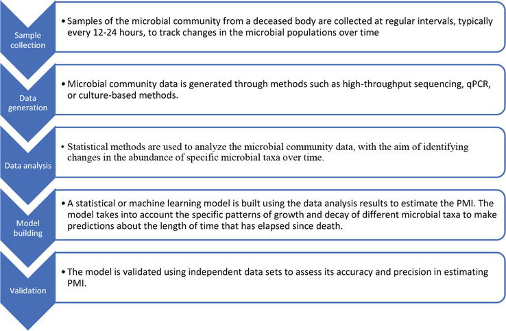
Microbial forensics considers functional changes in microbial communities after decomposition in addition to taxonomic variations. Researchers have conducted studies to characterize the microbial succession and establish a microbial clock for PMI estimation [2, 33]. By sampling the microbial communities present on a decomposing body and analyzing their composition and abundance, forensic scientists can compare the microbial profile to reference data collected from known PMI cases. This comparison allows them to estimate the time since death based on the similarities and differences in microbial succession. In 2014, Pechal et al. [26] developed a framework shown in Figure 1 to estimate post mortem interval from bacterial community. In real-world forensic investigations, this paradigm offered a conceptual roadmap for combining microbiological succession data into the computation of PMI. Their research identified seven families (Fusobacteriaceae, Clostridiales Incertae Sedis XI, Aerococcaceae, Micrococcaceae, Campylobacteraceae, Comamonadaceae, Moraxellaceae, Bacteroidetes) and four phyla (Proteobacteria, Actinobacteria, Firmicutes) as potentially significant predictors of estimating physiological. Additionally, the use of statistical models and machine learning techniques has improved the precision and dependability of PMI calculation utilizing microbial forensics. Predictive models can be created to estimate PMI with more precision by incorporating several parameters, such as taxonomic abundance, functional profiles, environmental conditions, and weather data [14, 27, 28, 34, 35, 36]. As additional information about the dynamics of microbial communities during decomposition is produced and included in the study, these models keep becoming better. Although microbiological forensics has a lot of potential as a method for PMI estimate, there are a number of obstacles and restrictions that need to be overcome.
Figure 1.
Framework to estimate post mortem interval from bacterial community [26].
5. Investigating the temporal changes in microbial communities during decomposition
Microbial postmortem interval (PMI) assessment must take into account the temporal changes in microbial populations during decomposition. Researchers can improve PMI estimation techniques and obtain important insights into the breakdown process by examining the dynamic changes in microbial populations over time. This field has used a variety of technologies, including culture-dependent and culture-independent techniques, as well as advanced molecular biology methods including DNA sequencing, metagenomics, and metabolomics.
A basic strategy for comprehending the temporal dynamics of microbial communities during decomposition is time-series sampling. In order to characterize microbial populations at various phases of decomposition, samples must be taken at various points after death [37]. Researchers can pinpoint temporal patterns and changes connected to various PMI phases by tracking the composition, variety, and abundance of microbes over time [10, 38, 39]. Shotgun metagenomics and 16S rRNA gene sequencing are two examples of high-throughput sequencing methods that have revolutionized the study of microbial communities during decomposition. These techniques allow for the identification, measurement, and evaluation of the functional potential of various microbial species [40, 41]. Researchers can determine how the relative abundance of different taxa has changed over time by analyzing the sequencing data derived from time-series samples [10, 38, 39]. To establish links between microbial dynamics and the breakdown process, these results can be connected with PMI. Another effective approach for examining the temporal variations in microbial activity during decomposition is metabolomics, the study of small-molecule metabolites. Understanding microbial communities’ metabolic processes can be accomplished by examining the volatile organic compounds (VOCs) and other metabolites they create [24]. Researchers can better understand the metabolic activities of various microbial species during decomposition and their relationship to PMI by analyzing the temporal patterns of these metabolites. Another method used to comprehend the metabolic capability of microbial communities during decomposition is functional profiling. Researchers can uncover the functional genes and pathways involved in organic matter decomposition, nutrient cycling, and other pertinent processes by using metagenomic or metatranscriptomic techniques [41, 42]. The activity and functional changes of microbial communities over time can be learned from the temporal examination of functional profiles, which helps with PMI calculation.
Large-scale microbiological datasets created from time-series samples can be analyzed using bioinformatics and statistical analysis. The processing, quality control, taxonomic assignment, and functional annotation of microbial sequencing data are made easier by data analysis pipelines like QIIME, Mothur, and MG-RAST [43]. In order to find relevant temporal trends, microbiological markers, and create PMI prediction models, statistical approaches like multivariate analysis, linear regression, and machine learning algorithms are also used [10, 27, 44, 45]. The temporal changes in microbial communities during decomposition have been investigated using these methods in several research. In a time-series investigation on decomposing human remains, for instance, Metcalf et al. [2] found distinct changes in the microbial communities throughout time, with particular species predominating during various phases of decomposition. Dekeirsschieter et al. [46] conducted research on the volatile organic chemicals released during decomposition and isolated particular molecules linked to various stages of decomposition. Table 1 represent the association of prominent microbial taxa with animal models with different decomposition stages.
heart, spleen, liver, buccal cavity, blood and Brain
Higher abundance of Pseudomonas and Clostridials were found in Female cadavers while male cadavers have higher abundance of Clostridium, Clostridiales, and Streptococcus as decomposition progress.
6. Factors influencing the composition and succession of microbial communities during decomposition
Numerous elements that affect the dynamics of microbial colonization and activity have an impact on the composition and succession of microbial communities during decomposition. Accurately evaluating the microbial profiles linked to breakdown processes requires an understanding of these parameters. Environmental circumstances, corpse location, and interspecies interactions are only a few of the variables that might affect the composition and succession of microbial communities during postmortem decomposition [15]. The composition of microbial communities during postmortem decomposition is greatly influenced by environmental factors, including temperature, moisture content, soil type, soil texture and oxygen availability [55, 56]. The rate and course of decomposition are significantly influenced by temperature. According to Carter et al. [56], high temperatures hasten tissue breakdown by accelerating microbial activity and low temperatures slow down degradation and lower microbial community activity. The amount of moisture is also important since it influences the water availability required for microbial growth and enzymatic activity [56]. Areas with high moisture content may be more conducive to microbial colonization and breakdown [55, 56]. On the other hand, oxygen availability affects the kinds of microbial communities that are present during decomposition. In oxygen-rich environments, aerobic decomposition favors the growth of aerobic bacteria and fungi, whereas anaerobic decomposition favors the activity of anaerobic microbial groups, such as sulfate-reducing bacteria [48]. The composition of the microbial community during postmortem decomposition is also influenced by the body’s location. Different bodily sites have different environmental characteristics, such as temperature, moisture content, and oxygen levels, which affect microbial colonization and succession in different ways. In contrast, buried bodies may experience lower temperatures and lower oxygen levels, which would favor the activity of anaerobic microbial communities [57]. For instance, exposed body surfaces may experience higher temperatures and increased oxygen availability, favoring the growth of aerobic microorganisms. Additionally, the presence of nearby soil or water may introduce new microbial species, changing the makeup of the community [57].
Interspecies interactions are important in determining the dynamics of the microbial community during postmortem decomposition. Microbes interact in a variety of ways, including competition, collaboration, and predation, which can have an impact on the composition and succession of communities. Microbial species may become dominated by some groups and excluded by others due to competition for resources [40]. By facilitating the breakdown of complex organic molecules, cooperative interactions like cross-feeding and syntrophy can improve the efficiency of decomposition. Necrophagous insects and scavengers can also influence microbial communities by modifying the availability of nutrients and spreading microbes across various body regions [45]. The diversity of microbes is also significantly shaped by maggot activity throughout the decomposition stages. Maggots actively feed on decomposing organic matter as the larvae of flies, which has a significant effect on microbial dynamics. The mechanical disruption caused by their eating and movement of the decaying material increases the surface area available for microbial colonization and speeds up the decomposition of organic materials [56]. According to Pechal et al. [40], certain microbial groups can be inhibited or selected for by medications, embalming fluids, and other chemical agents, changing the breakdown process. The dynamics of the microbial community can also be impacted using antibiotics during life or the presence of naturally occurring antimicrobial substances in body tissues, which may change the rate of breakdown and the types of organisms involved [58].
7. Advancement in techniques employed in microbial PMI estimation
In order to estimate the time since death, microbiological PMI estimates entails examining the changes in microbial communities linked to decomposing remains. To investigate microbial composition, diversity, and functional activity during decomposition, numerous methods and procedures have been developed. Utilizing molecular biology tools like DNA sequencing, metagenomics, and metabolomics, these techniques cover both culture-dependent and culture-independent procedures. Advancements in microbial identification techniques have significantly contributed to our understanding of postmortem microbial communities and their implications in forensic investigations. One widely used technique is 16S rRNA gene sequencing, which targets the highly conserved region of the 16S rRNA gene to identify and classify bacterial and archaeal taxa. This approach provides valuable information about the composition and diversity of microbial communities associated with decomposition processes [59]. The development of high-throughput sequencing platforms, such as Illumina and Ion Torrent, has greatly enhanced the efficiency and speed of 16S rRNA gene sequencing, enabling comprehensive characterization of microbial communities present on decomposing cadavers [4].
Another advanced technique is metagenomic shotgun sequencing, which involves the sequencing of all the DNA present in a microbial sample, including both the microbial and host DNA [60]. This method offers deeper understanding of the microbial community, allowing for the identification of not only bacteria but also viruses, fungi, and other microorganisms. Metagenomic shotgun sequencing has been instrumental in revealing the functional potential of microbial communities during decomposition, providing insights into metabolic activities and pathways associated with postmortem processes [48]. Advancements in bioinformatics tools and databases have also played a crucial role in microbial identification. The development of specialized databases, such as the Ribosomal Database Project (RDP) and the Greengenes database, has facilitated accurate taxonomic assignment of microbial sequences obtained through high-throughput sequencing [33]. Additionally, advanced bioinformatics pipelines and algorithms have been developed to process large-scale sequencing data and perform taxonomic profiling, allowing for more accurate identification and characterization of microbial communities [61].
Other techniques, such as quantitative polymerase chain reaction (qPCR), have also seen advancements in the context of postmortem microbial identification. qPCR enables the quantification of specific microbial taxa by targeting conserved regions of their genomes. This technique has been used to assess the relative abundance of certain bacterial species associated with decomposition, providing quantitative data that can be used in estimating PMI [13]. The application of NGS technologies in postmortem microbial community analysis has provided valuable insights into the temporal dynamics of microbial succession during decomposition. By analyzing the changes in microbial composition over time, researchers have identified specific microbial taxa associated with different stages of decomposition. For example, studies have shown that certain bacteria, such as members of the genera Clostridium and Bacteroides, are more abundant during the advanced stages of decomposition when anaerobic conditions prevail. Moreover, NGS technologies have enabled the detection of low-abundance microorganisms that may have important forensic implications. These technologies are highly sensitive and can detect microbial taxa present in minute quantities. This sensitivity is crucial when dealing with postmortem samples where the microbial biomass may be low or unevenly distributed across different body sites. In addition to microbial identification, NGS technologies have facilitated the analysis of microbial functional potential. Metagenomic sequencing allows researchers to predict the functional capabilities of microbial communities based on the presence of specific genes or functional pathways. This information can provide insights into the metabolic activities of microbial communities during decomposition and their potential roles in various stages of PMI estimation. The use of NGS technologies for the identification and analysis of microbial communities associated with postmortem processes has significantly advanced the field of forensic microbiology. These technologies have provided a more comprehensive understanding of the complex microbial dynamics during decomposition, offering potential applications for estimating PMI more accurately.
8. Microbial communities presence at different body organs
Approximately 1–10 trillion microbes, including bacteria, archaea, and fungi, reside inside the human body. Prior to birth, the human body is free from any type of microbes, but after birth, a complex microflora develops as these microorganisms begin to colonize, and this colonization continues throughout life. Each individual has a unique microbial composition that undergoes various changes and fluctuations depending on factors such as lifestyle, genetics, and food intake [62]. The earlier belief of internal organs being sterile was challenged by the Human Microbiome Project (HMP) [63]. Although the abundance of microorganisms varies from one organ to another, the composition of the microbiome differs between bodily regions due to site-specific conditions. The HMP studied the microbiome of seven body sites, including the nasal passage, skin, gut, oral cavity, and urogenital region, in healthy individuals [63]. The prominent bacterial phyla differed among these sites, with Firmicutes and Bacteroidetes being most abundant in the gut, Actinobacteria, Firmicutes, and Proteobacteria being prominent on the skin, and Fusobacteria, Bacteroidetes, Firmicutes, and Proteobacteria being prominent in the oral cavity. During decomposition, the richness of microbial communities increases, but overall diversity decreases, meaning that the number of individuals of species decreases [30]. This decrease in diversity may be due to varying abilities of microbial species to respond to the environment, with some species able to reproduce while others may fail [30]. Microbes associated with the decomposition process have been the subject of numerous investigations [28]. These studies have explored microbial communities in different locations of the body, such as the skin, gut region, internal organs, and oral cavities. Several of these studies will be discussed in the following section.
Skin: The largest organ in the human body, the skin, is essential in defending against microbial invasion. The epidermis, however, becomes more permeable as a result of the breakdown process, allowing microorganisms to colonize and aid in the decomposition process. According to studies, the skin displays various microbial successions as it decomposes [45]. Metcalf et al. [2] examined mice carcass skin as well as grave soil and found association of Gammaproteobacteria in both cases. In addition, the association of the family Pseudomonadaceae was also found dominant over the mouse skin. However, considerable quantities of Moraxellaceae bacteria have been found associated with the skin of a swine carcass during the first 24 hours of decomposition by Pechal et al. [26] and the number decreases as the decomposition progresses.
Oral Cavity: Different microbial communities related to the human host can be found in the nasal and oral cavities. These microbial communities are important in these regions’ tissues’ degradation during decomposition. The Oral cavity contains around 1000 types of microbial taxa from phyla such as Aponeurophytes, Proteobacteria, Synergists, Firmicutes, Actinomycetes and Bacteroidetes [64]. The relative abundance of Actinobacteria was found decreasing with rise in PMI, which is similar to findings of other previous microbial studies of oral cavity in decomposing [28, 36]. Adserias-Garriga et al. [28] also described the successional changes in the oral cavity microbes in decomposing cadaver from early putrefaction stage to the skeletonized stage.
Gut: A complex microbial community that supports overall health and plays a critical role in nutrition metabolism is found in the gut. The release of nutrients from the disintegrating tissues causes the gut microbial community to undergo major alterations throughout decomposition. Gut region is mostly dominated by Bacteroidetes and Firmicutes with smaller populations of Proteobacteria and Actinobacteria. Several studies have investigated the microbial communities of gut region, especially from caecum region. For example, microbial communities from gut region of 12 human cadavers were evaluated in a study conducted by Hauther et al. [31] where three bacterial genera were quantified by qPCR and target gene was 16S rRNA. The relative abundance of Bacteroides and Lactobacillus were found to be decreasing exponentially with increase in PMI and was suggested that these microbial species could be used as quantitative indicators of PMI. Later, DeBruyn & Hauther [30] observed evident change in microbial species with PMI where decline in Bacteroidetes and increase in Clostridial (Clostridium, Anaerosphaera) and Gamma proteobacteria (Ignatzschineria and Wohlfahrtiimonas) was noted. These microbes help transform complex molecules into simpler ones, such as proteins and carbohydrates. Numerous volatile organic compounds (VOCs) are produced by the metabolic processes of gut bacteria and can be employed as PMI indicators.
Viscera: During decomposition, the internal organs, also referred to as viscera, support various microbial communities. While the external microbial taxa are involved in the skin decomposition, internal organs usually remain free from microbial species interaction in healthy individuals [15]. However, after the death of the individual, the immune system of body does not work and microbial community present in the gut region of the individual starts spreading to the other organs [15]. Microbes overpower the immune system and enters into the internal organs within the 24 hours of the death [65]. The rate of decomposition does vary from one organ to another depending on its role in biological activity. Among all the internal organs, microbial communities of brain, liver, spleen and heart have stable composition. The uterus or prostate, for instance, normally decays last and lack enzymatic activity, while the putrefaction in the gut and intestine starts within a few hours [66]. Burcham et al. [42] tracked translocation of Staphylococcus and Clostridium perfringens in internal organs during decomposition using whole body fluorescent imaging and culture-based techniques. Structural and functional changes in the microbial taxa during decomposition of caecum, heart, bone marrow and lungs were observed in Rat carcass. The observation of this study co-related to several other study of microbial communities during decomposition such as [36, 50, 67].
Other: Several studies have been conducted to explore the microbial communities of burial cadavers. Decomposing environment of burial cadaver are quite different than the cadavers decomposing in the exposed environment. Thus, the microbial communities of these cadavers may be different than the other bodies due to different temperature, oxygen availability, humidity etc., Receiving the cadavers which have been buried in the grave is not so rare in the Forensic legal medicine investigations. Procopio et al. [68] studied successional pattern of soil microbial community in buried pig carcasses using metabarcoding approach where 16SrRNA gene (Variable region 4) was sequenced. They observed significant increase in Firmicutes, Proteobacteria, and Bacteroidetes at specific PMIs as well as a decrease in Acidobacteria. Additionally, along with the change in composition of microbial communities on surface and internal organs of the cadaver, change in microbial communities of soil around the decomposition cadaver also occur. Cobaugh et al. [41], observed such changes in the microbiome of surrounding soil of human cadavers. Study conducted by Metcalf et al. [67] revealed that microbial diversity of soil surrounding cadaver is generalized across selective season, ecosystem and microbial taxa of host and exchange of microbial species between the host and the surround soil may occur.
The use of microbial communities to estimate postmortem interval (PMI) holds great promise for forensic investigations. However, there are several future directions and limitations that need to be addressed to enhance the reliability and applicability of this approach. One future direction is the integration of multi-omics approaches in studying postmortem microbial communities. This involves combining metagenomic sequencing with other “-omics” techniques such as metatranscriptomics, metaproteomics, and metabolomics. By incorporating these approaches, researchers can obtain a more comprehensive understanding of the functional activities and interactions within postmortem microbial communities. This integration can provide valuable insights into the specific metabolic processes and molecular mechanisms involved in decomposition, which can further improve the accuracy of PMI estimation. Another important aspect for future development is the establishment of standardized protocols and databases specific to postmortem microbial analysis. Standardization of sample collection, processing, and analysis methods is crucial to ensure reproducibility and comparability of results across different studies. Additionally, the creation of comprehensive and curated databases that catalog microbial taxa associated with decomposition and their temporal dynamics would greatly facilitate microbial identification and analysis. These databases could serve as valuable references for PMI estimation, enabling the comparison of microbial community profiles in different postmortem scenarios.
Despite the potential of using microbial communities for PMI estimation, there are limitations that should be addressed. One limitation is the influence of external factors on microbial communities, such as environmental conditions and the presence of scavengers or competing microorganisms. These factors can introduce variability in microbial succession patterns and make it challenging to establish universal correlations between microbial profiles and PMI [1]. Therefore, future research should focus on understanding and accounting for the impact of these external factors on postmortem microbial communities to improve the accuracy of PMI estimation. Another limitation is the lack of comprehensive longitudinal studies that monitor microbial succession from the time of death to advanced decomposition stages. Long-term studies that track microbial communities over extended periods will provide valuable insights into the temporal dynamics of microbial succession and the stability of microbial profiles for PMI estimation [26]. Such studies would allow for the development of more accurate models and algorithms that incorporate the complex interplay between microbial communities and decomposition processes. The future direction of using microbial communities for PMI estimation lies in integrating multi-omics approaches, establishing standardized protocols and databases, and conducting comprehensive longitudinal studies. Overcoming the limitations associated with external factors and the lack of long-term studies will be essential in further improving the accuracy and reliability of microbial-based PMI estimation, ultimately enhancing the capabilities of forensic investigations.
Despite its promise, microbial forensics in PMI estimation is still a developing field with several challenges to overcome. Standardization of methods, protocols, and data analysis techniques is necessary to ensure reproducibility and comparability of results across different studies and forensic laboratories [1]. Additionally, the influence of confounding factors, such as environmental conditions, the presence of personal care products, and variations in microbial communities among individuals, needs to be thoroughly investigated and accounted for [2]. Ethical considerations, including sample collection procedures and privacy concerns, should also be addressed to ensure responsible and ethical application of microbial forensics in PMI estimation. In conclusion, microbial forensics is an emerging field that shows great potential in PMI estimation. By harnessing the power of microbial communities associated with the decomposition process, researchers can gain valuable insights into the postmortem interval. However, further research is needed to validate these findings across different environmental contexts and refine the predictive models. The continued exploration of microbial succession patterns and their correlation with PMI holds great promise in enhancing forensic investigations and providing valuable information for the justice system.
Conflict of interest
Author declares no conflict of interest.
References
1.Carter DO. The importance of microbial communities in the estimation of the time since death. In: Estimation of the Time since Death. London, United Kingdom: Elsevier; 2020. pp. 109-139. DOI: 10.1016/b978-0-12-815731-2.00006-6
2.Metcalf JL, Wegener Parfrey L, Gonzalez A, Lauber CL, Knights D, Ackermann G, et al. A microbial clock provides an accurate estimate of the postmortem interval in a mouse model system. eLife. 2013;2:1-19. DOI: 10.7554/elife.01104
3.Carter DO, Yellowlees D, Tibbett M. Cadaver decomposition in terrestrial ecosystems. Naturwissenschaften. 2007;94:12-24. DOI: 10.1007/s00114-006-0159-1
4.Clarke TH, Gomez A, Singh H, Nelson KE, Brinkac LM. Integrating the microbiome as a resource in the forensics toolkit. Forensic Science International. Genetics. 2017;30:141-147. DOI: 10.1016/J.FSIGEN.2017.06.008
5.Goff ML. Early postmortem changes and stages of decomposition. In: Current Concepts in Forensic Entomology. Netherlands: Springer; 2010. pp. 1-24. DOI: 10.1007/978-1-4020-9684-6_1
6.Megyesi MS, Nawrocki SP, Haskell NH. Using accumulated degree-days to estimate the postmortem interval from decomposed human remains. Journal of Forensic Sciences. 2005;50:1-9. DOI: 10.1520/JFS2004017
7.Heaton V, Lagden A, Moffatt C, Simmons T. Predicting the postmortem submersion interval for human remains recovered from U.K. waterways. Journal of Forensic Sciences. 2010;55:302-307. DOI: 10.1111/j.1556-4029.2009.01291.x
8.Jarvis H, Marc O. Estimation of the Time since Death: Current Research and Future Trends. London, United Kingdom: Academic Press; 2020
9.Madea B. Is there recent progress in the estimation of the postmortem interval by means of thanatochemistry? Forensic Science International. 2005;151:139-149. DOI: 10.1016/J.FORSCIINT.2005.01.013
10.Metcalf JL. Estimating the postmortem interval using microbes: Knowledge gaps and a path to technology adoption. Forensic Science International. Genetics. 2019;38:211-218. DOI: 10.1016/j.fsigen.2018.11.004
11.Cai J, Guo Y, Zha L, Guo J. The role of the microbiome in PMI estimation. In: Forensic Ecogenomics: The Application of Microbial Ecology Analyses in Forensic Contexts. Tess valley, United Kingdom: Elsevier; 2018. pp. 113-131. DOI: 10.1016/B978-0-12-809360-3.00005-9
12.Dash HR, Das S. Thanatomicrobiome and epinecrotic community signatures for estimation of post-mortem time interval in human cadaver. Applied Microbiology and Biotechnology. 2020;104:9497-9512. DOI: 10.1007/s00253-020-10922-3
13.Benbow ME, Pechal JL, Lang JM, Erb R, Wallace JR. The potential of high-throughput metagenomic sequencing of aquatic bacterial communities to estimate the postmortem submersion interval. Journal of Forensic Sciences. 2015;60:1500-1510. DOI: 10.1111/1556-4029.12859
14.Pechal JL, Schmidt CJ, Jordan HR, Benbow ME. A large-scale survey of the postmortem human microbiome, and its potential to provide insight into the living health condition. Scientific Reports. 2018;8:1-15. DOI: 10.1038/s41598-018-23989-w
15.Javan GT, Finley SJ, Abidin Z, Mulle JG. The thanatomicrobiome: A missing piece of the microbial puzzle of death. Frontiers in Microbiology. 2016;7:225. DOI: 10.3389/FMICB.2016.00225/BIBTEX
16.Dickson GC, Poulter RTM, Maas EW, Probert PK, Kieser JA. Marine bacterial succession as a potential indicator of postmortem submersion interval. Forensic Science International. 2011;209:1-10. DOI: 10.1016/j.forsciint.2010.10.016
17.Haefner JN, Wallace JR, Merritt RW. Pig decomposition in lotic aquatic systems: The potential use of algal growth in establishing a postmortem submersion interval (PMSI). Journal of Forensic Sciences. 2004;49:330-336
18.Zimmerman KA, Wallace JR. The potential to determine a postmortem submersion interval based on algal/diatom diversity on decomposing mammalian carcasses in brackish ponds in Delaware. Journal of Forensic Sciences. 2008;53:935-941. DOI: 10.1111/J.1556-4029.2008.00748.X
19.Kaszubinski SF, Receveur JP, Nestle Bsc ED, Pechal JL, Benbow ME. Microbial community succession of submerged bones in an aquatic habitat. Journal of Forensic Science. 2022;67:1565-1578. DOI: 10.1111/1556-4029.15036
20.Cartozzo C, Simmons T, Swall J, Singh B. Postmortem submersion interval (PMSI) estimation from the microbiome of Sus scrofa bone in a freshwater river. Forensic Science International. 2021;318:110480. DOI: 10.1016/j.forsciint.2020.110480
21.Paczkowski S, Schütz S. Post-mortem volatiles of vertebrate tissue. Applied Microbiology and Biotechnology. 2011;91:917-935. DOI: 10.1007/S00253-011-3417-X
22.Dent BB, Forbes SL, Stuart BH. Review of human decomposition processes in soil. Environmental Geology. 2004;45:576-585. DOI: 10.1007/S00254-003-0913-Z
23.Gennard D. Forensic Entomology: An Introduction. Chichester, UK: John Wiley & sons; 2007
24.Vass AA. Review of: Soil analysis in forensic Taphonomy: Chemical and biological effects of buried human remains. Journal of Forensic Sciences. 2008;53:1484-1485. DOI: 10.1111/j.1556-4029.2008.00886.x
25.Ahmad NW, Lim LH, Dhang CC, Chin HC, Abdullah AG, Mustaffa WNW, et al. Comparative insect fauna succession on indoor and outdoor monkey carrions in a semi-forested area in Malaysia. Asian Pacific Journal of Tropical Biomedicine. 2011;1:S232-S238. DOI: 10.1016/S2221-1691(11)60161-5
26.Pechal JL, Crippen TL, Benbow ME, Tarone AM, Dowd S, Tomberlin JK. The potential use of bacterial community succession in forensics as described by high throughput metagenomic sequencing. International Journal of Legal Medicine. 2014;128:193-205. DOI: 10.1007/s00414-013-0872-1
27.Johnson HR, Trinidad DD, Guzman S, Khan Z, Parziale JV, DeBruyn JM, et al. A machine learning approach for using the postmortem skin microbiome to estimate the postmortem interval. PLoS One. 2016;11:1-23. DOI: 10.1371/journal.pone.0167370
28.Adserias-Garriga J, Quijada NM, Hernandez M, Rodríguez Lázaro D, Steadman D, Garcia-Gil LJ. Dynamics of the oral microbiota as a tool to estimate time since death. Molecular Oral Microbiology. 2017;32(6):511-516. DOI: 10.1111/omi.12191
29.Dong K, Xin Y, Cao F, Huang Z, Sun J, Peng M, et al. Succession of oral microbiota community as a tool to estimate postmortem interval. Scientific Reports. 2019;9:13063. DOI: 10.1038/s41598-019-49338-z
30.DeBruyn JM, Hauther KA. Postmortem succession of gut microbial communities in deceased human subjects. PeerJ. 2017;5:e3437. DOI: 10.7717/PEERJ.3437
31.Hauther KA, Cobaugh KL, Jantz LM, Sparer TE, Debruyn JM. Estimating time since death from postmortem human gut microbial communities. Journal of Forensic Sciences. 2015;60:1234-1240. DOI: 10.1111/1556-4029.12828
32.Martínez Aragonés ÁA, Martínez-Manzanares E, Tapia-Paniagua ST. Early post mortem interval estimation in a mouse model using molecular analyses of the gut thanatomicrobiome. Spanish Journal of Legal Medicine. 2022;48:107-114. DOI: 10.1016/J.REMLE.2022.02.002
33.Finley SJ, Benbow ME, Javan GT. Microbial communities associated with human decomposition and their potential use as postmortem clocks. International Journal of Legal Medicine. 2015;129:623-632. DOI: 10.1007/s00414-014-1059-0
34.Damann FE, Williams DE, Layton AC. Potential use of bacterial community succession in decaying human bone for estimating postmortem interval. Journal of Forensic Sciences. 2015;60:844-850. DOI: 10.1111/1556-4029.12744
35.Palazzo C, Pelletti G, Fais P, Giorgetti A, Boscolo-Berto R, Gaudio RM, et al. Application of aquatic decomposition scores for the determination of the post mortem submersion interval on human bodies recovered from the northern Adriatic Sea. Forensic Science International. 2021;318:110599. DOI: 10.1016/j.forsciint.2020.110599
36.Guo J, Fu X, Liao H, Hu Z, Long L, Yan W, et al. Potential use of bacterial community succession for estimating post-mortem interval as revealed by high-throughput sequencing. Scientific Reports. 2016;6:24197. DOI: 10.1038/srep24197
37.Hyde ER, Haarmann DP, Petrosino JF, Lynne AM, Bucheli SR. Initial insights into bacterial succession during human decomposition. International Journal of Legal Medicine. 2014;129:661-671. DOI: 10.1007/s00414-014-1128-4
38.Liu R, Wang Q , Zhang K, Wu H, Wang G, Cai W, et al. Analysis of postmortem intestinal microbiota successional patterns with application in postmortem interval estimation. Microbial Ecology. 2021;84(4):1087-1102. DOI: 10.1007/s00248-021-01923-4
39.Deel H, Bucheli S, Belk A, Ogden S, Lynne A, Carter DO, et al. Using microbiome tools for estimating the postmortem interval. In: Microbial Forensics. London, United Kingdom: Elsevier; 2019. pp. 171-191. DOI: 10.1016/B978-0-12-815379-6.00012-X
40.Pechal JL, Crippen TL, Tarone AM, Lewis AJ, Tomberlin JK, Benbow ME. Microbial community functional change during vertebrate carrion decomposition. PLoS One. 2013;8:1-11. DOI: 10.1371/journal.pone.0079035
41.Cobaugh KL, Schaeffer SM, DeBruyn JM. Functional and structural succession of soil microbial communities below decomposing human cadavers. PLoS One. 2015;10:1-20. DOI: 10.1371/journal.pone.0130201
42.Burcham ZM, Pechal JL, Schmidt CJ, Bose JL, Rosch JW, Eric Benbow M, et al. Bacterial community succession, transmigration, and differential gene transcription in a controlled vertebrate decomposition model. Frontiers in Microbiology. 2019;10:745. DOI: 10.3389/FMICB.2019.00745/BIBTEX
43.D’Argenio V, Casaburi G, Precone V, Salvatore F. Comparative metagenomic analysis of human gut microbiome composition using two different bioinformatic pipelines. BioMed Research International. 2014;2014:325340. DOI: 10.1155/2014/325340
44.Belk A, Xu ZZ, Carter DO, Lynne A, Bucheli S, Knight R, et al. Microbiome data accurately predicts the postmortem interval using random Forest regression models. Genes (Basel). 2018;9:104. DOI: 10.3390/GENES9020104
45.Javan GT, Finley SJ, Can I, Wilkinson JE, Hanson JD, Tarone AM. Human thanatomicrobiome succession and time since death. Scientific Reports. 2016;6:29598. DOI: 10.1038/srep29598
46.Dekeirsschieter J, Verheggen FJ, Gohy M, Hubrecht F, Bourguignon L, Lognay G, et al. Cadaveric volatile organic compounds released by decaying pig carcasses (Sus domesticus L.) in different biotopes. Forensic Science International. 2009;189:46-53. DOI: 10.1016/J.FORSCIINT.2009.03.034
47.Can I, Javan GT, Pozhitkov AE, Noble PA. Distinctive thanatomicrobiome signatures found in the blood and internal organs of humans. Journal of Microbiological Methods. 2014;106:1-7. DOI: 10.1016/J.MIMET.2014.07.026
48.Hyde ER, Haarmann DP, Lynne AM, Bucheli SR, Petrosino JF. The living dead: Bacterial community structure of a cadaver at the onset and end of the bloat stage of decomposition. PLoS One. 2013;8:e77733. DOI: 10.1371/journal.pone.0077733
49.Tuomisto S, Pessi T, Collin P, Vuento R, Aittoniemi J, Karhunen PJ. Changes in gut bacterial populations and their translocation into liver and ascites in alcoholic liver cirrhotics. BMC Gastroenterology. 2014;14:40. DOI: 10.1186/1471-230X-14-40
50.Javan GT, Finley SJ, Smith T, Miller J, Wilkinson JE. Cadaver thanatomicrobiome signatures: The ubiquitous nature of clostridium species in human decomposition. Frontiers in Microbiology. 2017;8:2096. DOI: 10.3389/fmicb.2017.02096
51.Hu L, Xing Y, Jiang P, Gan L, Zhao F, Peng W, et al. Predicting the postmortem interval using human intestinal microbiome data and random forest algorithm. Science and Justice. 2021;61:516-527. DOI: 10.1016/j.scijus.2021.06.006
52.Wallace JR, Receveur JP, Hutchinson PH, Kaszubinski SF, Wallace HE, Benbow ME. Microbial community succession on submerged vertebrate carcasses in a tidal river habitat: Implications for aquatic forensic investigations. Journal of Forensic Sciences. 2021;66:2307-2318. DOI: 10.1111/1556-4029.14869
53.Houtz JL, Receveur JP, Pechal JL, Benbow ME, Horton BM, Wallace JR. Characterization of the avian postmortem gut microbiome across space and time using 16S rRNA sequencing. Forensic Science International: Animals and Environments. 2022;2:100053. DOI: 10.1016/J.FSIAE.2022.100053
54.Zhao X, Zhong Z, Hua Z. Estimation of the post-mortem interval by modelling the changes in oral bacterial diversity during decomposition. Journal of Applied Microbiology. 2022;133(6):3451-3464. DOI: 10.1111/jam.15771
55.Carter DO, Yellowlees D, Tibbett M. Temperature affects microbial decomposition of cadavers (Rattus rattus) in contrasting soils. Applied Soil Ecology. 2008;40:129-137. DOI: 10.1016/J.APSOIL.2008.03.010
56.Carter DO, Yellowlees D, Tibbett M. Moisture can be the dominant environmental parameter governing cadaver decomposition in soil. Forensic Science International. 2010;200:60-66. DOI: 10.1016/J.FORSCIINT.2010.03.031
57.Iancu L, Junkins EN, Necula-Petrareanu G, Purcarea C. Characterizing forensically important insect and microbial community colonization patterns in buried remains. Scientific Reports. 2018;8(1):15513. DOI: 10.1038/s41598-018-33794-0
58.Javan GT, Finley SJ, Tuomisto S, Hall A, Benbow ME, Mills DE. An interdisciplinary review of the thanatomicrobiome in human decomposition. Forensic Science, Medicine, and Pathology. 2019;15:75-83. DOI: 10.1007/s12024-018-0061-0
59.Hill GT, Mitkowski NA, Aldrich-Wolfe L, Emele LR, Jurkonie DD, Ficke A, et al. Methods for assessing the composition and diversity of soil microbial communities. Applied Soil Ecology. 2000;15:25-36. DOI: 10.1016/S0929-1393(00)00069-X
60.Leung MHY, Wilkins D, Lee PKH. Insights into the pan-microbiome: Skin microbial communities of Chinese individuals differ from other racial groups. Scientific Reports. 2015;5:11845. DOI: 10.1038/srep11845
61.Liu R, Gu Y, Shen M, Li H, Zhang K, Wang Q , et al. Predicting postmortem interval based on microbial community sequences and machine learning algorithms. Environmental Microbiology. 2020;22:2273-2291. DOI: 10.1111/1462-2920.15000
62.Speruda M, Piecuch A, Borzęcka J, Kadej M, Ogórek R. Microbial traces and their role in forensic science. Journal of Applied Microbiology. 2022;132:2547-2557. DOI: 10.1111/JAM.15426
63.Huttenhower C, Gevers D, Knight R, Abubucker S, Badger JH, Chinwalla AT, et al. The human microbiome project HMP consortium structure function and diversity of the healthy human microbiome. Nature. 2012;486:207-214. DOI: 10.1038/NATURE11234
64.Dewhirst FE, Chen T, Izard J, Paster BJ, Tanner ACR, Yu WH, et al. The human Oral microbiome. Journal of Bacteriology. 2010;192:5002. DOI: 10.1128/JB.00542-10
65.Gevers W. Biochemical aspects of cell death. Forensic Science. 1975;6:25-29. DOI: 10.1016/0300-9432(75)90220-4
66.Schmitt A, Cunha E, Pinheiro J. Forensic Anthropology and Medicine: Complementary Sciences from Recovery to Cause of Death. Totowa, New Jersey: Humana Press; 2006. DOI: 10.1007/978-1-59745-099-7
67.Metcalf JL, Xu ZZ, Weiss S, Lax S, Van Treuren W, Hyde ER, et al. Microbial community assembly and metabolic function during mammalian corpse decomposition. Science. 1979;2016(351):158-162. DOI: 10.1126/science.aad2646
68.Procopio N, Ghignone S, Williams A, Chamberlain A, Mello A, Buckley M. Metabarcoding to investigate changes in soil microbial communities within forensic burial contexts. Forensic Science International. Genetics. 2019;39:73-85. DOI: 10.1016/J.FSIGEN.2018.12.002
Written By
Chitra Jangid and Jyoti Dalal
Submitted: 10 June 2023Reviewed: 15 June 2023Published: 03 August 2023