
Abstract
After the death of individuals, sequential degradative processes called autolysis and putrefaction are initiated in the tissues, processes that cause total or partial degradation, sometimes leaving remnants of tissue. Mummification is a process by which the degradative processes are slowed and, in some cases, stopped, allowing the tissue to be apparently preserved. There are two main types of mummification: natural and artificial; natural mummification is caused by conditions related to the environment in which the tissues are located. Within the artificial techniques, there is cadaveric preservation (embalming), a procedure currently used for health and scientific studies. To carry out studies of tissues in some degree of decomposition or mummified, it is necessary to submit the samples for a process called recovery and/or rehydration, by means of immersion techniques in hypotonic solutions, which facilitates the processing and subsequent analysis of the characteristics of these tissues, both macroscopically and microscopically.
Keywords
- decomposition
- mummification
- recovery
- rehydration
- analysis
1. Introduction
Tissues present a characteristic process of decomposition after the death of the individual; these processes are responsible for degrading the tissue in most cases until its complete disappearance [1]. However, depending on the multiple factors and conditions to which they are subjected, there are tissues in which the decomposition is altered; due to these changes, the tissues can be analyzed by subjecting them to some kind of recovery process.
The tissue preservation phenomena, one of which is the mummification process, corresponds to the dehydration of tissues, in which the whole body, segments, or tissues are apparently maintained in a certain state of preservation characterized by dehydration, hardening and a stiffened appearance of the tissues, a state that gives the impression that they have been preserved. This process delays or may halt the characteristic processes occurring during decomposition. Mummification can occur in two ways: spontaneous or natural and anthropogenic or artificial. The natural mummification process is influenced by factors such as environmental conditions, such as climate, air currents, chemical characteristics of the soil, factors that favor natural mummification [2]. In the process of artificial mummification, there are a series of procedures carried out on the corpse such as the addition of chemical fixatives in order to preserve the body in a certain state for as long as possible.
For postmortem analysis of tissues in initial state of decomposition or mummified, from a macroscopic or microscopic point of view, it is necessary to take the tissues and subject them to processes that allow them to return their structural integrity. Obtaining, as far as possible, results like those of a normal tissue, since the limiting factor of adequate results corresponds to the initial conditions in which the sample to be recovered is found. Conditions such as stage of tissue degradation, degree of mummification, more desiccated tissues are more difficult to recover, among many other factors.
2. Postmortem changes in tissues
When working with decomposing or mummified tissues, it is necessary to know the postmortem phenomena and how these are affected by various factors, such as body size and condition, tissue type, environmental temperature, soil porosity, chemical reactivity, pH, micro-organisms, and so on [1]. In the initial phase of postmortem phenomena, there are chemical changes in which enzymes play an important role, which due to their lytic activity produce the breakdown of macromolecules such as proteins, carbohydrates, and fatty acids, a phenomenon known as autolysis [3].
The breakdown of soft tissues is separated into three consecutive stages of enzymatic action, the first by tissue enzymes, the second by bacterial enzymes, and the third by insect action [2]. The tissue enzymes involved are hydrolytic and determine the self-digestion of the tissue [4]. The second stage in tissue degeneration is known as “putrefaction,” given by endogenous or exogenous bacterial enzymatic activity, establishing the process of liquefaction and gasification of the tissues involved [5].
The emanation of various types of gases allows the arrival of other organisms such as insects and scavengers, which contribute to the decomposition process by definitively degrading the tissues [6]. As tissue decomposition requires certain conditions specific to the cellular and tissue environment, such as availability of water, humidity, adequate temperature, salt concentration [1], any alteration within these parameters affects the changes produced by the enzymes and the subsequent action of the various microorganisms and insects involved in the process.
2.1 Preservative phenomena
Adipocira, also known as saponification, is a change involving the fatty acids present in the tissue, consisting of the breakdown and subsequent hydroxylation of triglycerides, which, as their melting point increases, are transformed into a kind of grayish, rather friable wax [7].
The mummification process is characterized by the desiccation of tissues in which the whole body, segments of it, or different types of tissues are maintained in a state of apparent preservation, stopping the characteristic processes occurring in the decomposition phenomena [8]. This process can occur in two ways: spontaneous or natural and anthropogenic or artificial. In natural mummification, circumstances such as environmental conditions stand out: certain types of climates and soil characteristics in relation to the concentration of various chemical components such as salts, which would favor natural mummification. On the other hand, the artificial mummification process involves a series of procedures carried out on the corpse such as the addition of preservative chemicals, via arterial perfusion or immersion, to preserve the body for as long as possible by means of the technique known as cadaveric preservation or embalming [9].
2.2 Mechanisms of mummification
Historically, the need has arisen to carry out studies with mummified bodies or tissues, from a medicolegal point of view to the study of the sociocultural characteristics of individuals [8]. As the mechanisms of mummification are varied and there are numerous factors that affect or influence the process in one way or another, to better understand them, it is necessary to categorize them [8].
The types of mummification can be classified as follows:
Artificial (anthropogenic) mummification.
Natural (spontaneous) mummification.
Augmented spontaneous mummification.
Indeterminate mummification.
The mechanisms of spontaneous mummification are the most important, since from the histopathological point of view, the changes that occur in tissues are related to environmental characteristics [8].
As mentioned above, for the enzymatic degradative activity of the cell and microorganisms to occur, tissues present a large amount of water, oxygen, and other substrates in their composition; therefore, a rapid dehydration of the tissue allows a decrease in the rate of degradation, due to little or no enzymatic activity. Environmental factors such as high temperature and humidity generate a rapid decomposition; on the contrary, a low environmental humidity produces a slow decomposition due to the evaporation of moisture from the corpse, which partially prevents the phenomena of autolysis and putrefaction [10]. The saline concentration of the soil or the environment favors desiccation, since it generates a process of cellular dehydration by osmosis, both in the tissue and in the microorganisms [11].
2.3 Desiccation
Desiccation is the spontaneous preservation process characteristic of mummification, which corresponds to the dehydration of tissues. The tissue loses water due to different factors, especially environmental ones, the most influential as previously mentioned correspond to extremely hot and dry climates, which favor natural mummification; an example of this is the high heat of desert climates that generates the rapid loss of water in soft tissues [1]. Environments with good ventilation or dry air currents would also allow mummification [12]. Another type of desiccation is that caused by freezing, since in extremely cold environments, the low temperature favors mummification mechanisms, due to the decrease in enzymatic activity [13]. Desiccation by chemical factors, such as different types of salts, favors mummification due to dehydration by water outflow or osmosis, in addition to the fact that the high salt concentration alkalinizes the initial acidic environment in which enzymes develop their activity, which causes a decrease in the enzymatic activity of the cells [8]. Another important chemical factor is the concentration of heavy metals in the soil, such as arsenic, mercury, and copper, among others, which completely inactivate the enzymatic action in the degradative processes [14].
2.4 Current embalming methods
Throughout history, there have been several methods of cadaveric preservation; these have varied, from evisceration and use of different components, such as spices, resins, and some types of salts to fill the cavities, to contemporary methods that use different chemical components such as alcohol, formaldehyde, and glycerin, among others in a mixture that is perfused to the corpse by arterial route, or the sample is left in immersion [9]. It should be noted that this process is used for funerary purposes to delay the degradative processes or for academic purposes of a more permanent preservation, for teaching purposes in Normal Anatomy, Pathology, or Forensic Medicine.
2.5 Recovery and rehydration
The macroscopic and microscopic study of tissues in initial stages of decomposition and mummified tissues should be very rigorous, and it should be considered that the expectations regarding the process cannot be very high, since each tissue presents its own characteristics, related to the cause of death, form of maintenance, conservation, and environmental conditions, among others, which are relevant factors that generate characteristic results for each sample, after recovery or rehydration.
As decomposed tissues are friable and mummified tissues are hard and brittle, both are very difficult to handle, process, and analyze. The study at the macroscopic level does not provide much evidence for the analysis of lesions or other characteristics, and on the microscopic aspect, due to routine histopathological processing, the results can be very variable. For these reasons, it is of the utmost importance to have recovery and rehydration protocols that give relatively adequate results, that is, that provide a morphology in which tissue characteristics can be identified to guide the recognition and analysis of forensic medical aspects, as elements of identification of the individual or lesions.
2.6 Rehydration solutions
Most of the rehydration solutions that recover tissues have as main components alcohol, formaldehyde and a salt that provides an adequate osmolarity. The alcohol used corresponds in most cases to ethanol in concentrations between 70° and 95° and is used for its diffusion capacity in the tissue, added to its fixative properties that are complemented with the fixative properties of formaldehyde. Some authors mention that the use of formaldehyde is irrelevant and remove it from their solutions [15]. Other authors mention the importance of using a medium similar in pH and osmolarity in the rehydrating solution, mixing ethanol with some buffer [16]. Within the classification of alcohol-formaldehyde solutions, mention should be made of Sandison’s solution, which stands out for providing good results in rehydration protocols and for being one of the most widely used [17].
Consequently, over time, a series of studies have attempted to determine the solution that allows optimal rehydration; however, the quality of the tissue, the type of tissue, and the conditions under which the tissue degrades and/or mummifies, among multiple other factors establish that the vast majority of the methods used provide different results when considering the main criteria that are evaluated in the tissue, such as its morphology and structural integrity, from the point of view of the identification of tissue and cellular structures [17].
Objectively, a rehydration solution would give the best results if it tried to emulate the usual conditions in which living tissue is found. Because of this, human serum began to be used due to its similarity to the internal environment in which the cells are found; however, its use began to be discouraged due to the presumption that in the microscopic analysis, it could generate cross-reactions in the immunohistochemical analysis, generating a nonspecific background that does not provide adequate information on certain tissue components. Therefore, variations to this methodology have been proposed, such as the use of heat-inactivated serum, which provides a rehydration that gives adequate immunohistochemical results [18].
One of the revolutions in rehydration corresponded to the use of commercial fabric softeners in rehydration protocols [19]. There is a wide range of softeners on the market, and in many cases, their components and concentrations are not detailed; however, most of them contain polyamides, some esters, and surfactant amines, which would give “reorganizing” properties to the tissue, giving good results in tissues of high hardness such as fibrocartilage [17].
In the recovery and rehydration process, the fundamental aspect corresponds to the concentration of the solutions, since the solution must be hypotonic, a characteristic that allows it to enter the tissues and cells, reorganizing the remnants that are altered, either by degradation or by mummification.
Another relevant point is the time in which the samples remain in the recovery or rehydrating solution; the time is determined in relation to the characteristics of the tissue to be recovered, whether it is very degraded or very dehydrated, the times can vary, being this a determining step in the processing. Excessive rehydration generates tissue rupture and cell lysis; on the contrary, deficient rehydration impairs the subsequent steps in tissue analysis [20].
An adequate recovery and rehydration can provide adequate results in the cellular and tissue structure, but it must be considered that there are variations due to the multiple circumstances to which the tissues were subjected.
The purpose of these procedures is not only to carry out studies on mummified tissues; however, an important field is the forensic aspect and postmortem analysis, especially in expertise related to tissue findings or exhumations, which allow to clarify injuries and even the cause of death, as well as the use of rehydrating solutions in the recovery of fingerprints with the subsequent or subsequent identification of individuals [21].
2.7 Results in tissue recovery and rehydration
As a reference, the tissues most commonly used for rehydration procedures correspond to skin and skeletal muscle, since they are usually found with greater availability in the body or body segments, with some degree of decomposition or mummified. Internal organs, due to autolysis and putrefaction processes, usually do not give good results in the recovery procedure [20].
Due to the above, it is important to manage a tissue recovery and rehydration protocol that provides adequate morphological results related to the objective of the study. As mentioned above, the solution should be hypotonic in its composition and preferably use low molecular weight molecules that allow diffusion to the tissues, such as sodium bicarbonate, ammonia solution, sodium chloride, and potassium hydroxide, among others.
It should be noted that a standardized protocol based on times and concentrations depends on the type of sample to be recovered or rehydrated; this is at the discretion of the researchers, since variations in the concentrations can generate a decrease or increase in the immersion times in which the sample is left in the solution. For example, more hardened samples require longer rehydration times.
Despite this, the literature describes times from 18 to 24 hours in the recovery and rehydration protocols [17]. Because of this, it is necessary to perform the procedure with the greatest possible dedication, considering in the first instance the macroscopic aspects of the sample. It should be noted that after recovery or rehydration, the sample should be preserved by fixation according to each investigator’s own protocols, suggesting fixation in 10% w/v formaldehyde and/or 10% w/v glycerin.
2.8 Macroscopic results
Next, some macroscopic results in the recovery and rehydration of tissues are presented, using the rehydration solution with standardized times, fundamentally based on the macroscopic changes that the samples had and the color and texture to the touch, when being evaluated in the passage of time subjected to immersion.
Figure 1 shows the sample of a human hand in the initial state of decomposition, in which a dark color is observed, including some larvae in the sample.

Figure 1.
Dorsal side of the hand in the initial state of decomposition.
Figure 2 shows the same sample after 30 hours, subjected to the immersion recovery process.
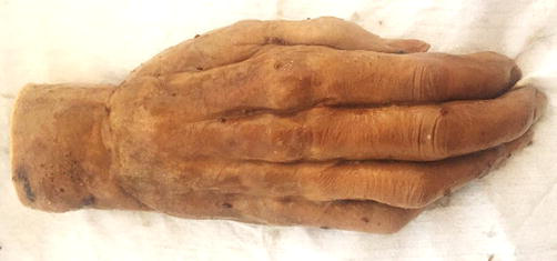
Figure 2.
Dorsal aspect of the hand, after 30 hours of recovery.
Figures 3 and 4 show the sample of a human hand mummified by desiccation; the dried tissues stand out; the texture of these was quite hard and presented a great rigidity.
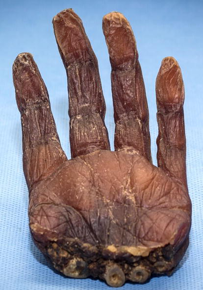
Figure 3.
Palmar side of the hand mummified by desiccation.
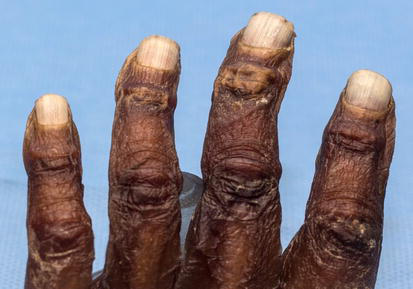
Figure 4.
Dorsum of the hand mummified by desiccation.
Figures 5 and 6 show the results of the rehydration process of the sample after 72 hours in immersion. Figure 7 shows that the protocol allows recovering the flexibility of the sample.
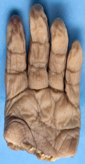
Figure 5.
Palmar side of the hand, after 72 hours of rehydration.

Figure 6.
Dorsum of the hand, after 72 hours of rehydration.
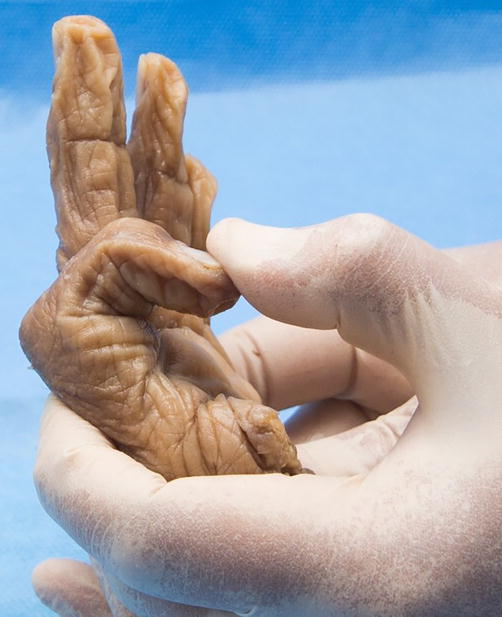
Figure 7.
Recovery of the sample flexibility.
2.9 Microscopic results
For the analysis of the samples at the microscopic level, skin samples were taken from a leg in a state of decomposition Figure 8, (red square).
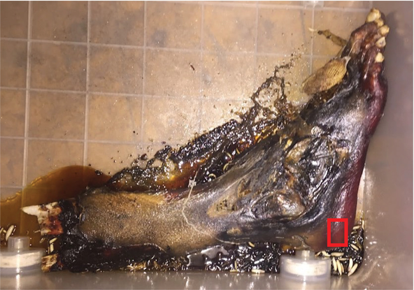
Figure 8.
Decomposing leg, red square corresponds to the area where the sample was taken.
Two skin samples were taken, one sample that was not subjected to the recovery process (Figure 9), and one sample that was subjected to the recovery process for 25 hours (Figure 10). Both samples were submitted to the routine histotechnical process. The samples were stained with Hematoxylin–Eosin. Figure 9 shows the skin without recovery, as a compact cut, where the layers of the skin are not evident. On the other hand, Figure 10 shows a tissue in which the skin layers are differentiated.
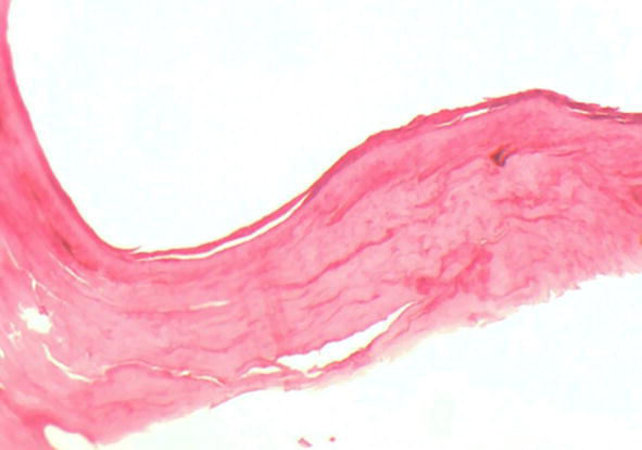
Figure 9.
Skin sample without rehydration protocol, H/E, 100×.
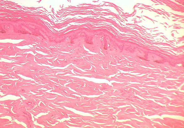
Figure 10.
Skin sample subjected to H/E rehydration protocol, 100×.
To differentiate the tissue components of the skin, such as the dermis and epidermis. Both samples were stained with Van Gieson’s trichrome technique. Figure 11 shows a sample that was not subjected to the rehydration process, which despite having differentiation of the tissue components, is very disintegrated. Figure 12 shows the sample subjected to the rehydration process. Here the layers of the skin can be seen, differentiating the epidermis from the connective tissue of the dermis.
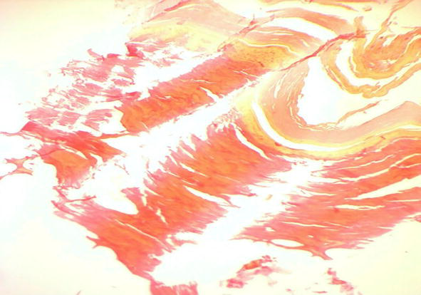
Figure 11.
Skin sample without rehydration protocol, Van Gieson, 100×.
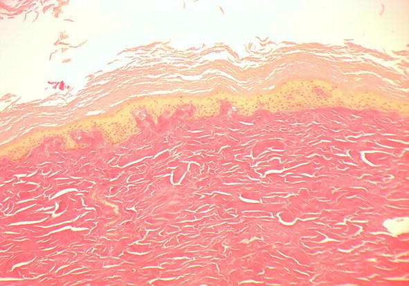
Figure 12.
Skin sample subjected to rehydration protocol, Van Gieson, 100×.
3. Conclusion
The study of tissues in the initial state of decomposition or mummified represents a challenge in postmortem analysis, since it is a complex subject due to the processes that allow the tissue to reach these states. This process is influenced by multiple factors, among which the characteristics of the type of tissue, as well as environmental factors, stand out.
The objective of the study and the expectations regarding the process of recovery and rehydration of the tissues is the first factor that must be considered, since it depends on the condition of the tissue and a previous evaluation must be made to determine concentrations and recovery time in the immersion solution. Considering from this aspect that not all tissues can be submitted to the recovery process, since it depends on the conditions in which the sample is found, tissues in certain states of decomposition or mummified can degrade due to an abrupt entrance of the rehydrating solution, which would alter the objective of the study in an important way.
There are many areas where these techniques can be used, one example is in post-mortem analysis. From a macroscopic point of view, it can provide information related to identification elements such as tattoos, marks, scars, lesions, and so forth. And in the microscopic field related to Forensic Pathology, it can provide information on vital or postmortem injuries and pathological conditions among other factors that have generated alterations in the tissue.
References
- 1.
Galloway A, Birkby W, Jones A, Henry T, Parks B. Decay rates of human remains in an arid environment. Journal of Forensic Sciences. 1989; 34 (3):607-616 - 2.
Aufderheide A. Soft tissue taphonomy: A paleopathology perspective. International Journal of Paleopathology. 2011; 1 :75-80 - 3.
Dent B, Forbes S, Stuart B. Review of human decomposition process in soil. Environmental Geology. 2004; 45 :576-585 - 4.
Fiedler S. Decomposition of buried corpses, with special reference to the formation of adipocere. Die Naturuissenschafter. 2003; 90 :291-300 - 5.
Vass A, Barshick S, Sega G, Caton J, Skeen J, Love J, et al. Decomposition chemistry of human remains: A new methodology for determining the postmortem interval of human remains. Journal of Forensic Sciences. 2002; 47 (3):542-553 - 6.
Campobasso CP, Di Vella G, Introna F. Factors affecting decomposition and Diptera colonization. Forensic Sciece International. 2001; 120 (1-2):18-27 - 7.
Schoenen D, Schoenen H. Adipocere formation--the result of insufficient microbial degradation. Forensic Science International. 2013; 226 :301-306 - 8.
Aufderheide A. Chapter 3 mechanisms of mummification. In: The Scientific Study of Mummies. Cambridge: Cambridge University Press; 2003. pp. 41-63 - 9.
Hanzlick R. Embalming, body preparation, burial, and disinterment. An overview for forensic pathologists. The American Journal of Forensic Medicine and Pathology. 1994; 15 (2):122-131 - 10.
Mann R, Bass W, Meadows L. Time since death and decomposition of the human body: Variables and observations in case and experimental field studies. Journal of Forensic Sciences. 1990; 35 :103-111 - 11.
Berkeley R, Campbell R. Nutrition, and the influence of environmental factors on microbial activities. In: Hawker LE, Linton AH, editors. Microorganisms Function, Form, and Environment. 2nd ed. Baltimore, Maryland: University Park Press; 1979. pp. 69-82 - 12.
Kaufmann B. Mummification in the middle ages. In: Spindler K et al., editors. Human Mummies a Global Survey of Their Status and the Techniques of Conservation. 1st ed. Vienna: Springer Vienna; 1996. pp. 231-248 - 13.
Spindler K, Wilfing H, et al. Human Mummies: A Global Survey of their Status and the Techniques of Conservation. New York: Springer-Verlag, Wien; 1996 - 14.
Arriaza B. Arseniasis as an environmental hypothetical explanation for the origin of the oldest artificial mummification practice in the world. Revista de Antropología Chilena. 2005; 37 :255-260 - 15.
Simmandl E. A contribution to the histology of the skin and of the muscle of an egyptian mummy. L'Anthropologie. 1928; 6 :56 - 16.
Fechner G, Petkovits T, Brinkmann B. Morphologische Identifizierung mumifizierter Gewebsspuren. Archiv für Kriminologie. 1988; 182 :154-158 - 17.
Mekota A, Vermehren M. 2005. Determination of optimal rehydration, fixation, and staining methods for histological and immunohistochemical analysis of mummified soft tissues. Biotechnic & Histochemistry. 2005; 80 (1):7-13 - 18.
Herman B. A simple method for softening and rehydration of dried and mummified tissues. Paleopath News. 1982; 39 :10 - 19.
Turner P, Holtom D. The use of a fabric softener in the reconstitution of mummified tissue prior to paraffin wax sectioning for light microscopical examination. Stain Technology. 1981; 56 :35-38 - 20.
Fulcheri E, Ventura L. Rileggendo tra antiche e nuove ricette per dare freschezza ai tessuti mummificati o disseccati. Pathologica. 2001; 93 :700-706 - 21.
Schmidt C, Nawrocki S, Williamson M, Marlin D. Obtaining fingerprints from mummified fingers: A method for tissue rehydration adapted from the archeological literature. Journal of Forensic Science. 2000; 45 (4):874-875