Open Access is an initiative that aims to make scientific research freely available to all. To date our community has made over 100 million downloads. It’s based on principles of collaboration, unobstructed discovery, and, most importantly, scientific progression. As PhD students, we found it difficult to access the research we needed, so we decided to create a new Open Access publisher that levels the playing field for scientists across the world. How? By making research easy to access, and puts the academic needs of the researchers before the business interests of publishers.
We are a community of more than 103,000 authors and editors from 3,291 institutions spanning 160 countries, including Nobel Prize winners and some of the world’s most-cited researchers. Publishing on IntechOpen allows authors to earn citations and find new collaborators, meaning more people see your work not only from your own field of study, but from other related fields too.
To purchase hard copies of this book, please contact the representative in India:
CBS Publishers & Distributors Pvt. Ltd.
www.cbspd.com
|
customercare@cbspd.com
Meniscal roots provide substantial stability to the knee against anteroposterior and rotational forces. Root injuries show similar biomechanic properties to total meniscectomy and are one of the preventable causes of early osteoarthritis. Various surgical methods were already described, and new techniques are being developed. Repair of meniscal root tears is almost always recommended, especially in acute traumatic cases. However, the surgical indications are controversial in cases where repair is not possible, in degenerative conditions, and in elderly patients. Along with this perspective, radiologic and clinical evaluation and current surgical techniques will be summarized, and treatment indications and outcomes will be discussed according to up-to-date data.
Interest in meniscal root tears is increasing as it is understood that injuries to this region directly affect the biomechanics of the knee and lead to progressive osteoarthritis. Meniscus root tears are defined as tears or avulsion injuries that are 1 cm away from the meniscus root attachment [1]. The prevalence of detecting meniscal root tears during arthroscopy has been reported as 9.1%, and meniscus root tears constitute 10–21% of all meniscus tears [2, 3]. While surgeries such as partial or subtotal meniscectomy were performed in the past, anatomical meniscus root repair is now recommended and new treatment options to prevent meniscus extrusion are sought [1]. Meniscal root injuries have been shown to be biomechanically equivalent to total meniscectomy [4, 5, 6]. Diagnosis and treatment of these tears are critical. Meniscal root injuries are considered a preventable cause of osteoarthritis in the knee. Delays in diagnosis and treatment may lead to irreversible consequences [7]. It has been reported that 78% of total knee replacement patients under the age of 60 had a medial meniscus posterior root tear [8]. Meniscal root tears can occur degeneratively, due to acute trauma, or iatrogenically. While lateral meniscus root tears are frequently observed in young patients, medial meniscus root tears are more common in older patients. It often occurs in the 4th and 5th decades. Although it is not a rare injury, delays and neglect in diagnosis and treatment can significantly reduce the patient’s knee functions and quality of life [9].
Meniscal roots are four strong structures that connect the meniscus horns to the tibial intercondylar region [10]. They contain main and complementary fibers that contribute significantly to the root attachment forces in the footprint areas and adhere to the bone by spreading like a fan [11]. When examined cross-sectionally, the connection of the meniscal root to the tibia consists of four layers: meniscus fibers, noncalcified fibrocartilage, calcified fibrocartilage, and bone [12].
The medial meniscal posterior root footprint is 9.6 mm posterior and 0.7 mm lateral to the medial tibial eminence. The center point of the footprint is located 3.5 mm lateral to the most proximal edge of the medial articular cartilage and 8.2 mm anterior to the most proximal part of the posterior cruciate ligament tibial attachment [13].
The lateral meniscal posterior root footprint is 1.5 mm posterior and 4.2 mm medial to the lateral tibial eminence. The midpoint of the footprint is located 4.3 mm medial to the most proximal edge of the lateral articular cartilage and 12.7 mm anterior to the most proximal part of the posterior cruciate ligament tibial attachment [13].
The anterior root footprint of the medial meniscus is inserted into the bone along the anterior aspect of the intercondylar crest of the tibia, through the anterior slope of the tibia. It has been reported that the center of the footprint is 18.2 mm anteromedial from the center of the tibial attachment of the anterior cruciate ligament and 27.5 mm anterolateral from the apex of the medial tibial eminence [14].
The lateral meniscal anterior root footprint is 5.0 mm anterolateral from the center of the anterior cruciate ligament tibial attachment, 14.4 mm from the lateral tibial eminence, and 7.1 mm from the nearest edge of the lateral joint cartilage of the tibia [14].
LaPrade classification, consisting of 5 types plus 1 variant type, has been defined for medial and lateral meniscus root tears (Table 1) [15].
Type 1
Partial root tear
Type 2
Complete radial root tear within 9 mm from bony root attachment. 2A: 0- < 3 mm from the center of the root attachment. 2B: 3- < 6 mm from the center of the root attachment. 2C: 6- < 9 mm from the center of the root attachment.
Type 3
Bucket handle tear with complete root detachment.
Type 4
Oblique or longitudinal tear with complete root detachment.
Type 5
Bony avulsion fracture of the root attachment.
Variant type
Complete lateral meniscal posterior root tear with intact meniscofemoral ligament(s).
Meniscal roots have an important function in transferring axial tibiofemoral loads from the meniscus body. The rupture of the meniscus attachment from the tibial plateau due to a meniscal root tear causes extrusion of the meniscus and changes in knee kinematics. This leads to unequal and abnormal distribution of the load transferred to the knee. Thus, the tibiofemoral contact area decreases and contact pressures increase [1]. The medial meniscus adheres to the capsular structures more rigidly and is less mobile than the lateral meniscus. Thus, a higher rate of injury is observed in the medial meniscus. Medial meniscal posterior root tears cause a 25% increase in contact pressures [6]. Increased contact pressures have negative effects on articular cartilage and may cause early osteoarthritis if not treated anatomically [16]. It has been reported that knee kinematics are also affected in lateral meniscus posterior root tears, and surgical repair normalizes the load distribution for both types of lesions [5]. In addition to axial load transfer, meniscal roots also play an important role in stabilizing the knee against rotational forces, especially after ligament damage [17]. Medial meniscus contributes to stabilization against anterior tibial translation [18]. Lateral meniscus plays an important role in stabilization against anterolateral rotational laxity in anterior cruciate ligament-deficient knees [19].
No prevalence has been reported for anterior meniscal root tears. Although rare compared to posterior root tears, iatrogenic injuries have been reported during intramedullary tibial nailing, anterior cruciate ligament reconstruction, and cyst resection [20, 21, 22]. They can be associated with tibial plateau and tibial eminence fractures [15]. If a tear is detected, it should be repaired with a suture anchor or transosseous methods.
The incidence of chondral defects with medial meniscus posterior root tears is 5.8 times higher. Medial meniscus posterior root tears are generally degenerative tears, are more likely to occur in middle-aged women, and account for 21% of medial meniscus posterior horn tears [23]. Additionally, iatrogenic damage due to nonanatomical tibial tunnel position during posterior cruciate ligament reconstruction has been reported [24]. Approximately 70% of cases occur in degenerative knees without any injury or with minor trauma such as squatting. The medial meniscal posterior root is the least mobile compared to other meniscus root attachments and therefore has the highest tear incidence [1]. Additionally, medial meniscus posterior root tear may occur in 80% of patients with spontaneous osteonecrosis of the knee (SONK) now known as subchondral deficiency fracture of the knee (SIFK) [25].
The incidence of lateral meniscus posterior root tears with anterior cruciate ligament tears is 10.3 times higher than medial meniscus posterior root tears [23]. In anterior cruciate ligament injuries, lateral meniscus posterior root tear occurs in 10–15% of cases [26]. Lateral meniscus root tear is detected in 8% to 9.8% of patients who underwent anterior cruciate ligament reconstruction [10]. In anterior cruciate ligament-deficient knees, the lateral meniscal posterior root contributes significantly to stabilization in both pivot activities and anteroposterior translation. Therefore, lateral meniscal posterior root tear should be suspected in pivot shift grade 3 knees [17].
Meniscus root tears lead to rapid joint degeneration; therefore, early diagnosis and appropriate treatment are crucial for achieving good outcomes. The clinical examination begins with a detailed patient history and physical examination. Although some radiographic findings raise clinical suspicion, the gold standard diagnostic method is MRI and arthroscopy [27]. Understanding the etiology and diagnostic findings and being clinically suspicious is an important step in making the correct diagnosis. Meniscal root tears do not cause symptoms like classical meniscus tears, and therefore, their diagnosis can be difficult and often be missed. Knee locking or giving way symptoms are reported in only 10–15% of patients with medial meniscus posterior root tear [28]. Similar to chronic meniscus tears, 70% of patients do not remember a direct event that initiated the injury [23]. The onset of pain may be subtle or sudden after a minor trauma [29]. A popping sound may be heard when squatting or getting up from a chair [28]. In the presence of anterior cruciate ligament injury, caution should be taken, especially in terms of lateral meniscus posterior root tears.
6.1.1 Risk factors
Varus malalignment, advanced age, high body mass index, and female sex are risk factors for medial meniscus posterior root tears [30]. Lateral meniscus posterior root tears usually occur after trauma and are most commonly associated with anterior cruciate ligament injuries. Other important risk factors are male gender, playing contact sports, having a medial meniscus tear, and being exposed to high-energy trauma [31].
6.2 Physical examination
The most common physical examination findings are pain at full knee flexion (66.7%), joint line tenderness (61.9%), and a positive McMurray test (57.1%) [29]. In medial meniscal posterior root tear, the extruded meniscus can be palpated along the joint line. Palpation of the extruded medial meniscus when full extension and varus stress are applied to the knee is considered a positive test. When normal alignment is achieved, extrusion disappears [32]. It has been stated that this test can be used in early diagnosis and also to check the stability of the repair in postoperative follow-up [32]. Since the place of physical examination is limited in diagnosis, advanced diagnostic imaging should be performed in case of suspicion.
6.3 Imaging
Plain radiographs, including the Rosenberg view, are used to detect degenerative changes rather than diagnosis. Since meniscus root tears are equivalent to total meniscectomy, they lead to rapid and progressive osteoarthritis. Kellgren-Lawrence or Ahlbäck classification and long-leg standing films are important for treatment planning and alignment evaluation. It has been reported that varus alignment of >5° has lower outcome scores than neutral alignment [33].
Diagnosis of meniscal root tears is difficult despite the use of advanced imaging techniques. MRI is considered the most reliable noninvasive test in the diagnosis of these lesions. Although previous studies reported a low specificity of 70%, the predictive value of MRI is over 90% in new studies [34, 35, 36]. There are 3 typical findings for meniscal root tears on MRI: Ghost sign characterized by the absence of the posterior horn of the meniscus in sagittal sections, vertical signal increase (truncation sign) in the meniscus root attachment area and > 3 mm extrusion in the medial meniscus in coronal sections, and linear signal increase perpendicular to the meniscus root attachment area (radial tear) in axial sections (Figures 1–3) [37]. In mid-coronal sections, meniscus extrusion of >3 mm has been associated with severe meniscus degeneration and meniscal root tears (Figure 4) [38]. Additionally, SONK, now known as SIFK, bone edema in the posteromedial part of the tibia or femoral condyles can be observed in cases with medial meniscus root tears [1].
Figure 1.
Ghost sign. Red arrow shows the absence of posterior horn of medial meniscus.
Figure 2.
Truncation sign. Red arrow shows the vertical signal increase of root tear adjacent to root stump.
Treatment of meniscal root tears is variable depending on the severity and timing of the injury, as well as the condition of the articular cartilage. Surgical repair should almost always be performed in appropriate cases. In light of increased awareness and current literature, conservative treatment is not preferred except for patients with surgical contraindications. Although there is no consensus on surgical contraindications, these can be defined as patients with advanced osteoarthritis (Kellgren–Lawrence 3–4), high BMI, and advanced coronal malalignment [39]. Patients with varus malalignment of >5° have slightly inferior results following meniscus root repair, yet significant improvements have been reported compared to their preoperative conditions [33]. Simultaneous open-wedge tibial osteotomy is recommended for patients with varus malalignment of >10°. In cases between 5° and 10° varus malalignment, there is no definitive indication [27]. In patients who prefer conservative treatment, activity modification, analgesic treatments, unloader brace. and physical therapy may be preferred. Partial or subtotal meniscectomy can be performed in patients with advanced degenerative changes that do not respond to conservative treatment and with mechanical symptoms such as locking. However, it should be known that the symptoms will improve temporarily and progressive osteoarthritis will develop.
Surgical repair aims to restore joint contact pressures and joint kinematics. It is aimed to increase the contact area, decrease the contact pressures, and delay the development of arthrosis by anatomic repair and correction of extrusion. The most commonly used surgical techniques for repairing meniscal root tears are suture anchors or transtibial techniques [1].
7.1 Author’s preferred technique for medial meniscus posterior root tears
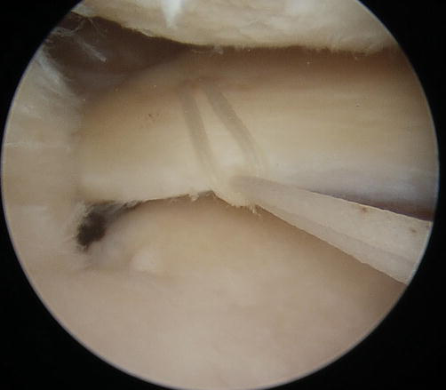
Knee arthroscopy starts with the standard anterolateral portal, and all knee joint compartments are observed. The anteromedial portal is used as the working portal. While making the anteromedial portal by outside-in technique, the reach of the spinal needle to the medial meniscal posterior root is checked (Figure 5). This step is important to provide clear access to the root from the anteromedial working portal if special low-profile instruments (root repair-specific suture passer, root tunneling guide, etc.) are not available. Careful examination and probing medial meniscus posterior root are critical, and pie crusting the deep medial collateral ligament is highly recommended for better visualization and larger working space (Figure 6). The center of the medial meniscal posterior root attachment is located and the cartilage surface is decorticated with the help of a curette. It must be checked whether the medial meniscus has been reduced to its anatomical location by pulling it with the help of a grasper. In cases where the meniscus is retracted or extruded, it must be loosened from the posterior capsule with the help of a shaver or radiofrequency device. A luggage-tag (simple cinch) suture is passed approximately 8 mm lateral to the medial meniscus posterior root stump with a suture passer (Figure 7). A second suture is passed in the form of a simple stitch, approximately 3 mm lateral to the stump (Figure 8). During this step, the first suture can be used as a traction suture for easier access to the meniscus root. While the threads are in the anteromedial portal, an anterior cruciate ligament tibial tunnel guide set at approximately 50° is used from the same portal and a passing pin is sent to the center of medial meniscal posterior root attachment from the anteromedial surface of the tibia (Figure 9). It is important not to damage the pes anserinus. If the position of the passing pin is anatomical, a 4.5 mm transtibial tunnel is drilled while protecting the pointed end of the pin from the surrounding tissues with the help of a curette. Meniscus root sutures are passed through this tunnel with the help of a suture shuttle, and the sutures are fixed to a screw with a washer as a fixation post on the anteromedial surface of the tibia with the knee flexed at 90° while reduction is checked arthroscopically (Figures 10-12). When the knee is flexed beyond 90°, there is an increase in tension in the posterior root of the medial meniscus due to femoral rollback and AP translation. If the meniscal root is tightened and fixed at lower flexion angles, further knee flexion may cause retraction of the improperly reduced meniscal root stump from the tibial tunnel end. While meniscus reduction is achieved, first traction is applied through luggage-tag suture, which clasps circumferential fibers of the meniscus; then, extrusion is resolved, and it is fixed to the fixation post screw. Secondly, with the simple suture on the medial side, the meniscus stump is reduced inside the tunnel and fixed in its anatomical position.
Probe examination of the root tear. MMPR: Medial meniscus posterior root. MFC: Medial femoral condyle.
Figure 7.
A luggage tag (simple cinch) suture.
Figure 8.
Second simple suture.
Figure 9.
A passing guide pin (red arrow) is introduced to the center of the medial meniscal posterior root attachment.
Figure 10.
Sutures are transferred through the tibial tunnel.
Figure 11.
Anatomic reduction of the medial meniscus posterior root.
Figure 12.
Fixation of sutures to a screw with washer.
7.2 Postoperative rehabilitation
After transtibial pullout meniscal root repair, the patient’s weight-bearing mobilization is restricted for 6 weeks. With the angle-adjustable knee brace, flexion is limited to 30° in the first 2 weeks, 60° in the next 2–4 weeks, and 90° in the next 4–6 weeks. After the 6th week, knee flexion above 90° and gradual weight bearing are allowed, depending on clinical examination and pain status. Squat movements that require knee flexion above 90° are restricted for the first 6 months. Different postoperative protocols are also mentioned in the literature.
Although there may be a short-term increase in subjective scores with partial meniscectomy in meniscus posterior root tears, there is a significant increase in degenerative changes within 5 years postoperatively. When partial meniscectomy and nonoperative treatment were compared, no significant difference was found in the final Tegner scores, IKDC and Kellgren–Lawrence grade [40]. However, in the group that underwent partial meniscectomy, 54% underwent total knee arthroplasty within an average of 4.5 years [40]. In cases followed conservatively, 87% of patients experienced clinical and radiographic deterioration within 5 years, while 31% of patients underwent total knee arthroplasty [9].
Clinical improvements and increases in subjective scores after meniscal root repairs have been reported in various meta-analyses [16, 41]. It has been reported that degenerative changes can be prevented in up to 84% of patients who underwent meniscus root repair [27]. There was no progression in osteoarthritis grade on the Kellgren–Lawrence grading scale. In another study, better clinical and radiological results were observed in the transtibial pullout meniscal root repair group compared to in partial meniscectomy at a 5-year follow-up. No patients in the root repair group underwent total knee arthroplasty during the follow-up [42]. Although advanced age is considered a relative contraindication for meniscal root repairs, it has been reported that there is no difference in the failure rate in patients over and under 50. Patient satisfaction increased in terms of pain, function, and activity level after repair [43].
Another important point in meniscus root repair is meniscus extrusion, which persists in postoperative follow-ups. It has been shown that meniscus extrusion does not improve at variable rates after surgery albeit its biomechanical effects are not fully understood. It has been reported in several studies that cartilage degeneration will increase as the level of meniscus extrusion increases, and extrusion of >3 mm may be the critical limit [38, 44]. Therefore, in cases of meniscus extrusion, an increase in axial load in the joint can be expected. Various surgical techniques such as centralization sutures and reduction with suture anchors have been described in the literature to prevent meniscal extrusion [45, 46].
Root tears are injuries that can potentially lead to severe consequences, causing pain and limitation of activities of daily living, which can reduce the quality of life. Clinical and biomechanical studies have shown that root tears behave like total meniscectomy and quickly lead to knee osteoarthritis and loss of function in patients. This course makes early diagnosis and treatment of this injury a must. Good clinical and radiological results can be obtained with anatomical root repair. Increasing awareness and advances in surgical technique reveal the necessity of meniscal root repair in patients with appropriate indications. Some causes of early-age osteoarthritis and total knee arthroplasty can be prevented by choosing anatomical root repair over partial meniscectomy and conservative treatment.
1.Pache S, Aman ZS, Kennedy M, et al. Meniscal root tears: Current concepts review. Archives in Bone and Joint Surgery. 2018;6:250-259
2.LaPrade RF, Floyd ER, Carlson GB, et al. Meniscal root tears: Solving the silent epidemic. Journal of Arthroscopic Surgery and Sports Medicine. 2021;2:47-57
3.Cinque ME, Chahla J, Moatshe G, et al. Meniscal root tears: A silent epidemic. British Journal of Sports Medicine. 2018;52:872-876
4.Padalecki JR, Jansson KS, Smith SD, et al. Biomechanical consequences of a complete radial tear adjacent to the medial meniscus posterior root attachment site: In situ pull-out repair restores derangement of joint mechanics. The American Journal of Sports Medicine. 2014;42:699-707
5.Schillhammer CK, Werner FW, Scuderi MG, et al. Repair of lateral meniscus posterior horn detachment lesions: A biomechanical evaluation. The American Journal of Sports Medicine. 2012;40:2604-2609
6.Allaire R, Muriuki M, Gilbertson L, et al. Biomechanical consequences of a tear of the posterior root of the medial meniscus. Similar to total meniscectomy. The Journal of Bone and Joint Surgery. American Volume. 2008;90:1922-1931
7.Cinque ME, Geeslin AG, Chahla J, et al. Two-tunnel transtibial repair of radial meniscus tears produces comparable results to inside-out repair of vertical meniscus tears. The American Journal of Sports Medicine. 2017;45:2253-2259
8.Choi E-S, Park S-J. Introduction clinical evaluation of the root tear of the posterior horn of the medial meniscus in total knee arthroplasty for osteoarthritis. Knee Surgery Related Research. 2015;27:90-94
9.Krych AJ, Reardon PJ, Johnson NR, et al. Non-operative management of medial meniscus posterior horn root tears is associated with worsening arthritis and poor clinical outcome at 5-year follow-up. Knee Surgery, Sports Traumatology, Arthroscopy. 2017;25:383-389
10.Brody JM, Lin HM, Hulstyn MJ, et al. Lateral meniscus root tear and meniscus extrusion with anterior cruciate ligament tear. Radiology. 2006;239:805-810
11.Ellman MB, Laprade CM, Smith SD, et al. Structural properties of the meniscal roots. The American Journal of Sports Medicine. 2014;42:1881-1887
12.Wang YJ, Yu JK, Luo H, et al. An anatomical and histological study of human meniscal horn bony insertions and peri-meniscal attachments as a basis for meniscal transplantation. Chinese Medical Journal. 2009;122:536-540
13.Johannsen AM, Civitarese DM, Padalecki JR, et al. Qualitative and quantitative anatomic analysis of the posterior root attachments of the medial and lateral menisci. The American Journal of Sports Medicine. 2012;40:2342-2347
14.Laprade CM, Ellman MB, Rasmussen MT, et al. Anatomy of the anterior root attachments of the medial and lateral menisci: A quantitative analysis. The American Journal of Sports Medicine. 2014;42:2386-2392
15.Feagin JA, Engebretsen L, LaPrade RF, et al. Meniscal root tears. The American Journal of Sports Medicine. 2014;43:363-369
16.Chung KS, Ha JK, Ra HJ, et al. A meta-analysis of clinical and radiographic outcomes of posterior horn medial meniscus root repairs. Knee Surgery, Sports Traumatology, Arthroscopy. 2016;24:1455-1468
17.Frank JM, Moatshe G, Brady AW, Dornan GJ, Coggins A, Muckenhirn KJ, et al. Lateral meniscus posterior root and meniscofemoral ligaments as stabilizing structures in the ACL-deficient knee: A biomechanical study. Orthopaedic Journal of Sports Medicine. 15 Jun 2017;5(6):2325967117695756
18.Levy IM, Torzilli PA, Warren RF. The effect of medial meniscectomy on anterior-posterior motion of the knee. The Journal of Bone and Joint Surgery. American Volume. 1982;64:883-888
19.Minami T, Muneta T, Sekiya I, et al. Lateral meniscus posterior root tear contributes to anterolateral rotational instability and meniscus extrusion in anterior cruciate ligament-injured patients. Knee Surgery, Sports Traumatology, Arthroscopy. 2018;26:1174-1181
20.LaPrade CM, James EW, Engebretsen L, et al. Anterior medial meniscal root avulsions due to malposition of the tibial tunnel during anterior cruciate ligament reconstruction: Two case reports. Knee Surgery, Sports Traumatology, Arthroscopy. 2014;22:1119-1123
21.Feucht MJ, Minzlaff P, Saier T, et al. Avulsion of the anterior medial meniscus root: Case report and surgical technique. Knee Surgery, Sports Traumatology, Arthroscopy. 2015;23:146-151
22.Ellman MB, James EW, LaPrade CM, et al. Anterior meniscus root avulsion following intramedullary nailing for a tibial shaft fracture. Knee Surgery, Sports Traumatology, Arthroscopy. 2015;23:1188-1191
23.Matheny LM, Ockuly AC, Steadman JR, et al. Posterior meniscus root tears: Associated pathologies to assist as diagnostic tools. Knee Surgery, Sports Traumatology, Arthroscopy. 2015;23:3127-3131
24.Laprade CM, Smith SD, Rasmussen MT, et al. Consequences of tibial tunnel reaming on the meniscal roots during cruciate ligament reconstruction in a cadaveric model, Part 2: The posterior cruciate ligament. The American Journal of Sports Medicine. 2015;43:207-212
25.Robertson DD, Armfield DR, Towers JD, et al. Meniscal root injury and spontaneous osteonecrosis of the knee: An observation. Journal of Bone and Joint Surgery. British Volume (London). 2009;91:190-195
26.Forkel P, Reuter S, Sprenker F, et al. Different patterns of lateral meniscus root tears in ACL injuries: Application of a differentiated classification system. Knee Surgery, Sports Traumatology, Arthroscopy. 2015;23:112-118
27.Homan MD, Braaten JA, Banovetz MT, et al. Meniscal root tears: Repair and salvage techniques. Journal of Cartilage and Joint Preservation. 2023;3:100098
28.Lee DW, Ha JK, Kim JG. Medial meniscus posterior root tear: A comprehensive review. Knee Surgery Related Research. 2014;26:125
29.Kim JH, Chung JH, Lee DH, et al. Arthroscopic suture anchor repair versus pullout suture repair in posterior root tear of the medial meniscus: A prospective comparison study. Arthroscopy. 2011;27:1644-1653
30.Hwang BY, Kim SJ, Lee SW, et al. Risk factors for medial meniscus posterior root tear. The American Journal of Sports Medicine. 2012;40:1606-1610
31.Praz C, Vieira TD, Saithna A, et al. Risk factors for lateral Meniscus posterior root tears in the anterior cruciate ligament-injured knee: An epidemiological analysis of 3956 patients from the SANTI study group. The American Journal of Sports Medicine. 2019;47:598-605
32.Seil R, Dück K, Pape D. A clinical sign to detect root avulsions of the posterior horn of the medial meniscus. Knee Surgery, Sports Traumatology, Arthroscopy. 2011;19:2072-2075
33.Ridley TJ, Ruzbarsky JJ, Dornan GJ, et al. Minimum 2-year clinical outcomes of medial meniscus root tears in relation to coronal alignment. The American Journal of Sports Medicine. 2022;50:1254-1260
34.Choi SH, Bae S, Ji SK, et al. The MRI findings of meniscal root tear of the medial meniscus: Emphasis on coronal, sagittal and axial images. Knee Surgery, Sports Traumatology, Arthroscopy. 2012;20:2098-2103
35.LaPrade RF, Ho CP, James E, et al. Diagnostic accuracy of 3.0 T magnetic resonance imaging for the detection of meniscus posterior root pathology. Knee Surgery, Sports Traumatology, Arthroscopy. 2015;23:152-157
36.De Smet AA, Mukherjee R. Clinical, MRI, and arthroscopic findings associated with failure to diagnose a lateral meniscal tear on knee MRI. AJR. American Journal of Roentgenology. 2008;190:22-26
37.Harper KW, Helms CA, Lambert HS, et al. Radial meniscal tears: Significance, incidence, and MR appearance. AJR. American Journal of Roentgenology. 2005;185:1429-1434
38.Costa CR, Morrison WB, Carrino JA. Medial meniscus extrusion on knee MRI: Is extent associated with severity of degeneration or type of tear? AJR. American Journal of Roentgenology. 2004;183:17-23
39.Lim HC, Bae JH, Wang JH, et al. Non-operative treatment of degenerative posterior root tear of the medial meniscus. Knee Surgery, Sports Traumatology, Arthroscopy. 2010;18:535-539
40.Krych AJ, Johnson NR, Mohan R, et al. Partial meniscectomy provides no benefit for symptomatic degenerative medial meniscus posterior root tears. Knee Surgery, Sports Traumatology, Arthroscopy. 2018;26:1117-1122
41.Feucht MJ, Kühle J, Bode G, et al. Arthroscopic transtibial pullout repair for posterior medial meniscus root tears: A systematic review of clinical, radiographic, and second-look arthroscopic results. Arthroscopy. 2015;31:1808-1816
42.Chung KS, Ha JK, Yeom CH, et al. Comparison of clinical and radiologic results between partial meniscectomy and refixation of medial meniscus posterior root tears: A minimum 5-year follow-up. Arthroscopy. 2015;31:1941-1950
43.LaPrade RF, Matheny LM, Moulton SG, et al. Posterior meniscal root repairs: Outcomes of an anatomic transtibial pull-out technique. The American Journal of Sports Medicine. 2017;45:884-891
44.Lee DH, Lee BS, Kim JM, et al. Predictors of degenerative medial meniscus extrusion: Radial component and knee osteoarthritis. Knee Surgery, Sports Traumatology, Arthroscopy. 2011;19:222-229
45.Koga H, Watanabe T, Horie M, et al. Augmentation of the Pullout repair of a medial meniscus posterior root tear by arthroscopic centralization. Arthroscopy Techniques. 2017;6:e1335-e1339
46.Nakagawa Y, Ozeki N, Koga H. A narrative review of lateral meniscus root tears and extrusion: Techniques and outcomes. Annals of Joint. 2022;7:15
Written By
Sancar Alp Ovali
Submitted: 15 October 2023Reviewed: 09 November 2023Published: 15 January 2024
 Open access peer-reviewed chapter