Open Access is an initiative that aims to make scientific research freely available to all. To date our community has made over 100 million downloads. It’s based on principles of collaboration, unobstructed discovery, and, most importantly, scientific progression. As PhD students, we found it difficult to access the research we needed, so we decided to create a new Open Access publisher that levels the playing field for scientists across the world. How? By making research easy to access, and puts the academic needs of the researchers before the business interests of publishers.
We are a community of more than 103,000 authors and editors from 3,291 institutions spanning 160 countries, including Nobel Prize winners and some of the world’s most-cited researchers. Publishing on IntechOpen allows authors to earn citations and find new collaborators, meaning more people see your work not only from your own field of study, but from other related fields too.
To purchase hard copies of this book, please contact the representative in India:
CBS Publishers & Distributors Pvt. Ltd.
www.cbspd.com
|
customercare@cbspd.com
Idiopathic intracranial hypertension (IIH), also known as pseudotumor cerebri, is a condition marked by increased cerebrospinal (CSF) pressure in the absence of secondary causes, such as brain tumors, venous sinus thrombosis, and meningitis. The exact cause of IIH is unknown, but the rise in intracranial pressure (ICP) is a defining characteristic leading to the clinical presentation of headaches, transient visual obscurations, pulsatile tinnitus, and retrobulbar pain. This text aims to discuss IIH and the effectiveness of a multi-disciplinary approach, emphasizing collaboration by neuro-ophthalmology, neurology, neurosurgery, radiology, oculoplastic, and weight loss teams to achieve clinical remission of IIH. Literature was reviewed to discuss aspects of IIH in the pediatric population, and our clinical experience was utilized to provide recommendations regarding the importance of a multi-disciplinary team. This chapter explores IIH in the pediatric population regarding its demographics, clinical manifestations, potential underlying pathophysiology, diagnostic steps, and the importance of a multi-disciplinary approach to management, as it is the most optimal. This allows patients to receive comprehensive care that will address more aspects of IIH while also maintaining long-term goals and remission.
Department of Ophthalmology, University of North Carolina, Chapel Hill, NC, United States
Bascom Palmer Eye Institute, University of Miami, Miami, FL, United States
Elizabeth Colvin
Campbell University, School of Osteopathic Medicine, Lillington, NC, United States
Huynh Duy
The Brody School of Medicine at East Carolina University, Greenville, NC, United States
Sarah Ro
Department of Family Medicine at the University of North Carolina, Chapel Hill, NC, United States
Carolyn Quinsey
Department of Neurosurgery at the University of North Carolina, Chapel Hill, NC, United States
Inga Shevtsova
Department of Internal Medicine, Billings Clinic, Billings, Montana, United States
Sriram Machineni
Albert Einstein College of Medicine, Bronx, NY, United States
*Address all correspondence to: mkostic@miami.edu
1. Introduction
Idiopathic intracranial hypertension (IIH), also known as pseudotumor cerebri, is a condition marked by increased cerebrospinal (CSF) pressure in the absence of secondary causes, such as brain tumors, venous sinus thrombosis, and meningitis [1]. The exact cause of IIH is unknown, but the rise in intracranial pressure (ICP) is a defining characteristic [2] leading to the clinical presentation of headaches, transient visual obscurations, pulsatile tinnitus, and retrobulbar pain [3]. Other symptoms include dizziness, neck, and back pain [1]. IIH patients commonly present with papilledema, vision loss, double vision, cranial nerve deficits, or nonspecific neurologic signs [4]. IIH is present in the adult and pediatric populations, impacting vision and quality of life.
Although symptoms of IIH can be debilitating, one of the priorities of care is to prevent any permanent vision loss. Lowering intracranial pressure becomes the focus once the risk of vision loss is mitigated. Currently, insufficient integration of multiple disciplines prevents early detection in some cases. Many IIH patients require neuro-ophthalmologic examination for proper diagnosis [5] but may instead be dismissed from primary care or emergency departments, allowing IIH to go undetected and worsen. While several investigators have reported on the importance of the multidisciplinary management approach of IIH, studies providing evidence of the superiority of multidisciplinary over monodisciplinary management are lacking. Due to the various presentations and categorization of IIH as a diagnosis of exclusion, swift diagnosis, and management of IIH in the pediatric population necessitates a multi-disciplinary approach. This text aims to discuss IIH and the effectiveness of a multi-disciplinary approach, emphasizing collaboration by neuro-ophthalmology, neurology, neurosurgery, radiology, oculoplastic and weight loss teams to achieve clinical remission of IIH.
IIH is regarded as a diagnosis of exclusion. Therefore, clinical presentation can be a defining indication of the underlying disorder. IIH has an incidence of 1–2 per 100,000 in the general population 4 with an incidence of 0.63–0.9 per 100,000 in the pediatric population (defined as <18 years old) [6, 7, 8]. In adults, IIH is disproportionately found in overweight women of childbearing age, in whom as much as 4–21 per 100,000 people are affected [4]. Although more predominant in the adult population, IIH is present in the pediatric population with differing demographics [9]. Pubescent status has been shown to play a role in the gender predilection of IIH in the pediatric population. Aylward et al. demonstrated that in the pre-pubescent population, there is nearly a 1:1 ratio of female to male incidence of IIH [8, 10]. However, female pubescent adolescents were more likely to present with IIH as compared to pubescent males 8.
Suspicion of IIH should be raised based on clinical complaints. Patients typically report headaches, transient visual obscurations, pulsatile tinnitus, and retrobulbar pain [3]. Headache is the most commonly described symptom associated with IIH, with one-quarter of patients reporting a constant, daily headache [1]. Pulsatile tinnitus may accompany the headache, which is described as a whooshing, whistling, or humming noise heard in the ear. It is believed to be caused by turbulent pulsatile blood flow that is perceived through the auditory system [1]. Neither headache nor pulsatile tinnitus are specific for IIH as they are clinically too broad, but they are clinical clues when combined with additional visual findings.
Visual symptoms include a wide range of deficits and are the main source of chronic morbidity in IIH [1, 11]. Visual symptoms may include transient visual obscurations, which are short (seconds to minutes) vision changes that can be described as fogginess, black, white, or gray-out, and episodes of brief visual sparkles or flashes different from a visual migraine aura [1, 12]. Often, papilledema presents as one of the cardinal signs of IIH [13]. Central vision loss is rare as the majority of vision loss in IIH begins in the periphery [1]. Constant or intermittent horizontal diplopia, which resolves with occlusion of either eye, may occur due to unilateral or bilateral cranial nerve VI (abducens) palsy. The palsy results as a false localizing sign of high ICP due to increased tension on cranial nerve VI. Early detection of IIH allows for early treatment to prevent intracranial pressure escalation, papilledema worsening, and loss of vision [1].
The typical clinical presentation of IIH involves a young, potentially overweight, post-pubescent female who complains of headaches and recent vision changes. A thorough investigation into current medications is required when a potential IIH patient presents as tetracycline usage has been shown to cause IIH [14, 15]. There is much debate regarding the link between hormonal oral contraceptives (OCP) and IIH. Contreras-Martin et al. showed that there is an association between IIH in women and pubescents taking OCPs; however, this has been refuted by Kilgore et al. [14, 16]. Therefore, we recommend a detailed history of the type of OCP (estrogen-containing versus non-estrogen-containing), how long the patient has taken the medication (i.e, was it recently started?), and the patient’s willingness to stop the OCP. With this information, the clinician should assess how likely the onset of IIH is correlated with OCP usage and if it is medically indicated for the patient to stop hormonal treatment. Further investigations are needed to definitively conclude if patients taking OCPs are at an increased risk of IIH.
IIH is correlated with numerous metabolic conditions, including obesity, polycystic ovarian syndrome (PCOS), and weight gain [17]. Therefore, questions regarding recent weight gain or rapid changes in weight can give a clinical clue as to whether the patient may be suffering from IIH. As pediatric rates of obesity continue to rise, there is an expected increase in the prevalence of IIH. When an overweight pediatric patient presents with recent onset vision changes, suspicion should be raised for IIH.
There are many theories regarding the underlying pathogenesis of IIH in adults and pediatric patients; however, not one theory is precise enough or all-encompassing. It is understood that the elevation in ICP is a main characteristic, but there are several mechanisms that may be responsible for the increase, as described by the following:
Increased CSF volume secondary to increased production or resistance to CSF outflow.
Loss of cerebral autoregulation resulting in elevated cerebral arterial pressure.
Increased cerebral venous pressure leading to increased CSF volume and decreased CSF outflow.
Abnormalities of vitamin A metabolism and others 2.
4.1 Popular theories: reduction in CSF absorption and transverse sinus stenosis
CSF reabsorption by arachnoid granulations may be reduced in IIH. This reduction may be attributed to either an increase in CSF outflow resistance or a reduction in the pressure gradient between the subarachnoid space and the superior sagittal sinus, leading to elevated ICP [18]. Outflow resistance of CSF is known to be elevated in posthemorrhagic hydrocephalus, malignant meningitis, and with high CSF protein in Guillain-Barré syndrome [2]. The reduction in CSF reabsorption may also be due to anatomical abnormalities in the cerebral venous sinus system, which are commonly seen in patients with IIH. This theory was explored by Ahmed et al., who utilized a Dural stent to improve papilledema and IIH symptoms. This resulted in 49 of the 52 patients being cured of all reported IIH symptoms [19].
4.2 Popular theories: obesity
In addition to the elevation of ICP, obesity is a consistent risk factor for the development of IIH. The risk of IIH increases as a function of body mass index (BMI) and weight gain over the following year. In addition, the risk of IIH-induced vision loss increases with increased BMI, the highest risk being a BMI >40 kg/m [20]. Fat distribution might play a significant role in IIH as truncal fat mass is correlated with LP pressure [21]. Central obesity increases intra-abdominal pressure, pleural pressure, cardiac filling pressure, and central venous pressure. Therefore, central obesity may lead to increased intracranial venous pressure, causing IIH [22]. Obesity has a link to IIH due to the resolution of symptoms with weight loss, preferentially adiposity from the truncal region [21]. Adipokine leptin, which regulated satiety in the hypothalamus, was found to be elevated in the serum and CSF of patients with IIH; however, further investigation is necessary to clarify if this cytokine plays a direct role in the dysregulation of ICP [23].
After a patient’s clinical presentation raises suspicion for IIH, certain measures should be taken to confirm the diagnosis. Currently, the Modified Dandy Criteria is the accepted diagnostic criteria for IIH [1, 13]. The Modified Dandy Criteria, as seen in Table 1, is composed of five criteria to separate IIH from secondary causes of elevated ICP.
1. Signs and symptoms of increased ICP
2. Absence of localizing findings on neurologic examination
3. Absence of deformity, displacement, or obstruction of ventricular system with otherwise normal neurodiagnostic studies, except for:
Evidence of increased CSF pressure (greater than 20 cm water)
Abnormal neuroimaging apart from empty sella turcica
Optic nerve sheath with filled-out CSF spaces
Smooth-walled non-flow-related venous sinus stenosis or collapse
4. Awake and alert
5. No other causes of increased ICP present with CSF opening pressure of 20–25 cm H2O, required at least one of the following:
Pulse-synchronous tinnitus (pulsatile tinnitus)
Cranial nerve VI palsy
Frisen Grade II papilledema
Echography for drusen negative and no other disc anomalies mimicking disc edema present
MRV (Magnetic Resonance Venography) with lateral sinus collapse/stenosis, preferably using ATECO technique
Partially empty sella on coronal or sagittal views and optic nerve sheaths with filled-out CSF spaces next to the globe on T2 weighted axial scans
Table 1.
Modified dandy criteria.
Papilledema, or optic nerve swelling, is a characteristic finding on examination of a patient with suspected elevated intracranial hypertension. Per the Modified Dandy criteria in Table 1, a patient may be diagnosed with IIH if Frisen Grade II papilledema is found with additional diagnostic criteria. The Frisen grading scale is explored below in Figure 1.
Figure 1.
Frisen grading scale of papilledema with characteristic findings on examination [24].
After a clinical examination, evaluation of suspected IIH should always start with neuroimaging to rule out any secondary causes of elevated intracranial pressure. A gadolinium-enhanced MRI and venography are the imaging modality of choice for neuroimaging. A number of radiographic features suggestive of IIH can be seen via neuroimaging, including transverse sinus stenosis, empty sella, distension of the perioptic subarachnoid space, vertical tortuosity of the intra-orbital optic nerve, intraocular protrusion or gadolinium enhancement of the prelaminar optic nerve, and posterior scleral flattening, all of which are shown in Figure 2 [17, 25].
Figure 2.
Common radiographic features suggestive of IIH on magnetic resonance imaging and venography. (A) Bilateral transverse sinus stenosis; (B) empty sella; (C) optic nerve head edema; (D) posterior scleral flattening [17, 25].
Following neuroimaging, a lumbar puncture can help identify an elevated opening pressure, which is defined as greater than 20 cm water per the Modified Dandy Criteria [25]. The overall goal of the lumbar puncture is to confirm if there is an elevated ICP [8]. However, there is variability within pediatric patients and their opening pressures. Therefore, some patients may be susceptible to IIH at a lower threshold than others. If the clinical manifestations are indicative of IIH but the opening pressure is within a normal range, we recommend pursuing treatment based on the clinical presentation outlined in the Modified Dandy criteria [26].
Additional imaging may include Optical coherence tomography (OCT) and automated perimetry. These tests not only provide further evidence of optic nerve involvement but also allow for better monitoring of edema of the retinal nerve fiber layer (RNFL) and retinal pigment epithelium and Bruch’s membrane as well as visual field over time [27]. Visual field (VF) testing, as seen in Figure 3 can allow for monitoring of progressive peripheral vision loss [28]. Figure 4 details the differences between a non-edemic nerve, as seen on fundus photography, versus an edematous nerve and how edema is manifested on an OCT [27].
Figure 3.
Visual field defects in idiopathic intracranial hypertension. (a) Enlarged blind spot. (b) Nasal step. (c) Biarcuate scotoma. (d) Severe visual field constriction [28].
Figure 4.
Panel of images demonstrates comparison between papilledema and normal optic disc by fundus photography and SD-OCT of RNFL. (A) Fundus photography shows marked swelling of optic disc with obscuration of peripapillary major retinal vessels in papilledema compares to (B) normal optic disc with sharp margin. Also depicting in fundus photography are (white dashed line in A, B) plane of cross-sectional tomogram of RNFL and (green circular dashed line in A, B) plane of circular tomogram of peripapillary RNFL. (C) Cross-sectional and (E) circular tomogram of swelling optic disc by SD-OCT demonstrates marked thickening of RNFL, area between red and purple line, compares to (D, F) tomograms from normal optic disc. (G, H) comparison of RNFL thickness to normal range for patient age (green area). (H) Measurement of RNFL is within normal range for age. On the contrary, (G) swelling optic disc RNFL thickness is well above normal range. However, validity of RNFL thickness measurement can be limited by poor automated segmentation in marked swelling optic disc. RNFL, retinal nerve fiber layer; SD-OCT, spectral-domain optical coherence tomography. Figure and caption from Malhotra et al. [27].
We believe a multi-disciplinary approach to IIH treatment and management provides the best outcomes for patients. In a non-multidisciplinary setting, barriers to expedited care can cause irreversible blindness. Coordination of multiple specialties involved in IIH can potentially improve successful weight loss, allow fast-track referrals to specialty clinics, and increase long-term remission of IIH for these patients. Each discipline, as outlined in Figure 5, has a unique role in expediting the treatment and continued management of a patient with IIH. Each discipline within the IIH team must be approachable and accessible, understanding the gravity of the diagnosis. At our institution, we have established an IIH team that encompasses these disciplines, and their roles will be further explored below.
Figure 5.
Diagram of all disciplines we incorporate into the management of a patient with IIH.
6.1 Neuro-ophthalmology
Any patient with diagnosed or highly suspected IIH must be seen by a neuro-ophthalmologist. Treatment (surgical versus non-surgical interventions) varies depending on the extent of papilledema and vision loss at the time of presentation. The underlying goal is to avoid surgical intervention and minimize vision loss while treating the symptoms of IIH. Pediatric patients typically begin with non-surgical interventions, which include acetazolamide or topiramate to help reduce ICP. In office imaging, including fundus photos, OCT, retinal nerve fiber layer (RNFL), ganglion cell layer (GCC), and VF are all taken at initial presentation to establish a baseline, especially if the patient has never been evaluated before. An immediate referral to radiology is placed to ensure an MRI is conducted within 48 hours of presentation to rule out any secondary causes of the ICP.
As early as the first presentation with IIH, weight management is discussed. We utilize a healthy eating poster, seen in Figure 6, that outlines what unhealthy food choices are and healthy replacement options. A referral to a weight loss clinic is placed, and the patients will subsequently see the same poster to ensure consistency in resources for weight loss.
Figure 6.
Healthy food poster that we utilize in clinic.
Pediatric patients in our clinic will then be followed for varying durations depending on the severity of symptoms and interventions pursued. Any pediatric patient who presents with Frisen grades II–IV papilledema is seen every 2–4 weeks. If a pediatric patient presents with Frisen Grade V papilledema, they are immediately hospitalized for further consultation with neurosurgery.
6.2 Neurosurgery
Surgical interventions like optic nerve sheath fenestration, CSF shunting, or venous sinus stenting are reserved for severe cases of IIH with immediate risk of permanent vision loss [29] or instances where full medical management is unsuccessful/not tolerated. There are several surgical options for individuals with IIH, but surgical management varies between adults versus pediatric patients. Making the decision to pursue surgical management is a very delicate decision that must incorporate collaboration with the entire IIH team.
In pediatric patients who do pursue surgical intervention, ventricular peritoneal shunt is the standard surgical treatment. This is most often placed in the frontal location since these patients often have slit or small ventricles, and placement and revision are more straightforward with frontal vs. occipital placement. There do not exist guidelines for specific hardware such as programmable valves [30].
Shunt placement surgery is often well tolerated with a 1-night hospital stay. However, infection and hardware failure rates need to be a consideration [31]. Further, reliance on a VP shunt forever changes the patient’s healthcare and can be a confounder factor that makes it difficult for patients to get care. Lastly, a plan should be in place for possible shunt failure and where the patient should be evaluated, as shunted IIH is often not well evaluated in the emergency room where other types of shunted hydrocephalus would be evaluated [32].
Venous stenting is not well studied in children and could be considered in a case-by-case patient anatomy and clinical context. Optic nerve sheath fenestration is a surgical procedure performed to decompress the optic nerve to alleviate papilledema. The procedure creates a window in the optic nerve sheath to release CSF from the subarachnoid space, subsequently decreasing the pressure on the optic nerve [33]. At our institution, optic nerve sheath fenestration is completed by the oculoplastic team in consultation with the neuro-ophthalmologist and neurosurgery team.
6.3 Weight loss clinic
Currently, the standard of care mostly focuses on weight loss counseling and medical therapy [34]. Patients under this management approach can sometimes be on acetazolamide for years with the prospect of continuing this for the rest of their lives. Reliance on medical therapy is common and has been shown to lower intracranial pressure and relieve most symptoms [34]. While clinical remission is observed in successful weight loss, [35] most patients with IIH struggle to maintain a consistent weight loss plan. In addition, patients attempting self-directed weight loss are less likely to be successful and likely to recur with weight gain afterward [20]. Therefore, we believe the inclusion of a weight loss clinic can be vital for IIH patients to create a weight loss program, ensuring proper methods for weight loss and continued weight stability after remission. Figure 7 is the algorithm that is utilized to structure weight loss management at our institution.
Figure 7.
Weight management algorithm.
6.4 Radiology
An immediate referral to radiology is placed for magnetic resonance imaging of the brain and orbits with venography. Per our IIH team, radiology ensures the necessary imaging is completed within 48 hours of referral. With suspected IIH patients, radiologists must assess for transverse sinus stenosis, empty sella, optic nerve edema, and posterior scleral flattening. If any of these are found, neuro-ophthalmology is called to expedite the relay of findings.
6.5 Neurology
Neurology is incorporated into the IIH team in order to help treat residual headaches that are refractive to acetazolamide or topiramate treatment or that are consistent with an underlying migraine disorder. In addition, neurologists help conduct and manage a lumbar procedure in patients with suspected IIH.
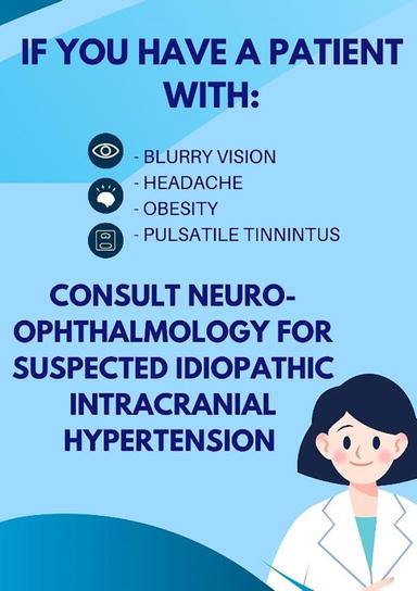
We believe an important step to mitigating vision loss from IIH is incorporating clinical education across multiple disciplines for the earliest signs and symptoms of IIH. We provide educational resources and tools for Emergency Room physicians, pediatricians, optometrists, neurologists, and primary care physicians at our institution. These educational resources attempt to mitigate the misdiagnosis of IIH as migraine symptoms. A major resource we utilize is Figure 8. Regardless of income or insurance status, anyone with IIH symptoms should not succumb to vision loss. We stand by a united goal to treat preventable blindness for all social groups through a multi-disciplinary team that helps expedite treatment and maintain vision.
Figure 8.
Our poster that is utilized as an educational resource in various medicaldisciplines to raise awareness of IIH symptoms.
This chapter explores IIH in the pediatric population regarding its demographics, clinical manifestations, potential underlying pathophysiology, diagnostic steps, and the importance of a multi-disciplinary approach to management, as it is the most optimal. This allows patients to receive comprehensive care that will address more aspects of IIH while also maintaining long-term goals and remission.
This is an example of one of our patients and our clinical approach to a patient with IIH:
Case 1 is a 15-year-old female with PMH of pre-diabetes, hypertriglyceridemia, eosinophilic esophagitis, class II Obesity (BMI 120% of 95th percentile, BMI 33.4 kg/m2) who was referred by her optometrist with severe headaches, blurred vision, and papilledema. On exam, her BCVA was 20/20 in both eyes and color vision was full. She has a maternal great-aunt who had IIH and has had a long history of consuming ultra-processed foods, daily sweetened beverages, and a sedentary lifestyle. OCT RNFL were 124 μm the right eye and 144 μm in the left eye. MRI of the brain was unremarkable. The patient did not want a lumbar puncture done at the time. She was started on acetazolamide 500 mg daily. Weight loss was recommended, and she started with a registered dietician and a pediatric weight management physician. Her headaches and blurred vision resolved. Her papilledema resolved to within normal limits after 3 months of acetazolamide. Her symptoms resolved after 6 months of therapy, and her acetazolamide was switched to topiramate1. In 1 year, her BMI went from 33.4 kg/m2 to 30.2 kg/m2, and lost a total of 15 lbs (Figures 9 and 10).
Figure 9.
Growth chart of BMI over time for Case 1.
Figure 10.
OCT RNFL and GCC trends for Case 1.
References
1.Ahmad SR, Moss HE. Update on the diagnosis and treatment of idiopathic intracranial hypertension. Seminars in Neurology. 2019;39(6):682-691. DOI: 10.1055/s-0039-1698744
2.Johanson CE, Duncan JA3, Klinge PM, Brinker T, Stopa EG, Silverberg GD. Multiplicity of cerebrospinal fluid functions: New challenges in health and disease. Cerebrospinal Fluid Research. 2008;5:10-10. DOI: 10.1186/1743-8454-5-10
3.Giuseffi V, Wall M, Siegel PZ, Rojas PB. Symptoms and disease associations in idiopathic intracranial hypertension (pseudotumor cerebri): A case-control study. Neurology. 1991;41(2 (Pt 1):239-244. DOI: 10.1212/wnl.41.2_part_1.239
4.Kilgore KP, Lee MS, Leavitt JA, et al. Re-evaluating the incidence of idiopathic intracranial hypertension in an era of increasing obesity. Ophthalmology. 2017;124(5):697-700. DOI: 10.1016/j.ophtha.2017.01.006
5.Degnan AJ, Levy LM. Pseudotumor cerebri: Brief review of clinical syndrome and imaging findings. American Journal of Neuroradiology. 2011;32(11):1986-1993. DOI: 10.3174/ajnr.A2404
6.Gillson N, Jones C, Reem RE, Rogers DL, Zumberge N, Aylward SC. Incidence and demographics of pediatric intracranial hypertension. Pediatric Neurology. 2017;73:42-47. DOI: 10.1016/j.pediatrneurol.2017.04.021
7.Gordon K. Pediatric pseudotumor cerebri: Descriptive epidemiology. Canadian Journal of Neurological Sciences. 1997;24(3):219-221. DOI: 10.1017/s031716710002182x
8.Gaier ED, Heidary G. Pediatric idiopathic intracranial hypertension. Seminars in Neurology. 2019;39(6):704-710. DOI: 10.1055/s-0039-1698743
9.Brara SM, Koebnick C, Porter AH, Langer-Gould A. Pediatric idiopathic intracranial hypertension and extreme childhood obesity. The Journal of Pediatrics. 2012;161(4):602-607. DOI: 10.1016/j.jpeds.2012.03.047
10.Aylward SC, Waslo CS, Au JN, Tanne E. Manifestations of pediatric intracranial hypertension from the intracranial hypertension registry. Pediatric Neurology. 2016;61:76-82. DOI: 10.1016/j.pediatrneurol.2016.04.007
11.Corbett JJ, Savino PJ, Thompson HS, et al. Visual loss in pseudotumor cerebri. Follow-up of 57 patients from five to 41 years and a profile of 14 patients with permanent severe visual loss. Archives of Neurology. 1982;39(8):461-474. DOI: 10.1001/archneur.1982.00510200003001
12.Friedman DI. Headaches due to low and high intracranial pressure. Continuum (Minneap Minn). 2018;24(4, Headache):1066-1091. DOI: 10.1212/CON.0000000000000623
13.Wall M, Corbett JJ. Revised diagnostic criteria for the pseudotumor cerebri syndrome in adults and children. Neurology. 2014;83(2):198-199. DOI: 10.1212/01.wnl.0000452039.32455.3e
14.Kilgore KP, Lee MS, Leavitt JA, Frank RD, McClelland CM, Chen JJ. A population-based, case-control evaluation of the association between hormonal contraceptives and idiopathic intracranial hypertension. American Journal of Ophthalmology. 2019;197:74-79. DOI: 10.1016/j.ajo.2018.09.014
15.Walters BN, Gubbay SS. Tetracycline and benign intracranial hypertension: Report of five cases. British Medical Journal (Clinical Research Edition). 1981;282(6257):19-20. DOI: 10.1136/bmj.282.6257.19
16.Contreras-Martin Y, Bueno-Perdomo JH. Idiopathic intracranial hypertension: Descriptive analysis in our setting. Neurologia. 2015;30(2):106-110. DOI: 10.1016/j.nrl.2013.08.009
17.Wang MTM, Bhatti MT, Danesh-Meyer HV. Idiopathic intracranial hypertension: Pathophysiology, diagnosis and management. Journal of Clinical Neuroscience. 2022;95:172-179. DOI: 10.1016/j.jocn.2021.11.029
18.Mollan SP, Ali F, Hassan-Smith G, Botfield H, Friedman DI, Sinclair AJ. Evolving evidence in adult idiopathic intracranial hypertension: Pathophysiology and management. Journal of Neurology, Neurosurgery and Psychiatry. 2016;87(9):982-992. DOI: 10.1136/jnnp-2015-311302
19.Ahmed RM, Wilkinson M, Parker GD, et al. Transverse sinus stenting for idiopathic intracranial hypertension: A review of 52 patients and of model predictions. American Journal of Ophthalmology. 2011;32(8):1408-1414. DOI: 10.3174/ajnr.A2575
20.Subramaniam S, Fletcher WA. Obesity and weight loss in idiopathic intracranial hypertension: A narrative review. Journal of Neuroophthalmology. 2017;37(2):197-205. DOI: 10.1097/WNO.0000000000000448
21.Hornby C, Botfield H, O'Reilly MW, et al. Evaluating the fat distribution in idiopathic intracranial hypertension using dual-energy X-ray absorptiometry scanning. Neuroophthalmology. 2017;42(2):99-104. DOI: 10.1080/01658107.2017.1334218
22.Sun WYL, Switzer NJ, Dang JT, et al. Idiopathic intracranial hypertension and bariatric surgery: A systematic review. Canadian Journal of Surgery. 2020;63(2):E123-E128. DOI: 10.1503/cjs.016616
23.Hornby C, Mollan SP, Botfield H, O'Reilly MW, Sinclair AJ. Metabolic concepts in idiopathic intracranial hypertension and their potential for therapeutic intervention. Journal of Neuroophthalmology. 2018;38(4):522-530. DOI: 10.1097/WNO.0000000000000684
24.Swiston C, Seay M. Case Report of Idiopathic Intracranial Hypertension and Frisen Scale Papilledema Grading. Utah: Moran CORE; 2018. Available from: https://morancore.utah.edu/section-05-neuro-ophthalmology/idiopathic-intracranial-hypertension-iih-and-papilledema-grading/
25.Friedman DI, Liu GT, Digre KB. Revised diagnostic criteria for the pseudotumor cerebri syndrome in adults and children. Neurology. 2013;81(13):1159-1165. DOI: 10.1212/WNL.0b013e3182a55f17
26.Suh SY, Kim S. IIH with normal CSF pressures? Indian Journal of Ophthalmology. 2013;61(11):681-682. DOI: 10.4103/0301-4738.119416
27.Malhotra K, Padungkiatsagul T, Moss HE. Optical coherence tomography use in idiopathic intracranial hypertension. Annals of Eye Science. 2020;5:3-10. DOI: 10.21037/aes.2019.12.06
28.Kedar S, Ghate D, Corbett JJ. Visual fields in neuro-ophthalmology. Indian Journal of Ophthalmology. 2011;59(2):103-109. DOI: 10.4103/0301-4738.77013
29.Higgins JNP, Cousins C, Owler BK, Sarkies N, Pickard JD. Idiopathic intracranial hypertension: 12 cases treated by venous sinus stenting. Journal of Neurology, Neurosurgery, Psychiatry. 2003;74(12):1662-1666. DOI: 10.1136/jnnp.74.12.1662
30.Hanak BW, Bonow RH, Harris CA, Browd SR. Cerebrospinal fluid shunting complications in children. Pediatric Neurosurgery. 2017;52(6):381-400. DOI: 10.1159/000452840
31.Kestle JRW, Riva-Cambrin J, Wellons JC, et al. A standardized protocol to reduce cerebrospinal fluid shunt infection: The hydrocephalus clinical research network quality improvement initiative. Journal of Neurosurgery Pediatrics. 2011;8(1):22-29. DOI: 10.3171/2011.4.PEDS10551
32.Greener DL, Akarca D, Durnford AJ, Ewbank F, Buckland GR, Hempenstall J. Idiopathic intracranial hypertension: Shunt failure and the role of obesity. World Neurosurgery. 2020;137:e83-e88. DOI: 10.1016/j.wneu.2020.01.040
33.Adesina O, Patel BC. Optic nerve decompression. In: StatPearls. Treasure Island, FL: StatPearls Publishing; 2023. Available from: https://www.ncbi.nlm.nih.gov/books/NBK538300/
34.Wall M, Kupersmith MJ, Kieburtz KD, et al. The idiopathic intracranial hypertension treatment trial: Clinical profile at baseline. JAMA Neurology. 2014;71(6):693-701. DOI: 10.1001/jamaneurol.2014.133
35.Ang JL, Teo KZ, Fraser CL. Weight loss in idiopathic intracranial hypertension: A retrospective review of outcomes in the clinical setting. Journal of Neuroophthalmology. 2021;41(4):e458-e463. DOI: 10.1097/WNO.0000000000001107
Written By
Maja Kostic, Elizabeth Colvin, Huynh Duy, Sarah Ro, Carolyn Quinsey, Inga Shevtsova and Sriram Machineni
Submitted: 08 September 2023Reviewed: 13 September 2023Published: 01 December 2023
 Open access peer-reviewed chapter