 Open access peer-reviewed chapter
Abstract
Non-auditory stimulation (NAS) is a potential complication in cochlear implants (CIs) that can impact both the effectiveness of sound transmission and the quality of life for users. This issue can often be mitigated through remedial CI device programming strategies. In some cases, the symptoms of NAS are persistent irrespective of typical audiological interventions. To develop an intervention for NAS that is tailored to the auditory system and surrounding structures of an individual CI user requires a transdisciplinary approach. This chapter proposes a model for transdisciplinary, patient-centred care of CI users who suffer from persistent NAS complications from intracochlear electrical stimulation. The model combines aspects of anatomy, radiology, computational modelling and audiology to gain an understanding of the parameters that give rise to the occurrence of NAS and to provide an avenue for investigating novel intervention strategies. Addressing unintended facial nerve stimulation by a CI is used to demonstrate the application of the model.
Keywords
- cochlear implants
- facial nerve stimulation
- computational modelling
- person-centred care
- transdisciplinary team
1. Introduction
The cochlear implant (CI) or ‘bionic earʼ is a technological intervention intended to treat severe to profound sensorineural hearing loss [1]. Often, hearing impairment is a result of cochlear hair cell loss where up to 80% of inner hair cells may be lost as a result of aetiological factors, such as medication or trauma [2]. More significant hearing loss resulting in profound deafness can only be treated by surgically implanting a CI. When the origin of deafness is sensorineural, a CI can stimulate the auditory nerve electrically to induce action potentials (APs) that travel along the auditory nerve to elicit a sensation of sound. The electrically induced APs are indistinguishable from naturally elicited APs, but there are a number of differences. Notably, the dynamic range between the stimulation threshold and saturation rates at which APs are generated (the firing rate) in response to increases in electrical current level is much smaller than that of natural neural excitation. CI speech perception in quiet may be close to normal, but it may deteriorate in noise [2].
While designs vary across CI companies [3], all commercial CIs have the same components. Externally, the CI system consists of a microphone, speech processor and transmitter [4]. Internally, the CI consists of a surgically implanted receiver-stimulator and the electrode array within the cochlea. The speech processor acts as an artificial cochlea by taking the sound input from the microphone and emulating some of the processing known to take place in the healthy cochlea. The speech processor translates the acoustic signal into a format that may be used to stimulate the surviving auditory nerve fibres in the inner ear or cochlea
Cochlear implants vary in electrode design and a variety of speech processing strategies exist. For example, electrode array designs differ in the number of electrode contacts, the spacing between electrodes, the positioning (e.g., lateral wall and perimodiolar designs) and the length of the electrode array [5]. Speech processors may, for example, focus on delivering temporal fine structure [6] or on accurate representation of sound spectra [7].
Cochlear implant outcomes are highly variable [8], with some CI users achieving open set speech recognition, and some having virtually no benefit other than being in contact with their sound environment. Variability in the hearing outcomes with a CI device is a direct result of interpersonal differences in the auditory systems of individual CI users and the integration of the CI technology with this unique system. Despite different designs that attempt to address different issues, outcomes are similar across electrode designs and speech processors [3]. For example, current focusing and current steering methods are used to mitigate the effect of current spread [9, 10], some electrode designs target deep insertions to ensure that the low-frequency region of the cochlea is stimulated [11] and electrode array positioning is either perimodiolar or against the cochlear lateral wall [12]. These different designs have not solved the problem of variable outcomes across CI users [13]. The same problems remain across implant designs: programming (or mapping) of the device for optimal hearing through optimal electrical stimulation parameter settings is unique for each user [14, 15]; neural survival patterns vary across CI users [16] but electrode arrays cannot be positioned to make provision for this; and patterns of current spread in each CI user is uniquely a function of individual anatomy [17]. To complicate matters further, programming of the CI device for adults can be done interactively, but mapping for babies or CI users with multiple disabilities needs to be done with objective methods, for example, based on electrically-evoked compound action potential (eCAP) measures [18]. Objective methods are needed for individualised mapping [19, 20]. Cochlear implant companies supply software platforms to streamline mapping and to some extent make the mapping process objective [21], but this is of use mostly for cases without additional complications.
Standard management and care approaches become inadequate once there is additional complexity, for example, where inner ear malformations exist [22], when the electrode array is not ideally placed, when neural survival is compromised, when the electrical environment deviates from the expected, for example when bone conductance is affected due to disease [23] or when non-auditory stimulation (NAS) causes complications that compromises hearing performance and/or quality of life [24].
When a CI user experiences sub-optimal performance or complications that cannot be resolved through conventional interventive mapping strategies, a multi- and transdisciplinary approach that incorporates computational modelling as an advanced analytic and diagnostic tool may be required to effectively investigate and ideally address the issue. This approach is person-specific, taking into account the unique characteristics of each CI user’s cochlear environment, as well as other factors that may impact hearing outcomes.
This chapter describes a transdisciplinary approach that encompasses three disciplines or domains within CI management, differentiated here as the clinical, medical sciences and engineering domains. The aim of a transdisciplinary approach is to actively integrate and synthesise knowledge across various knowledge fields to address complex problems and produce novel solutions. This enables a person-centred care approach in the management of complex CI cases that is underpinned by the translation of computational models to clinical application.
2. The disciplines in CI management
2.1 Clinical domain
The standard model of care for CI users is typically practised in the clinical domain illustrated in Figure 1. The standard of care is person-centred, which aims to recognise the unique needs, preferences, values and circumstances of each CI user. A clinical model of care is typically an interdisciplinary model, where different members of the care team share information and insights across their respective disciplines.

Figure 1.
Illustration of care, centred in the clinical domain.
Within the clinical domain, a CI team consists of a variety of clinical practitioners that need to operate in four interdisciplinary areas. The assessment of hearing and management of hearing loss is primarily dealt with by audiologists. The surgical implantation of the CI device and maintaining auditory health is the joint responsibility of the ear, nose and throat (ENT) surgeon, radiologist, neurologist and paediatrician in paediatric cases. Counselling and supporting the CI user and their family members to optimise quality of life is the task of a psychologist and social worker, while the auditory (re)habilitation through auditory training is managed by an audiologist and/or speech-language therapist.
When a person is first diagnosed with a hearing loss that indicates CI candidacy, the person is usually referred to a CI team for clinical evaluation. The evaluation includes at least an ENT surgeon and an audiologist. Often, evaluation for candidacy also involves a psychologist and a social worker to appraise whether personal circumstances will support the decision to implant [25], along with audiological measures [26] and imaging that may include either computed tomography (CT) or magnetic resonance imaging (MRI) or both [27]. Imaging allows evaluation of the anatomic suitability for receiving a CI and may inform a yes/no decision and selection of the ear to be implanted.
Surgical implantation of the internal components of the CI device is performed by the ENT surgeon with in theatre support from an audiologist and/or a specialised clinical engineer made available by the CI company. Objective neurophysiological tests are routinely performed in-theatre as initial assessment of the neural responses to electrical stimulation and to verify the integrity of the device. For example, electrocochleography (ECochG) can be used to monitor the auditory nerve’s response to electrode insertion and activation. This can provide real-time feedback to the surgeon about the electrode’s positioning and its potential impact on the auditory nerve.
Subsequent activation and programming of the CI device are carried out by an audiologist. Thereafter, the CI user will have regular follow-up visits to an audiologist for updating and maintaining the CI user’s map. The professional team may also include a speech-language therapist that assists with (re)habilitation with the CI device [28]. Initially, CI stimulation sounds distorted [29] and the postlingual hearing-impaired CI user needs to relearn to hear, while children with congenital deafness need to learn to hear with their CI(s) [30]. Facilitating this process is known as auditory training. (Re)habilitation and auditory training, which focus on improving speech perception, along with ongoing follow-up appointments and psychosocial support to address emotional aspects, are required to optimise CI outcomes. Lifelong management and CI device upgrades ensure access to the latest technology, aiming to enhance hearing outcomes and improve the quality of life for CI users.
An array of tools is available in the clinical domain for managing and maintaining hearing performance. From a surgical and medical practitioner perspective, this includes diagnostic tools to determine the aetiology of hearing loss, for example, genetic testing, assessment of the medical history of a patient to determine factors such as exposure to loud noise, ototoxicity, infections and underlying medical conditions, imaging that may help to identify structural abnormalities, tumours or nerve hypoplasia and physical examination. In cases where hearing loss is associated with balance problems, balance and vestibular testing may also form part of the assessments that may be conducted.
From an audiology perspective, the most important tool in the management of CI hearing performance is the CI device programming software provided by each of the CI companies. Besides mapping, this software also allows neurophysiological evaluations, such as measuring the eCAP and assessing the integrity of the device by measuring the electrical impedances of the electrode contacts. Pure tone audiometry is frequently used to monitor the unaided and aided hearing thresholds to assess both the progression of hearing loss and the aided benefit that a person receives from a hearing aid, CI or hybrid device. However, pure tone audiometry does not necessarily translate to speech understanding, and hence, tools to evaluate hearing performance as reflected by speech understanding are also employed. These include listening test batteries targeted at CI users, for example, the minimal auditory capabilities (MAC) test battery [31] and the basic auditory skills evaluation (BASE) battery [32]. Neurophysiological testing that assesses the functional level of the auditory pathway includes electrically evoked auditory brainstem responses (eABRs) and ECochGs. ECochGs can help identify conditions, such as endolymphatic hydrops (Meniere’s disease) and assist in determining the site of lesion in auditory neuropathy spectrum disorder (ANSD), which may affect the success of cochlear implantation.
The default interaction between the care provided by the clinical team and the CI user, if no complications are experienced, is shown by the arrows in Figure 1. However, if complications arise, a second step assessment is introduced in the management strategy. Figure 2 illustrates an array of additional tests and evaluations that may be employed in the assessment phase.

Figure 2.
Extended care that includes an assessment step for complicated CI cases.
2.2 Medical sciences domain
Much of what is known about the pathology of the auditory system originates from the medical sciences domain. Medical sciences are crucial for guiding clinical interventions related to hearing loss, particularly in the context of CIs. The success of CIs relies on contributions from diverse medical science fields, including anatomy, histology and medical imaging. These contribute significantly to the foundational knowledge base, offering insight into the structure and function of the various components of the auditory system and providing tools to assess this system in order to facilitate clinical decision-making. Figure 3 illustrates some of the expertise that the medical domain offers to inform and support the development of CI technology and the care and management of CI users.

Figure 3.
The medical sciences domain offers expertise to inform and support the development of CI technology and the care and management of CI users.
A detailed morphological (qualitative: form and structure) and morphometric (quantitative: size and proportions) description of the cochlea, cochlear nerve and surrounding structures, provided by anatomists is necessary for the design of CI electrode arrays that would minimise implant trauma while providing an effective interface with the remaining neural structures and contribute to hearing preservation. Knowledge about the morphology is essential for surgeons to locate the cochlea during the implantation procedure and to minimise the risk of damage to surrounding structures, specifically including the facial nerve which is in close proximity to the scala tympani [33]. Understanding the anatomical variations among individual patients’ cochleae and possible abnormalities is important for selecting appropriate electrode arrays [34] and customising implant procedures. Cadaver studies are an important facet of anatomy as this form the basis for characterising the detailed morphology and morphometry of the auditory system and also facilitate testing and refinement of new CI devices.
Histology provides insights into the cellular and histopathological changes that occur in the cochlea due to hearing loss. This information is invaluable for understanding the potential impact of long-term hearing loss on the cochlear structures and similarly, the ontogenesis of electric hearing over time. Histological studies can also guide researchers and clinicians in designing electrodes that effectively interface with the remaining auditory nerve fibres.
Medical imaging, specifically CT scans and MRI, provide visualisations of the inner ear anatomy, even though the resolution is low relative to the dimensions of the cochlea. Micro-computed tomography (μCT), of which the radiation levels are too high for safe clinical use, is often used in CI research to provide high-resolution images of cadaver temporal bones so that the variations in cochlear morphology and morphometry may be studied. Cadaver studies also allow researchers to correlate imaging data with actual anatomical structures in order to validate the accuracy of measurements from imaging data. Imaging, both clinical and scientific, is crucial for preoperative planning, allowing surgeons to appreciate the general anatomy of the cochlea and surrounding structures and to assess the patient’s individual cochlear anatomy [35]. Imaging provides a means to identify anatomical abnormalities and to plan the optimal trajectory for electrode insertion. Post-operative imaging provides information about the location of the electrode array within the cochlea and may also provide information about progressive changes in the cochlea and rest of the auditory system.
2.3 Engineering domain
Engineers are at the core of the design, development and improvement of CI technology, which involves several engineering disciplines, for example, biomedical engineering, electronic engineering, computer and software engineering, materials science and mechanical engineering. Engineers design and develop the hardware and software components of CI systems. This includes the external speech processor, the wireless, transcutaneous link between the external processor and the implanted system, the implanted electrode array and the powering of the implant. The development of sophisticated signal processing algorithms within the external processor that aims to enhance and optimise the quality of sound delivered to CI users is an ever-evolving engineering task. Careful design of the power supply and power management strategies are important to ensure that users can enjoy continuous hearing without frequent expensive battery replacements [36].
Apart from the development of the technology, engineers are also involved in research that probes the intricacies of the integration between biology and technology. Computational modelling is an effective tool to probe the biophysical interactions between CIs and the complex biology of the human auditory system. While untangling the factors influencing the relation between stimulus and perception in CI users is challenging because of the complexity and inaccessibility of the auditory system, computational models offer a pathway for examining how specific parameters influence aspects of hearing. The aim of a computational model is to create a digital twin of the auditory system of a specific CI user that would allow researchers to probe this system in a way that would involve excessively invasive procedures if conducted on the CI user (Figure 4).

Figure 4.
Engineering domain, where computational models of a CI users’ cochleae are designed and implemented. The computational model aims to be a digital twin of the CI user so that invasive test results and insights from the CI user may be gained. The arrow from the model to the CI user represents the validation route where predictions from the model are evaluated against data measured from the CI user.
There are two main types of models in CI research: models that describe the periphery, and therefore the biophysical interface between the implant and the auditory system and models that describe processing in the central auditory nervous system and perception of sound. Both of these types of models may use a physiologically-based approach, a phenomenological approach or a combination of the two. Because of the complexity of the complete auditory system, a large number of models have been created to describe distinct characteristics of this system. Often a number of different models, each describing a specific component or aspect, are combined to capture the characteristics of the auditory system more completely.
One example is modelling of the electrically stimulated auditory periphery that requires at least two models for a complete description: one to describe the volume conduction effects when stimulation current is injected into the cochlea (and surrounding tissue) and one to describe the response of the auditory nerve fibres to the injected current. Finite element volume conduction (FEVC) models of the cochlea attempt to capture a detailed three-dimensional (3D) description of the cochlea, auditory nerve and in many cases, the surrounding tissues and structures [17, 37, 38, 39, 40, 41]. The parameters that describe the anatomy and geometry of a particular CI user’s cochlear structures are extracted from medical imaging data (CT or MRI scans). Different tissues in the model are assigned different electrical properties, for which the values are derived from experimental data originating in the medical sciences domain. Such a model can be used to simulate the spread of electrical current through the tissues when current is injected through the CI electrodes.
The response to the stimulation current at the output of the FEVC model is predicted by an auditory nerve fibre (ANF) model. An ANF model may take several forms, for example, it may be conductance-based, such as the Hodgkin-Huxley [39, 42, 43] or Schwartz-Eikhoff [44, 45, 46] models that comprise a set of differential equations, or it may be phenomenological [47, 48, 49] or a combination of the two [50]. ANF models are commonly developed in programming languages, such as MATLAB or Python. In combination, the FEVC and ANF models, referred to as compound models, allow prediction and analyses of the neural response to electrical stimulation [17, 23, 40, 51, 52, 53]. From these, researchers can infer how the perception of sound may be affected by different intertwined factors such as the pattern of neural survival, the location of the electrode array relative to the surviving nerve fibres and different stimulation strategies. Simulation results are typically validated by comparing them to experimental data obtained from CI users.
2.4 The need for a transdisciplinary model of care
The previous sections expanded on the roles that each of the three domains play in providing medical care, understanding the anatomy and physiology of the auditory system and how this relates to the physical characteristics of its structures and developing the various components of CIs and their supporting technologies. While the clinical model of care has been successful in providing essential services and support to CI users, a transdisciplinary team that combines the tools and techniques from the different domains may provide a deeper understanding of the complex relationship among factors that affect the unique hearing experience of individual CI users. This becomes particularly important when addressing CI complications such as NAS, where identifying the root cause might not be straightforward, or when multiple factors contribute to an issue.
Figure 5 depicts a transdisciplinary model that includes the clinical, medical sciences and engineering domains.

Figure 5.
A diagrammatic representation of model-based person-centred care through a six-stage transdisciplinary approach.
The model consists of a six-stage approach centred around the CI user and a digital twin of their electrically stimulated hearing system. The digital twin is created as a computational model of the cochlea, cochlear nerve and surrounding structures and allows the team to investigate a particular CI user’s auditory system from an invasive viewpoint. If a CI user experiences NAS, as illustrated in Figure 5, the first stage in the approach is clinical care where a CI user’s clinical team will identify the issue, and then proceed to intervene according to the standard models of care. If the care model is not effective in mitigating the issue, the second stage is entered, where the transdisciplinary team considers the different facets of the case and formulates data collection requirements. To be able to create a computational model of the CI user’s cochlea, morphological and morphometric data must be measured for the specific individual in the third stage to complement data that was already collected during the care stage. The fourth stage involves parameterisation of the relevant data to create a person-specific computational model of components of the CI user’s auditory system. The fifth stage involves creating the model and validating it against empirical data obtained from the medical history of the user, as well as from clinical tests, experiments and image data. The model is then used to investigate possible solutions to alleviate or mitigate the complication. Interventions that may have been designed or identified are tested in stage six. The process is iterative as a first iteration might not yield desired results. It is also important to realise that it may be impossible to devise an effective solution for a particular person. However, the model provides an avenue for understanding the underlying causes of the complication, thereby informing the care strategy for this and future CI users.
In the remainder of the chapter, the application of the transdisciplinary model will be unpacked within the context of facial nerve stimulation (FNS).
3. Application of the model: a practical example
To demonstrate the application of the model of care through the complication of FNS, it is necessary to provide a conceptual framework of FNS through which the reader may appreciate the complexities of and the necessity for the transdisciplinary approach.
3.1 Facial nerve stimulation
3.1.1 What is FNS, and what does it do?
FNS occurs when electrical current from the CI intended to stimulate the ANFs, spreads to and excites the nearby labyrinthine segment of the facial nerve (FN) [24, 54, 55, 56]. Symptoms of FNS in CI users vary from user discomfort in the form of facial tingling to twitching of the facial muscle to severe, painful and disfiguring facial spasms. Other symptoms that may present are referred pain in the form of headaches, loss of taste, and sensory and secretomotor symptoms. Symptoms could negatively affect the functionality of the CI, may present directly after CI device activation or after a period of use and, in users with bilateral CIs, may present on both or only one side of the face [54, 57, 58, 59]. In extreme cases, symptoms can become so severe that explantation and reimplantation may be required [60].
3.1.2 How prevalent is FNS?
Using the PRISMA algorithm on an initial pool of over 1000 CI and FNS-related articles, Van Horn et al. [60] provide a systematic review of the rate of and factors associated with FNS in CI users followed by an assessment of FNS management strategies. In their review and meta-analysis of 37 articles representing 5936 CI users, the reported FNS rate ranged from 0.68–43%, with a cumulative incidence rate of 5.6%, or almost one in every 18 CI users. Cochlear implant users with otosclerosis showed an overall FNS rate of 26% (range, 6.25–75%) having a significantly higher odds ratio (OR) to non-otosclerosis (OR = 13.73, 95% confidence interval [CI] 3.57–52.78, p < 0.01). Users with lateral wall arrays are more likely to experience FNS with an OR = 3.92, 95% CI 1.6–10.47, p < 0.01 and those with cochlear malformations showed an overall FNS rate of 28% (range, 5.3–43%).
3.1.3 What factors affect, aggravate or increase the risk of FNS?
Factors that appear to affect the occurrence of FNS include cochlear ossification (post-meningitis and otosyphilis), otosclerosis, osteoporosis, temporal bone fracture, a narrow bony cochlear nerve canal and cochlear malformations [55, 56, 57, 61, 62, 63, 64]. Unfortunately, these factors also often result in inadequate loudness perception by the CI user, which then requires an increase in stimulus current level, thereby aggravating the FNS because the increased stimulus current level increases the spread or leakage of current to the facial nerve [55]. The type of CI array also proved a significant factor in FNS occurrence. Straight, lateral arrays are situated closer to the outer wall of the cochlea and thus closer to the facial nerve, whereas pre-curved, perimodiolar arrays with modiolar facing contacts lie in closer proximity to and direct the current towards the modiolus and are thus less likely to leak current towards the facial nerve [59].
Another risk factor in FNS is cochlear-facial dehiscence, an opening between the cochlea and labyrinthine segment of the FN [65]. When a CI electrode is activated, electrical current from the electrode may spread through the opening to the labyrinthine segment of the FN and cause FNS. Factors that may contribute to cochlear-facial dehiscences include local thinning of the otic capsule (such as in cases of meningitis and otosclerosis), topographic anatomy of the cochlea and impingement of the otic capsule upon the labyrinthine segment of the FN causing narrowing [66].
Intraoperative electrophysiology testing, such as eABR, can be performed during the surgical procedure to assess the function of the auditory. Abnormal findings, such as the detection of facial nerve (CN VII) and vestibular potentials on eABR testing, may indicate a higher likelihood of post-operative NAS [67].
3.2 Application of model-based person-centred care
Managing complications with CI stimulation is multifaceted and requires a comprehensive approach that considers all factors, including medical history, hearing needs and CI program settings. By utilising a multi- and transdisciplinary computational model-based approach, the objective is to create a person-specific map that maximises the individual’s hearing outcomes while minimising complications. In this section, a qualitative narrative of the management of five CI users, presenting with FNS, is presented to demonstrate how a transdisciplinary model of care is applied at the University of Pretoria, South Africa.
3.2.1 Case studies
Case 1. In a study by Badenhorst et al. [23], a CI user received a Med-El C40+ standard electrode array in the right ear at age 4 years and 4 months following a meningitis infection. The left cochlea was implanted 3 years later with a Med-El Pulsar ci100 short array electrode because of severe ossification of the scalae. A few years later this implant caused acute headaches and pain after which it was explanted and replaced with a dummy electrode for possible future reimplantation in the ossifying left cochlea. A year after the explantation, the user experienced FNS with the remaining right implant. FNS also presented in the left cochlea upon reimplantation with a Med-El compressed array.
Case 2. The study by Van der Westhuizen et al. [53] reports on a CI user for whom hearing was lost after gentamicin injection in the middle ear following a retrosigmoid vestibular neurectomy for intractable vertigo in Ménière’s disease. The person received a Cochlear Nucleus implant with a CI24RE Contour Advance electrode in the right ear. While image analysis revealed electrodes 16 and 17 to be closest to the FN, the CI user experienced severe FNS on most of the electrodes in the array.
Case 3. The origin of this CI user’s deafness is unknown and started at around 4 to 5 years of age. The right ear was implanted, and the user experienced FNS since initial stimulation. The first implant was removed and was replaced with a Cochlear Nucleus implant with a CI24RE Contour Advance electrode. The FNS persisted, causing epiphora (watery, red eye) and activation of the orbicularis oculi muscle on the side of the implant.
Case 4. This CI user’s hearing was lost following a tuberculosis meningitis infection. The right ear was implanted with a Cochlear Nucleus implant with CI24RE Contour Advance electrode. The CI user experienced FNS on all electrodes.
Case 5. This CI user’s biological mother contracted rubella during pregnancy. The CI user was diagnosed with bilateral hearing loss at age four and started to use a hearing aid in the right ear at age nine and only 10 years later started to use a hearing aid in the left ear. This CI user received a cochlear nucleus implant in the left ear with a CI422 electrode as an adult and experienced no FNS complications in this ear. Eight years later, this CI user was implanted with a Cochlear Nucleus implant with a CI632 electrode in the right ear. Severe FNS is experienced on all electrodes implanted on the right side.
3.2.2 Care
From the brief descriptions of the FNS cases above, it is evident that the origins and management of FNS are intricate. For all the above cases, not only is the quality of sound provided by the implant affected by FNS but also the quality of life of the CI users. Despite technological advances in cochlear implantation, the interpersonal variability in factors that affect hearing outcomes, for example, aetiology of deafness and neural survival patterns implies that auditory (re)habilitation may require a range of different management strategies.
Before the need for a transdisciplinary approach becomes evident, there are a number of standard interventions that are typically attempted within the clinical domain (Figure 2). The most common non-invasive intervention strategy for FNS involves reprogramming or remapping of the CI by either increasing stimulation pulse widths or increasing interphase gaps to reduce stimulation levels [60, 68]. In a recent systematic review [60], FNS management strategies were reported in 28 of the 37 included studies, with a cumulative sample of 259 CI users with FNS. All studies reported the resolution of FNS through remedial CI device programming strategies, but the exact success rate of these programming strategies was not reported [60]. Reprogramming of the CI device has been attempted for all five cases described above but failed to mitigate the adverse effects of FNS. Upon onset of FNS in case 1, a new map was programmed that initially did not cause FNS, but it returned over time because of steadily increasing current levels that were required to maintain functional hearing.
To counteract the larger current spread associated with higher current levels (and associated FNS), electrical pulse width increase may present a solution. A disadvantage of this approach may be in a processor, where the time duration of a stimulus cycle (cycling through all electrodes that need to be activated) is fixed, which (e.g.) is the case in a SPEAK processing strategy. The number of spectral maxima used in the processor relates to the spectral content of the acoustic signal that is encoded in the electrical stimuli. SPEAK uses between six and eight maxima, and the spectral representation becomes sparse if the number of maxima becomes too small. For case 4, the map that rendered the best hearing (though still unsatisfactory) could accommodate only four maxima due to large pulse widths.
Lowering the stimulation levels of offending electrodes to current levels just below the stimulus level inducing the FNS symptoms while still achieving auditory stimulation [55] was also attempted in combination with deactivation of one or more electrode contacts. Deactivation of electrodes on multichannel arrays is relatively common in clinical practice, although this tends to compromise the quality of hearing and speech perception outcomes, especially in cases where multiple electrodes of the upper basal and/ or middle turns of the cochlea need to be deactivated to manage FNS [60, 68]. In cases, where both lowered stimulation levels and electrode deactivation were required to manage severe FNS, all five CI cases reported insufficient loudness to offer a comfortable listening experience. This is in accordance with the literature that states remedial CI programming strategies to be useful in most cases to reduce FNS symptoms to some extent, though sound quality and auditory performance are often affected, especially in cases that require significantly decreased levels of electrical stimulation or aggressive electrode deactivation [54, 60].
Alternative stimulation modes and triphasic pulse stimulation have also been documented as non-invasive intervention strategies for FNS [68, 69]. For example, for case 4, after the conventional extra-cochlear stimulation modes MP1 and MP2 were explored with increased pulse widths and decreased rate, intra-cochlear stimulation modes, common ground, bipolar+3, and bipolar+5 were tested. However, the intracochlear modes failed to provide sufficient loudness growth because of the close proximity of the active and reference electrodes [70]. The largest electrical dynamic range for the most active channels was achieved with a pseudomonopolar map. However, even with this map, loudness was compromised and did not improve with pulse widths beyond 200 μs. Similar challenges were experienced in the mapping of the other four CI cases, with none of the conventional FNS intervention strategies rendering a satisfactory outcome.
If none of the non-invasive strategies prove successful in managing the FNS, there are invasive strategies that may be considered. The first is botulinum toxin (Botox) treatment [71], but the inability to use facial muscles or facial expressions may lead to reduced quality of life [53]. For case 1, Botox injections were administered as a temporary measure to alleviate the effect of the FNS. The treatment partially inhibited the FNS, but at the cost of little to no facial expression while the CI user still reported perceiving contraction of the facial muscles. Botox injections were administered in case 4, but because of the risk of affecting the motor neuron pathways required for eating and drinking, the treatment was conservative. The FNS was less visible, but the user was still aware of the muscle contractions, similar to the experience of case 1.
In some severe FNS cases, where typical audiological interventions such as reprogramming of the CI and deactivation of electrodes do not alleviate the FNS symptoms, explantation and reimplantation of the CI device may be necessary [60, 68, 72, 73]. Case 3 had the first implant removed due to persistent FNS. The CI user was re-implanted with a perimodiolar electrode array that placed the electrodes further away from the FN because of its modiolus-hugging intracochlear location. The strategy of changing the type of electrode array from a lateral wall type to a perimodiolar type at reimplantation has been documented as an effective approach to eliminate FNS [74], though this approach did not resolve FNS for case 4. Case 1 had the CI on the left removed due to acute headaches and pain, which is indicative of NAS. The left side was re-implanted with a compressed array that could only be partially inserted because of ossification. Reimplantation was, however, not effective to mitigate complications in this CI user. FNS was experienced shortly after activation.
Removal of the CI and its intracochlear electrode array can also cause explantation trauma. Since explantation is often carried out a number of years after initial implantation, reactive tissue may have formed around the electrode, which may increase the risk for explantation trauma [75] and may, therefore, compromise the outcomes that may be achieved with a subsequent implant. For this reason, explantation should be a last resort.
In a transdisciplinary team, where computational modelling expertise is available, the model presented in Figure 5 should be applied before explantation is considered, and if the latter is seen as the only option, to predict the chances of success of reimplantation given what can be extrapolated through the tools of a transdisciplinary team and specifically computational modelling.
3.2.3 Assessment
When the methods available for mitigating FNS in the clinical domain (Figure 2) have been exhausted, the transdisciplinary team needs to assess the available information and data that have been collected for the CI user. The transdisciplinary team comprises a core team that includes members having expertise in the relevant domains, as well as the clinical team of the specific CI user. The team may, thus, be geographically distributed, necessitating an agreement on a remote operation strategy.
The assessment will determine how computational modelling will be used to inform management of the case. In some cases, a complete model will be constructed to assess a CI user’s unique situation, while it may be possible to extrapolate from observations that were made in previous modelling studies that investigated other cases.
The primary inputs required to continue with a model-based approach are good quality imaging data that may be complemented by anatomical, neuroanatomical and neurophysiological data originating from the intersection between the clinical and medical domains, the CI user’s medical history and device programming information from the clinical domain, and data from speech perception tests, CI device integrity tests, neurophysiological tests and psychoacoustic tests originating from the intersection between the clinical and engineering domains.
3.2.4 Measurement
3.2.4.1 Imaging
The foundation for the construction of the 3D computational models of a specific CI user’s cochlea and surrounding structures is clinical images, for example, CT. These images are used to quantify the dimensions of cochlear structures. The human cochlea is as unique as a fingerprint [76], and it has been shown that person-specific modelling of the cochlea needs to take these variations into account [17].
Multi-slice computed tomography (MSCT), cone-beam computed tomography (CBCT) and magnetic resonance imaging (MRI) are part of the routine pre-operative evaluation of a CI candidate, while only CT-based modalities are viable for post-operative assessment due to the metal artefacts that obscure the cochlea in MRI scans.
For computational models, post-operative CT images are the most crucial as these provide information on the morphology of the individual cochlea, as well as the intracochlear location of the electrode array, the depth of insertion and possibly insertion trauma should the array appear not to follow the trajectory of the scala tympani [77]. If post-operative CT imaging is not available, it has to be acquired, for example. in case 3, where the process could not continue before the images were available.
It is also worth noting that imaging modalities may differ depending on the availability of scanner technology. Figure 6 shows a mid-modiolar slice through the CT images for each case. Cases 1, 2 and 4 had MSCT scans available, while cases 3 and 5 had CBCT images available. The advantage of MSCT is that Hounsfield units may be used to track changes in bone density over time should multiple scans be available. This characteristic of MSCT was utilised for case 1, where the progression of changes in the bone density could be measured over time. However, the grey values in CBCT are at most moderately correlated with Hounsfield units and are dependent, among others, on the specific brand of the scanner [78]. This might make it difficult to track changes in bone density should the only image data be CBCT. On the other hand, CBCT is considered a low-radiation dose alternative to MSCT with the added advantage of superior image resolution and metal artefact reduction [79], which are beneficial to capture morphometric data for the cochlea.

Figure 6.
Panels for cases 1 to 5. Two-dimensional mid-modiolar slices through the cochleae of cases 1 to 5 show the variation in image quality. Cases 1, 2 and 4 had MSCT scans, while cases 3 and 5 had CBCT scans. The bright areas in the images are the metal electrode contacts. The scan for case 2 is particularly affected by artefacts. Landmarks panel. A landmark set that may be used to describe the bony shape of the cochlea. Closed dots indicate landmarks that should be visible on clinical CT images, while open dots indicate landmarks that have to be derived from high-resolution images such as micro-CT scans (not used for live humans) to augment the clinical scans.
3.2.4.2 Landmarking
To measure the morphometric characteristics of the cochlea and facial nerve, landmarking is used to quantify the shape of the cochlea. Landmarks are a 3D constellation of discrete anatomical locations described by Cartesian coordinates. They are points of correspondence on each specimen that are identifiable and distinguishable in every cochlea. The landmarks shown in Figure 6 have been selected to describe the shape of the bony aspects of the cochlear canal for the purpose of 3D modelling. A minimum set of landmarks is required for the construction of 3D cochlear landmarks and for cochlear modelling; at least, the lateral spiral (LS points in Figure 6) must be distinguishable.
Considering the scans in Figure 6, case 1 shows no artefact and the lateral spiral and electrode position can be measured. Case 2 presents a scan, where the artefacts from the electrode array obscured the lateral spiral due to beam hardening. In this case, a parametric model that may be less accurate than a landmark-based model may be required [80]. While a parametric model may be constructed from a reduced landmark set, it could miss some of the person-specific local variations in cochlear shape. In case 3 no landmarks can be accurately measured and a parametric model will be required. The image for case 4, although blurry, allows the landmarks that describe the lateral (LS) and superior spirals (SS) to be delineated. Case 5 has little interference from electrode artefacts and here the superior, inferior, lateral, superolateral and inferolateral spirals can be measured. This scan can be used for 3D computational modelling of the implanted cochlea.
The measurements that need to be taken to reconstruct the FN are the length, width and angle of the labyrinthine, tympanic and mastoid segments of the FN as described in Badenhorst et al. [23]. Because the FN is enclosed in a bony canal, these measurements can all be made on the CT images of the CI users.
3.2.5 Parameterisation
The parameters in a computational model are values, settings or variables that define the characteristics, behaviour and properties of the model.
Model parameters that define the 3D structure of the model are the size and shape of the cochlear and surrounding structures and originate from the interface between medical sciences and engineering (Figure 5). Parameterisation of the landmark measurements obtained from the CT images involves the transformation of the measured coordinates to the coordinate system in which the models are constructed, as well as applying appropriate scaling factors. Electrical material properties are mostly obtained from the literature since it is not possible to measure these in a living CI user. However, the electrical properties of the bone surrounding the cochlea play a crucial role in the distribution of current outside the cochlea and is, therefore, an important parameter to include in FNS models. The only way to obtain an indication of the value of this parameter for a particular CI user is by means of imaging. While there is no direct relationship between Hounsfield units, bone mineral density and the electrical properties of bone, the trends in this parameter may be inferred from CT images taken at various time intervals. In the models, a decrease in the mineral content of the bone encapsulating the cochlea is represented by decreasing the bone impedance relative to the accepted value for this parameter in modelling studies [23, 38]. For case 1, several CT scans were available over a period of 18 months where a decrease in the bone density could be observed, which could then be included in the modelling study [23].
Model parameters that attempt to capture neural health, for example, the extent of neural degeneration, loss of auditory nerve fibres or other damage to the auditory nerve fibres, originate from neurophysiological testing that is performed at the intersection between the clinical and engineering domains (Figure 5). This includes eCAP measurements and eABR measurements, which provide an indication of the status of the auditory nerve [81, 82].
3.2.6 Modelling
Once the measurements and parameterisation are done for a specific CI user, the person-specific 3D finite element model of the cochlea, auditory nerve, facial nerve and surrounding tissues, all having different electrical properties [17], is constructed in COMSOL and positioned in a generic head model as shown in Figure 7 for case 1 [23]. The basal-to-middle turn of the cochlea’s scala tympani was partially ossified having an order of magnitude higher resistivity compared to the normal perilymph.

Figure 7.
Finite element volume conduction (FEVC) COMSOL model of a head, skull, brain, partially ossified cochlea, CI and facial nerve (Case 1).
Current spreads through the cochlea as shown in Figure 8 for a monopolar electrode that injects current. Notice the close proximity of the FN’s labyrinthine segment to the cochlea in the bottom-right of Figures 7 and 8. The bottom left panel shows how the current emanates from electrode 5. Most current then spreads through the partially ossified cochlea with some current passing through and possibly stimulating the FN (top frame).

Figure 8.
Bottom-right shows asemi-transparent view of a partially ossified cochlea, electrode and FN for a FEVC simulation in COMSOL for case 1. The top panel shows the current spread (red lines) through the cochlea upon activation of electrode 5 (E5), which is nearest to the FN. The bottom left panel, an enlargement of the section indicated, shows the current originating from the superior and inferior electrodes. As expected, the top panel shows the majority of the current spreading towards the right through the cochlea, but some current finds its way through the FN causing FNS.
Using a MATLAB model, ANFs are scaled and distributed within the cochlea based on the cochlear geometry and ANF morphology such as that shown in Figure 9 [39, 43, 51].
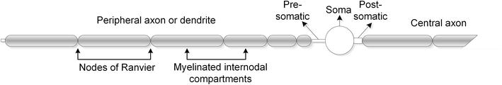
Figure 9.
Morphology of a computationally modelled auditory nerve fibre.
The distribution of the ANFs relative to the cochlear geometry and electrode array for case 1 is shown in Figure 10 with the soma and nodes represented by the markers. The FEVC current spread output and resulting electric potential distribution in the cochlea combined with the fibre node locations provide the potential at each node for any selected stimulus strategy and level. The individual nodal potentials then serve as input to the purely conductance-based computational ANF model in MATLAB to determine which auditory and/or facial nerve fibres will activate and produce a propagating action potential along the fibre [43].

Figure 10.
A MATLAB rendering of the ANF distribution within the cochlea for case 1 indicating the dendritic nodes of Ranvier, soma, electrode array and central axons.
From the model simulations, the onset of FNS in case 1 was attributed to a decrease in resistivity of the otic capsule, likely because of the ossification of the cochlear ducts. The study suggests that the left cochlea showed a larger increase in auditory stimulation and FNS compared to the right cochlea, possibly because of increased resistivity in the cochlear ducts and decreased resistivity of the otic capsule. This could result in a decreased dynamic range in the left cochlea, which in turn could lead to decreased thresholds of the auditory and facial nerve fibres, resulting in the observed FNS.
Similar person-specific modelling and simulations for case 2 were used to investigate the implementation of apical reference (AR) stimulation as a means of reducing FNS. In using the most apical electrode as the reference electrode, the current could be contained within the cochlea and directed away from the facial nerve fibre (FNF). The hypothesis of an increased pulse width reducing the effect of FNS was tested by simulating both AR and conventional monopolar (MP1 + 2) stimulation with phase durations of 25 μs and 300 μs. For MP1 + 2 stimulation, a number of electrodes showed FNF thresholds lower than the ANF thresholds at 25 μs, with the increase in pulse width barely increasing these FNF thresholds above the ANF thresholds. For AR stimulation, however, FNF thresholds were already above ANF thresholds at 25 μs, with the increase to 300 μs further increasing the threshold difference (dynamic range) while also decreasing both FNF and ANF thresholds. This supports the hypothesis that AR stimulation is a viable strategy to alleviate FNS.
3.2.7 Intervention
The last stage of the transdisciplinary model of care is the intervention stage, where the findings of the process need to inform the subsequent management of the CI user. It is important to note that it might be necessary to iterate through the six stages of the care model based on an improved understanding of the CI user’s situation from previous iterations. The following provides a brief summary of the intervention stage for the five case studies after the first iteration through the model details in this chapter.
For case 1, personal circumstances of the CI user and Med-El implants’ inability to accommodate the implementation of the AR stimulation strategy with its standard clinical software limited the intervention options. Though much was learned through the modelling process that can be applied in future cases, the only intervention possible at the time was counselling for the CI user to better understand what was happening in the cochleae, how that was causing severe FNS and why the standard care procedures of remapping do not work in this specific case.
For cases 2 and 3, the outcomes were similar as both CI users have been using their CIs for an extensive period. Both MP1 + 2 and AR stimulation were tested in the laboratory. AR stimulation resulted in a decrease in threshold and an increase in comfort levels as the model predicted and the users reported a reduction in FNS symptoms. The quality of sound with AR stimulation was, however, either poor or the CI users reported very little sound perception. Despite this, the study validated the model prediction that an AR strategy could reduce FNS.
Case 4 had not benefited from the CI as the FNS was present from initial stimulation. While a reasonable map could be fitted for this CI user, no speech perception of sound could be achieved with the AR stimulation mode.
To verify that the AR stimulation mode could provide a viable alternative stimulation mode, it was tested on two CI users who do not experience FNS. These CI users could perceive speech, though they reported their perception of sound to be much different from that provided by their original map with default parameter settings. This suggested that should a CI user be able to listen with this map, extensive auditory retraining may be required, especially if the CI user had become accustomed to another stimulation mode.
Case 5 experienced FNS almost from initial stimulation, and therefore did not have as much experience with the CI in the ear affected by FNS as cases 1 to 4. The CI user started auditory retraining with the AR map, which was effective in mitigating the FNS, but after approximately 5 weeks, the FNS returned. This suggests changes in the cochlear environment and sensitivity of the FN that have not been taken into consideration in the present models. This observation informs the next iteration through the care model to tease out the underlying mechanisms that are responsible for the FNS to reoccur.
It is worth noting that a non-invasive solution to FNS may or may not be attained through the application of a transdisciplinary model. However, the process provides a deep insight into a specific CI user’s auditory system that may provide grounds for decision-making and may inform counselling. The model also contributes to the knowledge base about the factors that cause a unique listening experience for each CI user.
4. Conclusions
A transdisciplinary model of care that incorporates expertise and tools from the domains of clinical care, medical sciences and engineering is of great value for managing complex FNS cases in CI users. As discussed in the previous sections, the challenges associated with FNS complications are multifaceted and require a comprehensive approach that goes beyond traditional disciplinary boundaries. A transdisciplinary model of care offers two main advantages above a purely clinical approach to FNS management.
Firstly, it embraces a holistic person-centred care approach. Complex cases in CI require an approach that focuses on the individual’s overall well-being and quality of life. A transdisciplinary model of care enables professionals from various disciplines to collaborate and provide a comprehensive assessment and tailored treatment plan. By considering medical, clinical and engineering perspectives together, the model considers the unique needs and limitations of each CI user more comprehensively than a single perspective approach.
Secondly, it offers a means to develop a detailed understanding of the effects of electrical stimulation on the particular CI user’s auditory system. By involving medical sciences and bioengineering, the model can leverage expertise in fields such as computational neurophysiology and anatomy, medical imaging, signal processing and control systems theory and medical device design to devise intervention strategies for a particular CI user, or if a satisfactory intervention is not possible, to provide an understanding of the underlying causes of the FNS that may be used to manage the CI user’s expectations.
It is important to acknowledge the challenges associated with implementing a transdisciplinary model of care in all CI clinics. Limited resources, geographical constraints and limited availability of experts from multiple disciplines can hinder the establishment of a transdisciplinary team. Telehealth and remote consultation technologies can be utilised to facilitate communication and collaboration among specialists located in different locations. In this way, CI users and their clinical teams may gain access to the advantages that a multidisciplinary team may offer.
Finally, while a purely clinical model of care remains the standard approach for CI users in many clinics, the inclusion of a transdisciplinary team can offer significant advantages when CI users experience complications with their CIs.
References
- 1.
Carlyon RP, Goehring T. Cochlear implant research and development in the twenty-first century: A critical update. Journal of the Association for Research in Otolaryngology. 2021; 22 (5):481-508 - 2.
Salvi R, Sun W, Ding D, Chen GD, Lobarinas E, Wang J, et al. Inner hair cell loss disrupts hearing and cochlear function leading to sensory deprivation and enhanced central auditory gain. Frontiers in Neuroscience. 2017; 10 :1-14 - 3.
Zeng FG. Trends in cochlear implants. Trends in Amplification. 2004; 8 (1):1-34 - 4.
Patrick JF, Busby PA, Gibson PJ. The development of the Nucleus®Freedom™ cochlear implant system. Trends in Amplification. 2006; 10 (4):175-200 - 5.
Ertas YN, Ozpolat D, Karasu SN, Ashammakhi N. Recent advances in cochlear implant electrode Array design parameters. Micromachines. 2022; 13 (7):1081, 1-20. Available from:https://www.mdpi.com/2072-666X/13/7/1081/pdf?version=1657700835 - 6.
Vermeire K, Punte AK, Van De Heyning P. Better speech recognition in noise with the fine structure processing coding strategy. ORL. 2010; 72 (6):305-311 - 7.
Donaldson GS, Nelson DA. Place-pitch sensitivity and its relation to consonant recognition by cochlear implant listeners using the MPEAK and SPEAK speech processing strategies. Journal of the Acoustical Society of America. 2000; 107 (3):1645-1658 - 8.
Moberly AC, Bates C, Harris MS, Pisoni DB. The enigma of poor performance by adults with cochlear implants. Otology & Neurotology. 2016; 37 (10):1522-1528 - 9.
Srinivasan AG, Landsberger DM, Shannon RV. Current focusing sharpens local peaks of excitation in cochlear implant stimulation. Hearing Research. 2010; 270 (1-2):89-100 - 10.
Roux J, Hanekom JJ. Effect of stimulation parameters on sequential current-steered stimuli in cochlear implants. Journal of the Acoustical Society of America. 2022; 152 (1):609-623 - 11.
Hochmair I, Hochmair E, Nopp P, Waller M, Jolly C. Deep electrode insertion and sound coding in cochlear implants. Hearing Research. 2015; 322 :14-23 - 12.
Risi F. Considerations and rationale for cochlear implant electrode design-past, present and future. Journal of International Advanced Otology. 2018; 14 (3):382-391 - 13.
Cosetti MK, Waltzman SB. Outcomes in cochlear implantation: Variables affecting performance in adults and children. Otolaryngologic Clinics of North America. 2012; 45 (1):155-171 - 14.
Shapiro WH, Bradham TS. Cochlear implant programming. Otolaryngologic Clinics of North America. 2012; 45 (1):111-127 - 15.
Chang CJ, Sun CH, Hsu CJ, Chiu T, Yu SH, Wu HP. Cochlear implant mapping strategy to solve difficulty in speech recognition. Journal of the Chinese Medical Association. 2022; 85 (8):874-879 - 16.
Zimmermann CE, Burgess BJ, Nadol J. Patterns of degeneration in the human cochlear nerve. Hearing Research. 1995; 90 (1-2):192-201 - 17.
Malherbe TK, Hanekom T, Hanekom JJ. Constructing a three-dimensional electrical model of a living cochlear implant user’s cochlea. International Journal for Numerical Methods in Biomedical Engineering. 2016; 32 (7):e02751, 1-23. Available from:https://pubmed.ncbi.nlm.nih.gov/26430919/ - 18.
Scorpecci A, D’Elia A, Malerba P, Cantore I, Consolino P, Trabalzini F, et al. Maps created using a new objective procedure (C-NRT) correlate with behavioral, loudness-balanced maps: A study in adult cochlear implant users. European Archives of Oto-Rhino-Laryngology. 2016; 273 (12):4167-4173 - 19.
Ali H, Noble JH, Gifford RH, Labadie RF, Dawant BM, JHL H, et al., editors. Image-guided customization of frequency-place mapping in cochlear implants. In: ICASSP, IEEE International Conference on Acoustics, Speech and Signal Processing – Proceedings. Brisbane, Australia; 2015. pp. 5843-5847 - 20.
Kurz A, Müller-Graff FT, Hagen R, Rak K. One click is not enough: Anatomy-based fitting in experienced cochlear implant users. Otology and Neurotology. 2022; 43 (10):1176-1180 - 21.
Wathour J, Govaerts PJ, Lacroix E, Naïma D. Effect of a CI programming fitting tool with artificial intelligence in experienced cochlear implant patients. Otology and Neurotology. 2023; 44 (3):209-215 - 22.
Sennaroglu L. Cochlear implantation in inner ear malformations – A review article. Cochlear Implants International. 2010; 11 (1):4-41 - 23.
Badenhorst W, Hanekom T, Gross L, Hanekom JJ. Facial nerve stimulation in a post-meningitic cochlear implant user: Using computational modelling as a tool to probe mechanisms and progression of complications on a case-by-case basis. Cochlear Implants International. 2021; 22 (2):68-79 - 24.
Broomfield S, Mawman D, Woolford TJ, O'Driscoll M, Luff D, Ramsden RT. Non-auditory stimulation in adult cochlear implant users. Cochlear Implants International. 2000; 1 (1):55-66 - 25.
Entwisle LK, Warren SE, Messersmith JJ. Cochlear implantation for children and adults with severe-to-profound hearing loss. Seminars in Hearing. 2018; 39 (4):390-404 - 26.
Reddy P, Dornhoffer JR, Camposeo EL, Dubno JR, McRackan TR. Using clinical Audiologic measures to determine cochlear implant candidacy. Audiology and Neurotology. 2022; 27 (3):235-242 - 27.
Vaid S, Vaid N. Imaging for cochlear implantation: Structuring a clinically relevant report. Clinical Radiology. 2014; 69 (7):e307-ee22 - 28.
Van Bogaert L, Machart L, Gerber S, Lœvenbruck H, Vilain A. Speech rehabilitation in children with cochlear implants using a multisensory (French Cued Speech) or a hearing-focused (Auditory Verbal Therapy) approach. Frontiers in Human Neuroscience. 2023; 17 :1-17 - 29.
Caldwell MT, Jiam NT, Limb CJ. Assessment and improvement of sound quality in cochlear implant users. Laryngoscope Investigative Otolaryngology. 2017; 2 (3):119-124 - 30.
Roman S, Rochette F, Triglia JM, Schön D, Bigand E. Auditory training improves auditory performance in cochlear implanted children. Hearing Research. 2016; 337 :89-95 - 31.
Owens E, Kessler DK, Raggio MW, Schubert ED. Analysis and revision of the minimal auditory capabilities (MAC) battery. Ear and Hearing. 1985; 6 (6):280-290. DOI: 10.1097/00003446-198511000-00002 - 32.
Shafiro V, Hebb M, Walker C, Oh J, Hsiao Y, Brown K, et al. Development of the basic auditory skills evaluation battery for online testing of cochlear implant listeners. American Journal of Audiology. 2020; 29 (3s):577-590 - 33.
Kruschinski C, Weber BP, Pabst R. Clinical relevance of the distance between the cochlea and the facial nerve in cochlear implantation. Otology and Neurotology. 2003; 24 (5):823-827 - 34.
Eshraghi AA, Ocak E. Cochlear implant electrode choice in challenging surgical cases: Malformation, residual hearing, ossification, or Reimplantation. Current Otorhinolaryngology Reports. 2017; 5 (4):315-322 - 35.
Walker N, Pham N, Ledbetter L. Cochlear implantation: Current and future roles of imaging before, during, and after implantation. Current Radiology Reports. 2023; 11 (7):97-107 - 36.
de Nobel J, Kononova AV, Briaire JJ, Frijns JHM, Bäck THW. Optimizing stimulus energy for cochlear implants with a machine learning model of the auditory nerve. Hearing Research. 2023; 432 :108741. Available from:https://www.sciencedirect.com/science/article/pii/S0378595523000539?via%3Dihub - 37.
Malherbe TK, Hanekom T, Hanekom JJ. Can subject-specific single-fibre electrically evoked auditory brainstem response data be predicted from a model? Medical Engineering and Physics. 2013; 35 :926-936 - 38.
Malherbe TK, Hanekom T, Hanekom JJ. The effect of the resistive properties of bone on neural excitation and electric fields in cochlear implant models. Hearing Research. 2015; 327 :126-135 - 39.
Badenhorst W, Hanekom T, Hanekom JJ. Analysis of a purely conductance-based stochastic nerve fibre model as applied to compound models of populations of human auditory nerve fibres used in cochlear implant simulations. Biological Cybernetics. 2017; 111 (5-6):439-458 - 40.
Hanekom T, Hanekom JJ. Three-dimensional models of cochlear implants: A review of their development and how they could support management and maintenance of cochlear implant performance. Network: Computation in Neural Systems. 2016; 27 (2-3):67-106 - 41.
Kalkman RK, Briaire JJ, Frijns JH. Stimulation strategies and electrode design in computational models of the electrically stimulated cochlea: An overview of existing literature. Network (Bristol, England). 2016; 27 (2-3):107-134 - 42.
Hodgkin AL, Huxley AF. A quantitative description of membrane current and its application to conduction and excitation in nerve. Journal of Physiology. 1952; 117 :500-544 - 43.
Rattay F, Lutter P, Felix H. A model of the electrically excited human cochlear neuron. I. Contribution of neural substructures to the generation and propagation of spikes. Hearing Research. 2001; 153 (1-2):43-63 - 44.
Schwartz JR, Eikhof G. Na currents and action potentials in rat myelinated nerve fibres at 20 and 30 degrees. Archives. 1987; 409 :569-577 - 45.
Frijns JHM, Mooij J, ten Kate JH. A quantitative approach to modeling mammalian myelinated nerve fibres for electrical prosthesis design. IEEE Transactions on Biomedical Engineering. 1994; 41 (6):556-566 - 46.
Frijns JHM, Mooij J, Schoonhoven R. Have mammalian myelinated nerve fibres diameter dependent nodal properties? A model study. In: Frijns JHM, editor. Cochlear Implants a Modelling Approach. Den Haag: CIP-Data Koninklijke Bibliotheek; 1995. pp. 69-92 - 47.
Takanen M, Bruce IC, Seeber BU. Phenomenological modelling of electrically stimulated auditory nerve fibers: A review. Network: Computation in Neural Systems. 2016; 27 (2-3):157-185 - 48.
Bruce IC, Erfani Y, Zilany MSA. A phenomenological model of the synapse between the inner hair cell and auditory nerve: Implications of limited neurotransmitter release sites. Hearing Research. 2018; 360 :40-54 - 49.
Tabibi S, Boulet J, Dillier N, Bruce IC. Phenomenological model of auditory nerve population responses to cochlear implant stimulation. Journal of Neuroscience Methods. 2021; 358 :109212, 1-12 - 50.
van Gendt MJ, Briaire JJ, Kalkman RK, Frijns JHM. A fast, stochastic, and adaptive model of auditory nerve responses to cochlear implant stimulation. Hearing Research. 2016; 341 :130-143 - 51.
Kalkman RK, Briaire JJ, Dekker DMT, Frijns JHM. The relation between polarity sensitivity and neural degeneration in a computational model of cochlear implant stimulation. Hearing Research. 2022; 415 :108413, 1-14. Available from:https://www.sciencedirect.com/science/article/pii/S0378595521002471/pdfft?md5=f7e5acecb2878bb928091aead5010391&pid=1-s2.0-S0378595521002471-main.pdf - 52.
Biesheuvel JD, Briaire JJ, Kalkman RK, Frijns JHM. The effect of stimulus level on excitation patterns of individual electrode contacts in cochlear implants. Hearing Research. 2022; 420 :108490, 1-12 - 53.
van der Westhuizen J, Hanekom T, Hanekom JJ. Apical reference stimulation: A possible solution to facial nerve stimulation. Ear and Hearing. 2022; 43 (4):1189-1197 - 54.
Ahn JH, Oh SH, Chung JW, Lee K-S. Facial nerve stimulation after cochlear implantation according to types of nucleus 24-channel electrode arrays. Acta Otolaryngologica. 2009; 129 (6):588-591 - 55.
Bigelow DC, Kay DJ, Rafter KO, Montes M, Knox GW, Yousem DM. Facial nerve stimulation from cochlear implants. American Journal of Otology. 1998; 19 (2):163-169 - 56.
Niparko JK, Oviatt DL, Coker NJ, Sutton L, Waltzman SB, Cohen NL. Facial nerve stimulation with cochlear implantation. VA cooperative study group on cochlear implantation. Otolaryngology and Head and Neck Surgery. 1991; 104 (6):826-830 - 57.
Kelsall DC, Shallop JK, Brammeier TG, Prenger EC. Facial nerve stimulation after nucleus 22-channel cochlear implantation. The American Journal of Otology. 1997; 18 (3):336-341 - 58.
Berrettini S, Vito de A, Bruschini L, Passetti S, Forli F. Facial nerve stimulation after cochlear implantation: Our experience. Acta Otorhinolaryngologica Italica. 2011; 31 (1):11-16 - 59.
Seyyedi M, Herrmann BS, Eddington DK, Nadol JB Jr. The pathologic basis of facial nerve stimulation in otosclerosis and multi-channel cochlear implantation. Otology & Neurotology. 2013; 34 (9):1603-1609 - 60.
Van Horn A, Hayden C, Mahairas AD, Leader P, Bush ML. Factors influencing aberrant facial nerve stimulation following cochlear implantation: A systematic review and meta-analysis. Otology and Neurotology. 2020; 41 (8):1050-1059 - 61.
Muckle RP, Levine SC. Facial nerve stimulation produced by cochlear implants in patients with cochlear otosclerosis. The American Journal of Otology. 1994; 15 (3):394-398 - 62.
Kempf HG, Tempel S, Johann K, Lenarz T. Complications of cochlear implant surgery in children and adults. Laryngo- Rhino- Otologie. 1999; 78 (10):529-537 - 63.
Rotteveel LJ, Proops DW, Ramsden RT, Saeed SR, van Olphen AF, Mylanus EA. Cochlear implantation in 53 patients with otosclerosis: Demographics, computed tomographic scanning, surgery, and complications. Otology & Neurotology. 2004; 25 (6):943-952 - 64.
Bahmer A, Adel Y, Baumann U. Preventing facial nerve stimulation by triphasic pulse stimulation in cochlear implant users: Intraoperative recordings. Otology & Neurotology. 2017; 38 (10):e438-ee44 - 65.
Song Y, Alyono JC, Bartholomew RA, Vaisbuch Y, Corrales CE, Blevins NH. Prevalence of radiographic cochlear–facial nerve dehiscence. Otology & Neurotology. 2018; 39 (10):1319-1325 - 66.
Schart-Morén N, Larsson S, Rask-Andersen H, Li H. Anatomical characteristics of facial nerve and cochlea interaction. Audiology and Neurotology. 2017; 22 (1):41-49 - 67.
Alnafjan F, Hasan Z, Sanli H, da Cruz MJ. Risk factors for facial nerve and other non-auditory side effects following cochlear implantation. Otology & Neurotology. 2021; 42 (8):e1022-e10e9 - 68.
Braun K, Walker K, Sürth W, Löwenheim H, Tropitzsch A. Triphasic pulses in cochlear implant patients with facial nerve stimulation. Otology & Neurotology. 2019; 40 (10):1268-1277 - 69.
Wolfe J, Schafer E. Programming Cochlear Implants. San Diego, CA: Plural Publishing; 2014 - 70.
Arenberg J. Threshold and channel interaction in cochlear implant users: Evaluation of the tripolar electrode configuration. The Journal of the Acoustical Society of America. 2007; 121 :1642-1653 - 71.
Langman A, Quigley S, Heffernan J, Brazil C. Use of botulinum toxin to prevent facial nerve stimulation following cochlear implantation. The Annals of Otology, Rhinology & laryngology Supplement. 1995; 166 :426-428 - 72.
Polak M, Ulubil SA, Hodges AV, Balkany TJ. Revision cochlear implantation for facial nerve stimulation in otosclerosis. Archives of Otolaryngology–Head & Neck Surgery. 2006; 132 (4):398-404 - 73.
Alharbi FA, Spreng M, Issing PR. Facial nerve stimulation can improve after cochlear reimplantation and post-operative advanced programming techniques: Case report. 2012 - 74.
Battmer R, Pesch J, Stöver T, Lesinski-Schiedat A, Lenarz M, Lenarz T. Elimination of facial nerve stimulation by reimplantation in cochlear implant subjects. Otology & Neurotology. 2006; 27 (7):918-922 - 75.
Asfour L, Risi F, Miah H, Roland JT Jr. Cochlear implant explantation: An in vitro model to evaluate electrode explant force and trauma. Cochlear Implants International. 2022; 23 (4):189-194 - 76.
Erixon E, Högstorp H, Wadin K, Rask-Andersen H. Variational anatomy of the human cochlea: Implications for cochlear implantation. Otology and Neurotology. 2009; 30 (1):14-22 - 77.
Van Wermeskerken GKA, Prokop M, Van Olphen AF, Albers FWJ. Intracochlear assessment of electrode position after cochlear implant surgery by means of multislice computer tomography. European Archives of Oto-Rhino-Laryngology. 2007; 264 (12):1405-1407 - 78.
Selvaraj A, Jain RK, Nagi R, Balasubramaniam A. Correlation between gray values of cone-beam computed tomograms and Hounsfield units of computed tomograms: A systematic review and meta-analysis. Imaging Science in Dentistry. 2022; 52 (2):133 - 79.
Scarfe WC, Farman AG. What is cone-beam CT and how does it work? Dental Clinics of North America. 2008; 52 (4):707-730 - 80.
Pietsch M, Aguirre Dávila L, Erfurt P, Avci E, Lenarz T, Kral A. Spiral form of the human cochlea results from spatial constraints. Scientific Reports. 2017; 7 (1):7500 - 81.
Bierer JA, Faulkner KF, Tremblay KL. Identifying cochlear implant channels with poor electrode-neuron interfaces: Electrically evoked auditory brain stem responses measured with the partial tripolar configuration. Ear and Hearing. 2011; 32 (4):436-444. DOI: 10.1097/AUD.0b013e3181ff33ab - 82.
Jahn KN, Arenberg JG. Identifying cochlear implant channels with relatively poor electrode-neuron interfaces using the electrically evoked compound action potential. Ear and Hearing. 2020; 41 (4):961-973. DOI: 10.1097/AUD.0000000000000844