Open Access is an initiative that aims to make scientific research freely available to all. To date our community has made over 100 million downloads. It’s based on principles of collaboration, unobstructed discovery, and, most importantly, scientific progression. As PhD students, we found it difficult to access the research we needed, so we decided to create a new Open Access publisher that levels the playing field for scientists across the world. How? By making research easy to access, and puts the academic needs of the researchers before the business interests of publishers.
We are a community of more than 103,000 authors and editors from 3,291 institutions spanning 160 countries, including Nobel Prize winners and some of the world’s most-cited researchers. Publishing on IntechOpen allows authors to earn citations and find new collaborators, meaning more people see your work not only from your own field of study, but from other related fields too.
To purchase hard copies of this book, please contact the representative in India:
CBS Publishers & Distributors Pvt. Ltd.
www.cbspd.com
|
customercare@cbspd.com
Despite homeopathy being a therapy that has been around for over 200 years, it still arouses controversy, both because of the high dilutions in which it is administered and because of the methodologies used in research, which are not always adequate to reach a definitive conclusion. Our group has been researching the action of single homeopathic products and homeopathic complexes on immune system cells and on tumor cells, both animal and human, for over 25 years. In this chapter, we intend to summarize the main results obtained in theses and dissertations, products of academic research with strict controls, all carried out by nonhomeopathic professionals, mainly by biologists and biochemists, whose conclusion is unquestionable. We may not know the detailed mechanisms of action of homeopathy, just as we do not know about most allopathic medicines, medicines that often work one way in a patient. In some people, the doses must be tested, or even sometimes have a paradoxical action in others. The results were surprising, and here we will present two of the investigated complexes, which have already been published with different names or acronyms, but which, in recent years, have been referred to as M1 (mixture 1) and M8 (mixture 8), letters and numbers used in a large double-blind study conducted.
*Address all correspondence to: dorlybuchi@gmail.com
1. Introduction
The growing popularity of complementary and alternative medicine in the public sector is reflected in the scientific community by an increase in the number of scientific papers attempting to assess the efficacy of therapeutic effects. Data on biological effects of ultra-dilutions are investigated with laboratory studies under carefully controlled conditions. The use of models with plants or animals allows the observation of direct responses, which eliminates subjectivity and speculation regarding to placebo effect, which is the main criticism in relation to studies with humans. The lack of conclusive studies that provide information on how homeopathy works, led us to test some homeopathic medicines, widely used by homeopathic physicians, using macrophages as biological models, as well as other cell types. To find out which medicine to look for, we held a meeting with homeopathic physicians with extensive clinical experience, asking which quick results suggested action on macrophages or other cells of the immune system. We were informed that some medications are even suggested as first aid, acting first on the symptoms, for example, pain and fever, and then they look for the “background medication.” These drugs were exactly the ones we were interested in. These medicines were tested in macrophages culture, individually and mixed. These cells responded to all products, and surprisingly differently for each product. The results with Mercurius solubilis were the only one of the published single drugs [1]. The more interesting result is that Merc sol stimulated different macrophages answer, modulating the liberation of cytokines and reactive species production. Lower dilutions increase interferon-gamma (IFNγ), while the higher ones augmented IL-4 production after in vitro treatment. It should be noted that IFNγ promotes Th1 cell responses and IL-4 leads to Th2-type humoral immunity and is involved in proliferation and differentiation of activated B-cells. That is, the same substance but in different dilutions present paradoxical responses, which before being published, were repeated several times.
Returning to the complexes, that is also too interesting, not all mixtures worked; for example, when we added Viscum album or Turmeric (Curcuma longa), the complexes seem to be inactivated. The homeopathic extracts used to produce M1 and M8 were specified by name and by doses in Table 1.
Composition
Matrix
Final concentration (v/v%)
M1
M8
Aconitum napellus
MT
20 d
20 d
0.1 × 10⁻¹⁹
Arsenicum album
6 d
18 d
18 d
0.1 × 10⁻¹⁷
Asa foetida
MT
20 d
20 d
0.1 × 10⁻¹⁹
Calcarea carbonica
8 d
16 d
16 d
0.1 × 10⁻¹⁵
Chelidonium majus
MT
20 d
-
0.1 × 10⁻¹⁹
Cinnamon
MT
20 d
-
0.1 × 10⁻¹⁹
Conium maculatum
5 d
17 d
17 d
0.1 × 10⁻¹⁶
Echinacea purpurea
MT
20 d
-
0.1 × 10⁻¹⁹
Gelsemium sempervirens
MT
20 d
-
0.1 × 10⁻¹⁹
Ipecacuanha
5 d
13 d
13 d
0.1 × 10⁻¹²
Phosphorus
12 d
20 d
20 d
0.1 × 10⁻¹⁹
Rhus toxicodendron
6 d
17 d
17 d
0.1 × 10⁻¹⁶
Silicea
12 d
20 d
20 d
0.1 × 10⁻¹⁹
Sulfur
12 d
24 d
24 d
0.1 × 10⁻²³
Thuja occidentalis
6 d
19 d
19 d
0.1 × 10⁻¹⁸
Table 1.
Summary of the main differences between cytokines production and the treatment with M1 and M8 complexes. Tumor necrosis factor alpha (TNFα), when in excess, is more reduced by M8, interferon-gamma (IFNγ) is more reduced by M1, and interleukin 10 (IL-10) is enhanced by M1 and reduced by M8.
It is known that cell cultures are particularly appropriate as an initial screening system in pharmaceutical research when possible modulatory effects of novel drugs are estimated. This experimental model seems to be particularly useful in evaluating the effects of homeopathic treatments, due to the great possibility of data for statistical analysis, without the disadvantages of clinical screening.
The immune system works perfectly by coordinating actions between cells and proteins to provide defense against infection. These cells and proteins do not form a single organ like the heart or liver. Instead, the immune system is dispersed throughout the body to provide rapid responses to a possible invasion and infection. The development of all immune system cells begins in the bone marrow with a hematopoietic (blood-forming) stem cell. Although all components of the immune system interact with each other, it is typical to consider two broad categories of immune responses: the innate immune system and the adaptive immune system.
Innate immune responses are carried out without prior additional “training” to do their jobs. These cells include neutrophils, monocytes, natural killer (NK) cells, and a set of proteins termed complement proteins. Innate responses to infection occur rapidly and reliably. Adaptive immune responses comprise the second category. These responses involve T-cells and B-cells, two cell types that require “training” or education to learn not to attack our own cells. The advantages of adaptive responses are their long-lived memory and the ability to adapt to new germs. Activation of macrophages represents one of the first events in the innate response. The mechanisms of innate and adaptive immunity are, however, interdependent. This intercommunication is also performed through macrophages, which participate in the production, mobilization, activation, and regulation of all effector cells of the immune system. They interact reciprocally with other cells, which causes the change of their properties, for specialized immunological functions. Macrophages have an important role in the secretion of various cytokines, in addition to act as antigen-presenting cells (APCs).
Cytokines are produced by many cell populations, but the main producers are helper T (Th) cells and macrophages, which predominantly produce proinflammatory cytokines when activated, involved in the upregulation of inflammatory reactions. The anti-inflammatory cytokines are a series of immunoregulatory molecules that control the proinflammatory cytokine response. These specific cytokines or antagonists would act to disrupt the hyperexcitability cycle, providing a new, nonopioid therapeutic approach for the treatment of pathological pain due to inflammation or injury. Several situations can lead to systemic inflammatory syndromes in the human body. Their common feature is a massive release of cytokines due to excessive activation of immune cells. Cytokine storm is usually understood to mean an exacerbated immune response characterized by the release of cytokines and other mediators. These mediators are part of an evolutionarily well-conserved innate immune response that is required for the efficient elimination of infectious agents and the repair processes immediately. In an appropriate inflammatory response, there is a balance between adequate cytokine production to clear invaders, on the one hand, and avoidance of a hyperinflammatory response follows. A balanced, “protective” inflammatory response consists of diverse mechanisms and involves activation of both pro- and anti-inflammatory pathways within the innate and the acquired immune systems. You can see a good review of storm cytokines in Jarczak and Nierhaus [2].
Our laboratory at the Federal University of Paraná used to research the action of pesticides (agriculture is one of the strong points in our state and in the country), fungal and lichen extracts on tumor cells, macrophages, and the immune system in general. At the end of the 1990s, the positive pulmonary response found in some patients with the use of homeopathic products aroused our curiosity. As we already had a cell culture laboratory, we consulted health homeopaths professionals, and we established protocols. Despite homeopathy being a medical and pharmaceutical specialty in our country, it is still a therapy that raises much debate, often confused with belief, as if it were a religion. It is common to hear expressions like “I believe” or “I don’t believe” in homeopathy.
Once we started to obtain quick and interesting answers, previous results were presented at conferences, and the questions asked by our peers led us to improve methodology to reduce possible variables. And curiosity led us to test different mixed products (homeopathic complexes) on cells and animals, but always produced according to the homeopathic pharmacopeia, by a specialized pharmacist. Highly diluted natural complexes comprise a combination of different compounds considered useful for a particular symptom or disease. The studies were double-blind, and positive and negative control groups were rigorously evaluated. The results were surprising with different products, but two of these complexes, due to their fast action, were studied more and became known by us as M1 and M8, symbols encoded in a double-blind study.
In general, the evaluations were carried out in the following order: (1) first, in vitro, where mice or humans cells were treated with different potencies of a drug and evaluated morphologically; (2) still in vitro, the tests were repeated for metabolic and biochemical evaluations in the potencies that presented the most interesting results; (3) then, tumor cells from mice and humans were evaluated in the presence of treated immune cells; (4) finally, the mice were treated in vivo with the drugs in the potencies that showed the best results; at the end of the treatment, their cells were collected to be evaluated morphologically and biochemically; (5) finally, tumor cells were inoculated into the mice, which were then treated, the tumors collected and evaluated.
The results presented here were obtained from theses, dissertations, and published articles. Therefore, equipment and technologies used in conventional research and fundamental software were used, such as Mirax Viewer Software, ImageJ (NIH) Software, and ImageJ’s Image Deconvolution plugin. The evaluations were done by light microscopy, confocal microscopy, transmission and scanning electron microscopy, flow cytometry, immunocytochemical, ultrastructural cytochemistry, invasion assay with transwell plates, human colon-rectal cancer cells stably transfected with the pNF-κB-hrGFP PlasmidStratagene was performed on HT-29 cells, histopathology, immunohistochemistry, ultrastructural pathology, slide scanning system, Microarrays and Gene Chips, microplate reader, enzyme-linked immunosorbent assay (ELISA), and many other routine biochemical techniques.
All homeopathic products we test must be shaken vigorously (succussion) before use. Whatever happens to the solution during shaking remains for a limited time.
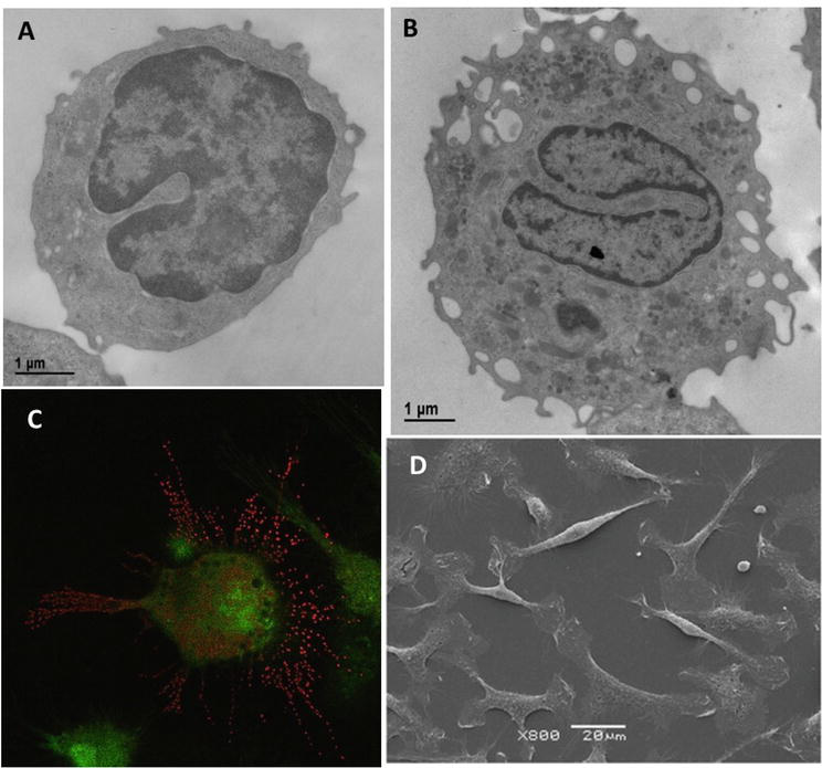
Cell culture is a good technique to evaluate metabolic and morphologic changing by treatment with different drugs. In our lab, it was well-established immune and tumor cell culture. Macrophages are cells from the immune system whose response usually appears quickly since they are part of our body's first line of defense. The majority, around 80% of cells in the culture, changed morphology after internal and external treatment to an activated cell appearance, as observed by light or electron microscopy. In Figure 1, it is possible to observe morphologic modifications that macrophages presented after just 24 hours of treatment with our homeopathic products using different methodologies.
Figure 1.
Macrophages were observed by transmission (A and B), confocal (C), and scanning (D) electron microscopy. In A: resting macrophage, nucleus with condensed chromatin, and few cytoplasmic vesicles indicating only basal metabolism. B: activated macrophage with many euchromatins, cytoplasmatic vesicles, and projections suggesting high metabolism. In C: treated macrophage treated stained with Acridine Orange, showing acid vesicles in red and DNA in green at confocal microscopy. In D: a culture of treated macrophages with activated aspect, most of them sprayed.
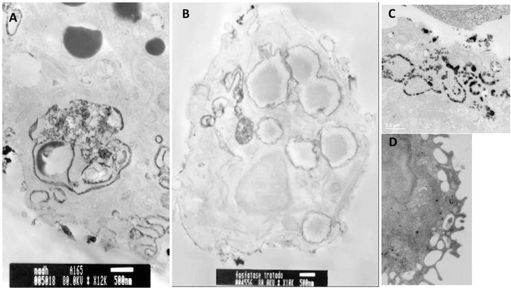
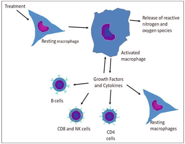
Studies in our lab demonstrated that homeopathic products activated macrophages both in vivo and in vitro. In addition, tumor necrosis factor-α (TNF α), when occurred excessive production in vitro, was significantly decreased [3]. It was observed by ultrastructural cytochemistry that NAD(P)H oxidase activity was increased as well as that of inducible nitric oxide synthase (iNOS), consequently producing reactive oxygen species (ROS) and nitric oxide (NO), respectively. NO inhibited cytochrome oxidase and peroxisomes activities. In response to stimuli, these cells underwent an activation that allowed them, among other functions, to acquire a great capacity to defeat microorganisms and some tumor cells. This can occur through oxygen or nitrogen-dependent mechanisms, in which reactive oxygen (ROS) and nitrogen (RNS) species are produced [4]. These diffuse and short-lived products have a role in antimicrobial defense, which are well-defined and signaled in the cell. For the protection of macrophages against these toxic products, ROS are stored in vesicles called lysosomes, which can be placed in contact with vesicles that contain the ingested material, the phagosomes, or endosomes. In Figure 2, it is possible to observe the cellular vesicles labeled by cerium chloride for different membrane cytoplasmatic enzymes. To make sure that the label is the result of cerium chloride precipitation after the activity of the respective enzyme, these cells were not stained with lead or uranyl. After treatment, the cells showed an increase of endosomal/lysosomal system, amplifying the capacity to phagocyte noninfective microorganisms and cell debris [5]. All these metabolic alterations were those we could measure. But we can ask, how many other alterations occur? How many alterations do we not have technology to measure yet and are occurring not only in macrophages but also in other cells? We tested in macrophages because we know that these cells are extremely sensitive to small modifications in their microenvironment, thus being able to successfully perform its various functions. In the scheme represented in Figure 3, we can summarize its main functions in the control of other cells of the immune system, modifying the inflammatory response, healing, cellular, humoral defense, etc.
Figure 2.
Ultrastructural images of macrophage enzymes. Treated macrophages showed an increase of endosomal/lysosomal system in transmission electron microscopy. The ultrastructural cytochemistry showed cells labeled with cerium chloride for different enzymes. In A, the vesicles showed NAD(P)H oxidase activity; in B, they were labeled for acid phosphatase; in C, the vesicles were labeled for Mg++ATPase; in D, cytochrome oxidase activity (the one that decreased the activity). These cells were observed without contrasting with lead, avoiding possible contaminants.
Figure 3.
After 60 days of infection, the lesions of the control BALB/c mice, treated orally with the vehicle, developed progressive centrally located crusts, exhibiting an ulcerative pattern. In the M1-treated group, the lesions caused by the parasites developed slower than the control, presenting reduced paw thickness. There was edema in the early lesions, followed by almost complete remission of the lesion.
Physical and chemical barriers, such as skin and mucosal surfaces, limit microorganisms to the body's outer surfaces, and when pathogens can break down, these barriers are usually destroyed by the immune system. Thanks to this system, animals can resist almost all types of microorganisms or toxins that tend to damage tissues and organs and even often protect us from infections and modified cells such as cancer cells. As part of the natural body response, inflammation is a complex process that includes a variety of cells and molecules such as the immune cells and cytokines as mediators. Its function is mainly focused on eliminating the injury cause, clearing out damaged cells and tissues, and initiating tissue repair. Therefore, inflammation is considered as a mechanism of innate immunity. Old names for cytokines are lymphokines, interleukins, and chemokines. An inflammatory cytokine or proinflammatory cytokine is a type of signaling molecule that is excreted from immune cells (not only) that promote inflammation and play an important role in mediating the innate immune response. Inflammatory cytokines are predominantly produced by and involved in the upregulation of inflammatory reactions. The anti-inflammatory cytokines are a series of immunoregulatory molecules that control the proinflammatory cytokine response. The main function of cytokine receptors is to convert an extracellular signal, such as a specific binding of a cytokine to a target cell, into an intracellular signal, such as the activation of an enzyme or a transcription factor that can trigger a response of the target cell. Treatment alone did not change cytokines production by cells, but when cells were stimulated with lipopolysaccharide (LPS) and then treated, IFN-γ and TNF-α production was decreased.
Tumor necrosis factor alpha (TNF-α) is an important inflammatory factor that acts as a master switch in establishing an intricate link between inflammation and cancer. TNF-α secretion can be induced by conserved structural elements common to microbial pathogens as well as by tumor cells. Several studies have focused on the transcriptional regulation of TNF-α, looking at transcription factors that bind to the responsive element sites within the TNF-α promoter. NF-κB is a transcription factor that plays crucial roles in inflammation and immunity. Many proinflammatory stimuli can activate NF-κB, mainly through IKK-dependent phosphorylation and degradation of the IκB inhibitory proteins. When NF-κB translocates to the nucleus, it activates the transcription of target genes, including cytokines like TNF-α, chemokines, and antiapoptotic factors [6]. We have used a reporter cell line, HT29-pNF-κB-hrGFP, to find out if M1, M2, and M8, in the presence or absence of TNF-α stimulus, have any effect on NF-κB activity. The reporter cell line HT29-pNF-κB-hrGFP is routinely used to screen natural or synthetic compounds that interfere and/or modulate NF-κB activity. HT29 cells were stimulated for 24 h with a proinflammatory cocktail. GFP (green fluorescent protein) positive cells were sorted. We have observed that only M1 has decreased NF-κB activity on TNF-α stimulated HT29-pNF-κB-hrGFP cells. Thus, we tested in vitro different M1 concentrations (10, 20, ad 30%) and they all presented the same effect on NF-κB activity [3]. We have observed that only M1 has decreased NF-κB activity (Figure 4), but it was not downregulated by M2 and M8. However, we have observed TNF-α reduction by these highly diluted tinctures.
Figure 4.
HT-29 cell line (human carcinoma colon-rectal cells) transfected with plasmid pNF-κB-hrGFP showed a reduction in NF-kb complex activation after 48 h of treatment with M1 (In collaboration with institute Pasteur—Montevideo—Uruguay).
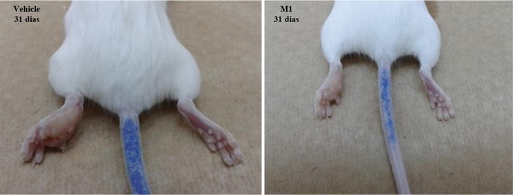
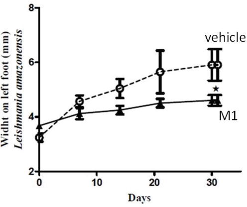
Leishmaniasis is a disease that produces high morbidity but low mortality and results in stigma-producing deformities. In the Americas, it is caused mainly by Leishmania sp, and requires treatment. The parasite is of great medical and veterinary public health significance, for it infects numerous mammal species, including humans. The parasites enter mammalian hosts through the bite of sandflies and replicate intracellularly. Cutaneous Leishmaniasis, the most common form of the disease, causes ulcers on exposed parts of the body, leading to disfigurement, permanent scars, stigma, and in some cases, disability. Modulatory effects were well observed in experimental infection, both in vivo and in vitro, by L. amazonensis, controlling infection progression and limiting its dissemination [7]. The animals were infected with parasites, and after 60 days, when the lesion was well-established, the animals were treated orally for 30 days. The treatment did not allow parasitic evolution as in the control animals. In Figure 5, it is possible to observe the difference in leishmaniosis lesions between animals treated or not. Probably among the slight metabolic changes that occur in the macrophages of treated mice is the increase in the production of Interleukin-10 (IL-10), considered an anti-inflammatory cytokine, the increase of ROS and NOS, and the increase in the number of natural killer cells (NK cells) by treatment in vitro and in vivo with M1 are responsible for this impressive result. After the well-established lesion, the mice were treated with M1 for 30 days and we compare the evolution during the treatment. The evolution of lesions during this 30 days on the footpad of mice paws infected with the parasite Leishmania amazonensis can be seen in Figure 6. Note that M1 decreases the progression of the lesion throughout the treatment and shows a statistical difference after 30 days (* p < 0.05). Each point represents the average of five animals per group.
Figure 5.
Leishmaniosis amazonensis lesion: after 60 days of infection, the lesions of the control BALB/c mice, treated orally with the vehicle, developed progressive centrally located crusts, exhibiting an ulcerative pattern. In the M1-treated group, the lesions caused by the parasites were developed slower than the control, presenting reduced paw thickness. There was edema in the early lesions, followed by almost complete remission of the lesion.
Figure 6.
Evolution of lesions on the footpad of mice paws infected with the parasite Leishmania amazonensis: the mice were treated with M1 for 30 days and compared here with the control group that was treated with water (H2O). Note that M1 decreases the progression of the lesion throughout the treatment and shows a statistical difference after 30 days (* p < 0.05). Each point represents the average of five animals per group.
Beyond increasing ROS and NOS, the M1 complex also increased the number of natural killer (NK) cells and their activity. Cells of the innate immune system recognize pathogens and tissue injury. The processes are rapid, nonspecific, and include responses such as phagocytosis, cell locomotion, killing of pathogens or cells, and cytokine production. These innate immune mechanisms are usually very effective in the elimination of invading pathogens because NK cells can recognize the target cell without the need to be activated by prior immunization or stimulation by contact with antigen-presenting cells as occurs in T-cells to become efficient in their response. Since their identification in 1975, NK cells have been classified as lymphocytes based on their morphology, expression of lymphocyte markers, and their common origin from lymphoid progenitor cells in the bone marrow. NK cells, however, are generally considered components of the innate immune defense because they do not need antigen-specific receptors on their surface to carry out their activities. Their derivation from either lymphoid or myeloid lineages was debated early in their discovery. Research showed that NK cells can be derived from common lymphoid progenitors, and some studies have shown that progenitors expressing myeloid antigens can also develop into NK cells. However, alternative views have been proposed, including the existence of a common myeloid-lymphoid progenitor and this process depending on which cytokines they are exposed. The notion that myeloid precursors previously known to give rise to monocyte/macrophage and Dendritic Cells (DCs) are also capable of NK-cell differentiation puts the recent findings in a new therapeutic perspective. Therefore, NK cells are innate immune effector cells [8, 9].
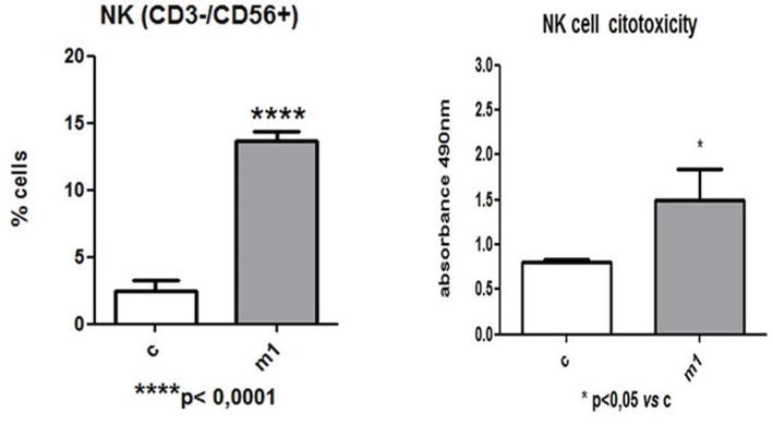
Our group also performed assays to evaluate in vitro effects of M1 in a coculture model between human mononuclear cells obtained from leukoreduction chambers (LRS chambers) after plateletpheresis procedure and melanoma cells. When mononuclear cells were analyzed by flow cytometry, an increase in CD3−/CD56+ natural killer cell population was observed in the treated group. Finally, after treatment, when isolated from mononuclear cultures, and co-cultured with human metastatic melanoma cell lineage, those natural killer cells from M1 treated group revealed a significantly increased cytotoxicity against melanoma cells (Figure 7) [3]. This occurred only with M1 treatment.
Figure 7.
Detection of CD3 and CD56 markers (natural killer cells) by flow cytometry in a population of mononuclear cells after coculture with 1205Lu tumor cells. After co-cultivation, mononuclear cells previously treated (48 h) with the M1 complex showed a statistically significant increase in the percentage of CD3−/CD56+ (NK) cells.
The immune system, a set of defense and healing mechanisms of our body, is highly competent and controlled, performing its functions without compromising the rest of the body. However, many autoimmune diseases, immunodepression syndromes, and cancer may be the result of disorders in the immune system, where the action of this decisively determines the patient’s prognosis, and an inadequate or insufficient immune response can mean the loss of the body’s fight against the disease. In these cases, why not rehabilitate it and stimulate it to perform its function, guiding it to eliminate the disease? Unlike the drugs used by allopathic medicine, which act directly on the physiological processes related to disease symptoms, homeopathic medicines promote the individual improvement of a general health state, stimulating the immune system to trigger appropriate responses for each situation. Thus, homeopathic treatment allows the individual to restore health and prevent disease without, however, producing the side effects experienced by many of the conventional treatments. The incredible adaptability and intelligence of the immune system, which keeps us healthy, despite considerable adversities, is crucial. It is important to note that the existence of this force that works to keep us as healthy as possible was perceived and accepted before we know the cells and molecules that make up the immune system.
Cytokines are soluble, low molecular-weight proteins that mediate cell-to-cell communication. They can modulate the host immune response toward cancer cells and induce apoptosis. Cytokine-based immunotherapy has been a promising area of research and is currently an area of much interest, mainly due to the large amount of side effects. That is why there is a lot of discussion about the burden and bonus balance of its use. You can see a good revision in [10]. Cancer cells, despite their phenotypic characteristics acquired by genetic and epigenetic alterations, do not act alone in the development of the disease. Cancer-associated fibroblasts are involved in all the processes leading to physiological changes that allow cancer cells to become malignant, such as the production of extracellular matrix molecules and its remodeling, providing survival signals, and promoting cancer cell invasion and proliferation. Gonçalves and Potrich [11] used molecular biology techniques and standard functional assays to assess the changes related to the metastatic phenotype. The findings of this study indicate that these products reprogram, molecularly and functionally, melanoma cells in vitro, modulating their metastatic phenotype [11]. Guimaraes et al. [12] described the results of an experimental laboratory validation of the potential of peritoneal macrophages, challenged with a complex homeopathic medication (CHM/M8), to stimulate the immune effectiveness of mesenteric lymph node lymphocytes. This new form of immunomodulatory therapy is based on Hahnemann’s ancient homeopathic techniques, which use diluted substances that are vigorously shaken (succussed) during preparation. The results of this kind of treated coculture were fast and treated macrophages and lymphocytes exhibited a greater degree of interaction than did control cells. Evidence of tumor cells in apoptosis induced by stimulated lymphocytes, apparently prevented tumor cells could use their multiple mechanisms to escape of the immune system. Such findings are likely to be responsible for the attenuation of tumor growth and lung colonization previously observed in vivo [12, 13, 14].
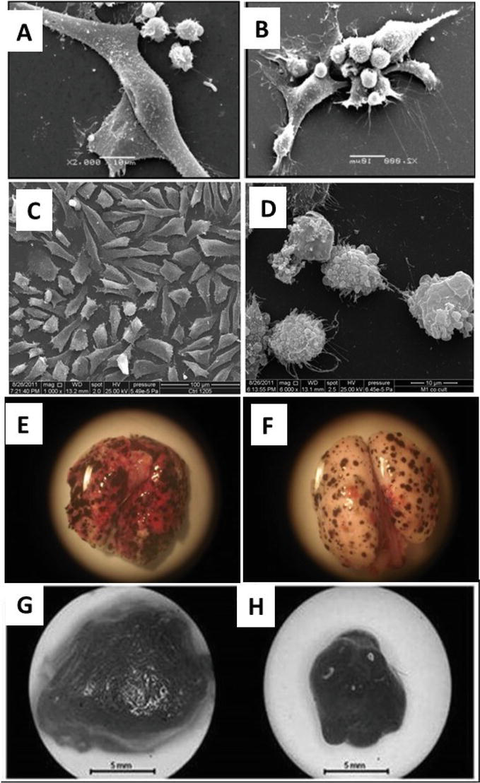
If we collected the supernatant of treated macrophage culture and added it to lymphocytes culture, these lymphocytes could destroy melanoma cells [12]. It is important to note that those lymphocytes could not do this before the treatment. When melanoma cells were injected into mice veins to cause lung melanoma nodules, and the animals inhaled M8 for only 2 weeks, the number of nodules was reduced [13]. When the tumor cells were inoculated in the dorsal subcutaneous region, a solid tumor mass was developed, but after 2 weeks of inhalation, the tumor mass was smaller than the control one, showing that inhalation for only two weeks did not allow tumor mass to grow in the same velocity (Figure 8) [14]. If you think that the animals did not have defense against the millions of tumor cells inoculated, it was an amazing and fast defense answer. More about answers of immune system cells and inflammation after treatment with homeopathy in [15].
Figure 8.
Images of mice and human melanoma cells, and mice tumor nodules and tumor mass after treatment with the homeopathic complexes. At left-A, C, E, and G control groups; images at right-treated groups. A and B: the big cell melanoma cells in culture; the small one, lymphocytes. C and D: human melanoma cells in culture, and in D dying cells after adding treated lymphocytes; E and F lungs with melanoma nodules; G and H tumor mass. A–D, cells observed at scanning electron microscopy; E–H at light microscopy.
The activation of macrophages represents one of the first events in the innate response. The mechanisms of innate and adaptive immunity are, however, interdependent. This intercommunication is also carried out through macrophages, which participate in the production, mobilization, activation, and regulation of all effector cells in the immune system. They interact reciprocally with other cells, which causes their properties to change, for specialized immune functions. Macrophages play an important role in the secretion of various cytokines in addition to acting as antigen-presenting cells. The results of our research have shown that homeopathic medicines stimulate the immune system to trigger appropriate responses for each situation and do not produce side effects. The development of therapies capable of modulating the inflammatory process, without suppressing the desirable effect of its physiological aspects, could be an interesting alternative to obtain a better efficacy of the tissue response against external aggressors. We are sure that much more studies will be needed to verify the effects of homeopathy on diseased organisms. But we are also sure that highly diluted drugs that act on isolated cells in a cell culture are not having a placebo effect, since careful controls were performed during all experiments.
The harmonic results obtained in research during these years allowed us to conclude that, in general, highly diluted products trigger quick and effective responses by living organisms, whether cells or animals, or people. M1 and M8 are homeopathic complex medicines with immunomodulatory properties, without toxicity or mutagenic effects. This homeopathic immunotherapy can restore the immune system to recognize tumors or infected cells; thus, it can be used to help some diseases without acting on a specific molecular target and without toxicity, since self-healing is stimulated through immune system. Here in Table 2 we show the composition of M1 and M8 complexes. Therefore, the M1 and M8 complexes can be good candidates for complementary therapy to conventional treatments. Complementary does not mean one or the other, but that if you complement the conventional treatment with these products, you surely increase the chances of rehabilitation.
Table 2.
Final composition of M1 and M8 (MT = Mother Tincture).
Homeopathy is a medical and pharmaceutical specialty and must be submitted to standard evaluation. Therefore, taking together all the results published, by using standard assays and methodologically reproducible tests verified by statistical analyses, we could demonstrate by scientific evidence that those homeopathic complexes presented effects on cellular and molecular levels. It is important to note that homeopathy is just another kind of treatment. These results show the importance of basic research in obtaining new knowledge about homeopathic medicine, using techniques and methods accepted by conventional medicine.
We are deeply grateful to all the people who have passed through our laboratories at Universidade Federal do Paraná, Brazil, whether for a simple internship, a postgraduation, or a postdoc. They had carried out the research carefully, with love, and often with the opposition from the academic community. They really are special people. We are also grateful to the research funding agencies CNPq, CAPES, and Paraná Tecnologia, to the Federal University of Paraná (UFPR) for unconditional support, and to the Electronic Microscopy Center of UFPR.
The authors declare that they have no competing interests.
References
1.Oliveira SM et al. Mercurius solubilis: Actions on macrophages. Homeopathy. 2011;100:228-236. DOI: 10.1016/j.homp.2011.05.005
2.Jarczak D, Nierhaus A. Cytokine storm-Definition, causes, and implications. International Journal of Molecular Sciences. 2022;23:11740. DOI: 10.3390/ijms231911740
3.De Oliveira CC et al. Developments on drug discovery and on new therapeutics: Highly diluted tinctures act as biological response modifiers. BMC Complementary and Alternative Medicine. 2011;11:101. Available from: http://www.biomedcentral.com/1472-6882/11/101
4.De Oliveira CC et al. Canova, a Brazilian medical formulation, alters oxidative metabolism of mice macrophages. Journal of Infection. 2006;52(6):420-432
5.Lopes L et al. Phagocytosis, endosomal/lysosomal system and other cellular aspects of macrophage activation by Canova medication. Micron. 2006;37(3):277-287. DOI: 10.1016/j.micron.2005.08.005
6.Gilmore TD. The Rel/NF-kappaB signal transduction pathway: Introduction. Oncogene. 1999;18(49):684. DOI: 10.1038/sj.onc.1203237
7.Nascimento KF et al. M1 homeopathic complex trigger effective responses against Leishmania (L) amazonensis. Cytokine. 2017;99:80-90. DOI: 10.1016/j.cyto.2017.07.001
8.Laskowski TJ et al. Natural killer cells in antitumour adoptive cell immunotherapy. Nature, Reviews, Cancer. 2022;22:558. DOI: 10.1038/s41568-022-00491-0
9.Wu SY et al. Natural killer cells in cancer biology and therapy. Molecular Cancer Review. 2020;19:120. DOI: 10.1186/s12943-020-01238-x
10.Rallis SK et al. Cytokine-based cancer immunotherapy: Challenges and opportunities for IL-10. Anticancer Research. 2021;41:3247-3252. DOI: 10.21873/anticanres.15110
11.Goncalves JP, Potrich FB, et al. In vitro attenuation of classic metastatic melanoma-related features by highly diluted natural complexes: Molecular and functional analyses. International Journal of Oncology. 2019;55:721-732. DOI: 10.3892/ijo.2019.4846
12.Guimaraes FS et al. Stimulation of lymphocyte anti-melanoma activity by co-cultured macrophages activated by complex homeopathic medication. BMC Cancer. 2009;9:293. DOI: 10.1186/1471-2407-9-293
13.Guimaraes FS et al. In vitro and in vivo anticancer properties of a Calcarea carbonica derivative complex (M8) treatment in a murine melanoma model. BMC Cancer. 2010;10:113. Available from: http://www.biomedcentral.com/1471-2407/10/113
14.Andrade LF et al. Inhalation therapy with M1 inhibits experimental melanoma development and metastases in mice. Homeopathy. 2016;105(1):109-118. DOI: 10.1016/j.homp.2015.08.007
15.Bellavite P et al. Immunology and homeopathy. 2. Cells of the immune system and inflammation. Evidence-based Complementary and Alternative Medicine. 2006;3(1):13-24. DOI: 10.1093/ecam/nek018
Written By
Dorly de Freitas Buchi, Edvaldo S. Trindade and Carolina C. de Oliveira
Submitted: 25 May 2023Reviewed: 30 May 2023Published: 29 July 2023
 Open access peer-reviewed chapter