Open Access is an initiative that aims to make scientific research freely available to all. To date our community has made over 100 million downloads. It’s based on principles of collaboration, unobstructed discovery, and, most importantly, scientific progression. As PhD students, we found it difficult to access the research we needed, so we decided to create a new Open Access publisher that levels the playing field for scientists across the world. How? By making research easy to access, and puts the academic needs of the researchers before the business interests of publishers.
We are a community of more than 103,000 authors and editors from 3,291 institutions spanning 160 countries, including Nobel Prize winners and some of the world’s most-cited researchers. Publishing on IntechOpen allows authors to earn citations and find new collaborators, meaning more people see your work not only from your own field of study, but from other related fields too.
This chapter provides an extensive discussion of the pulsed laser ablation in liquid (PLAL) method for synthesizing nanoparticles. It covers the production of various types of nanoparticles, such as metal, semiconductor, and metal-oxide nanoparticles, and the impact of laser parameters on their properties, such as size, shape, composition, and crystallinity. The chapter also delves into the physical and chemical processes involved in PLAL, including nucleation, growth, and coalescence, and how they can be controlled to achieve tailored nanoparticle synthesis. Additionally, it examines the challenges and limitations of PLAL, such as particle aggregation, contamination, and reproducibility, and strategies for improving nanoparticle stability and dispersibility. This chapter is a valuable resource for researchers and scientists in the laser synthesis of nanoparticles, emphasizing the significance of pulsed laser parameters in achieving desired nanoparticle properties.
Faculty of Materials and Manufacturing Technologies, Malek Ashtar University of Technology, Tehran, Iran
Reza Shoja Razavi*
Faculty of Materials and Manufacturing Technologies, Malek Ashtar University of Technology, Tehran, Iran
*Address all correspondence to: shoja_r@yahoo.com
1. Introduction
Pulsed laser ablation in liquid (PLAL) is divided into top-down methods in the synthesis of nanoparticles. The PLAL method is performed under ambient conditions and does not require temperature or pressure. Nanoparticles can be synthesized using the PLAL process, which has an almost unlimited domain. The laser source is known as the most significant parameter in the synthesis of nanoparticles by the PLAL. In most of the research conducted on the synthesis of nanoparticles by the PLAL method, nanosecond (ns) pulsed lasers have been applied. Millisecond (ms), microsecond (μs), femtosecond (fs), and picosecond (ps) pulsed lasers have also been used by researchers in the synthesis of nanoparticles. Lasers with different pulse widths due to variable interactions with liquids and targets give researchers various nanostructure synthesis choices.
Liquid is the second key parameter in the PLAL process after the laser source. In various research, liquids such as water, alcohol, acetone, sodium dodecyl sulfate (SDS), polymers, liquid nitrogen, liquid helium, have been used for the synthesis of nanostructures, which has expanded the application of the PLAL process in various fields. Furthermore, the research examined the effect of external factors on the PLAL process using electric field-assisted pulsed laser ablation in liquid (EF-PLAL) and electrochemistry-assisted laser ablation in liquid (EC-PLAL). It has been shown that changing these external factors can easily alter nanomaterials’ morphology, synthesis, and structure. Nanostructure morphology and synthesis can be controlled by considering the above three factors (laser, liquid, and external factors) in the PLAL process.
This part reviews the study’s references and examines the PLAL process’s mechanisms, the laser’s effect, the ablation environment, and target composition parameters. Finally, the conclusions in this field are provided.
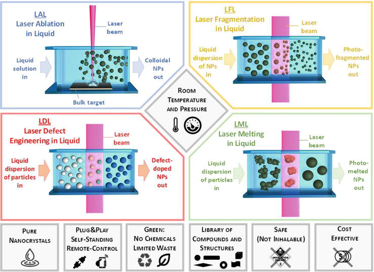
Over the past few decades, a new field of research called laser synthesis of colloids (LSC) has been created to produce nanoparticles [1]. According to Figure 1, LSC can be classified by four different methods: laser ablation in liquid (LAL), laser fragmentation in liquid (LFL), laser melting in liquid (LML), and laser defect-engineering in liquid (LDL) [2, 3]. In the following, these methods are briefly introduced.
Figure 1.
The schematic of laser synthesis classification of colloids into LAL, LFL, LDL, and LML methods [2].
LFL and LML were developed as techniques derived from LAL. These techniques use particle dispersion in liquids as raw materials and may also be classified as laser processing in liquids or laser processing of colloids (LPC). The goal is to shrink the colloidal particles, whereas the LML goal is to increase the scattered particles’ size or deformity through colloids’ laser radiation. LFL refers to the fragmentation of micro- and nanometer particles into smaller particles up to 2 nm [4]. The fragmentation is caused by the absorption of laser light and the evaporation of the photothermal or the Coulomb explosion with the size of the specific particles. In contrast, the LML refers to the melting and intertwined agglomerated nanoparticles in the sub-micrometer and micrometer spheres [5].
The use of controlled laser fluence (poor laser fluence) of the laser beam to radiation to colloid aiming to change the atomic structure of nanoparticles by creating defects without changing the size of nanoparticles is called LDL. Like the LFL and LML methods, the LDL method irradiates scattered particles with lower laser fluence. It does not intend to shrink particles like LFL or melt particles like LML. Therefore, LDL summarizes all studies that aim to change the properties and density without altering the particle size. In LDL, defects not existing in the primary particles must be inserted into the final product [2].
This study focuses mainly on PLAL, which developed in the 1990s. This method has recently attracted increasing attention. Nanoparticles produced by this method make promising applications in the fields of catalysts [6, 7], optics [8, 9], biomedical [10, 11], polymer composites [12, 13], thin layers [14], and additive manufacturing [15, 16, 17]. It has been proven that this method can be economical and scalable at a multi-gram production rate [18, 19]. In addition, it allows nanomaterials to be produced from unstable phases. However, conventional chemical methods cannot obtain non-stable phases [20, 21].
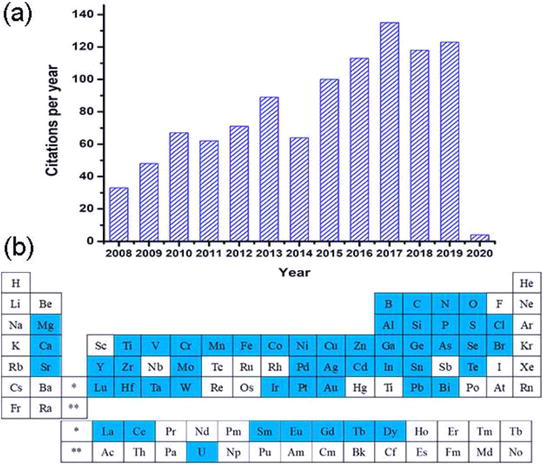
Given these benefits, research into nanoparticle production using the PLAL method has been highly regarded in the past 10 years. The number of articles cited from 2008 to 2020 is presented in Figure 2(a). Also, many of the periodic table elements specified in Figure 2(b) are produced and studied by the PLAL method [22].
Figure 2.
(a) Citations from 2008 to 2020 using the keyword nanoparticle synthesis by PLAL and (b) elements specified in the periodic table with the PLAL method [22].
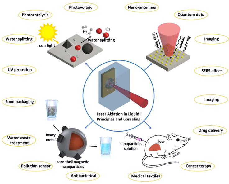
Figure 3 shows the most common applications of the PLAL method in biomedical, catalysts, and sensors. Water, soil, and atmospheric pollution caused by human activity’s spreading of toxic chemicals is becoming a serious problem worldwide. Given the significant advances in nanotechnology, there is an urgent need to develop green and economical approaches to innovative biomedical goals without environmental issues. Nanomaterials made with PLAL can successfully be used in biomedical, sewage treatment systems, and energy sources (e.g., solar cells and hydrogen production systems), and for producing clean and sustainable materials [23].
Figure 3.
Four important areas for PLAL [23].
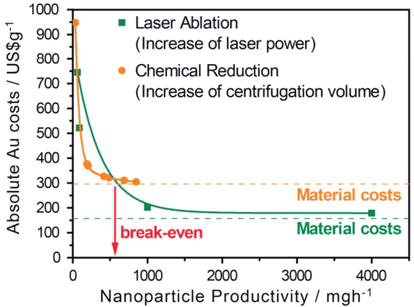
Therefore, the PLAL method as an alternative synthesis path for nanoparticle production in liquids has proven its ability to produce high-purity colloidal nanoparticles for various materials. The basis of synthesis with this method is using a pulsed laser. The PLAL method can create complex structures, such as core-shell structures, which are difficult to synthesize by typical chemical synthesis [3]. Also, researchers have increased the efficiency and reproducibility of colloidal nanoparticle production on a liter using a strong laser system [18, 19, 24]. Given the benefits, companies such as GmbH and Strem in the United States produce colloidal nanoparticles using the PLAL method [25, 26]. Figure 4 compares the production efficiency of colloidal nanoparticles in PLAL and the chemical method performed by Jendrzej et al. [3, 19]. Accordingly, if gold nanoparticle production exceeds 550 mg.h−1, PLAL synthesis is more economical than chemical synthesis.
Figure 4.
Comparison of the costs of producing gold colloidal nanoparticles by PLAL and chemical methods [19].
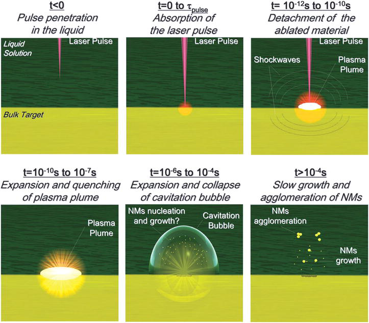
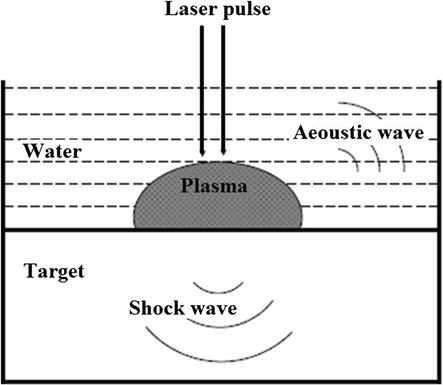
The PLAL mechanism contains several physical processes. The main steps of PLAL are shown in Figure 5. This process begins with the absorption of the laser pulse by the target. Then, a plasma plume containing the ablated material expands into the surrounding liquid and is accompanied by a shock wave. This expansion is due to the high pressure and temperature inside the plume. The plasma plume cools during expansion and releases energy into the liquid. This phenomenon creates a bubble caused by cavitation that expands in the liquid. Then, this bubble, due to cavitation, collapses on a time scale of hundreds of μs with the release of the second shock wave [27].
Figure 5.
The schematic of the main steps in the PLAL method [27].
During the PLAL process, the metal target absorbs the laser photon energy and creates heating and ionization in the area under radiation. Laser energy stimulates the interatomic bonds of the metal target, causing them to break at the threshold energy level. According to bremsstrahlung, free electrons absorb the input laser photons and induce more ionization in the target matter. In addition, melting, evaporation, and plasma processes co-occur. These interactions cause the material to be removed from the solid target surface through vapors, liquid droplets, and solid fragmentation. The amount of ablated area depends on the absorbed energy (E). PLAL process parameters to estimate the ablated amount by the relation of E1/3∝ τe, E1/2∝ τa, E2/3∝ La, and τa˃˃τ1 are related to each other, where in this relation, τ1, La, τa, and τe are the laser pulse time, ablation depth, ablation process time, and the electronic temperature during the ablation process, respectively [28].
The atomization and ionization process produces dense, energetic, non-equilibrium plasma at supersonic speed. Because of its rapid expansion, plasma acts as a piston against the surroundings (liquid and target). It creates shock waves moving toward solid and liquid target in opposite directions. This shock wave increases the temperature and pressure inside the plume [28].
Giacomo et al. [29] showed that the shock wave created by the NS pulse may last several hundred microseconds in the water and up to a few millimeters before the collapse during cavitation. These researchers found that the external shock wave (toward the liquid environment) did not play an essential role in cavitation.
The plasma plume cools during expansion and releases energy into the liquid environment. The plasma plume turns off after 10−8 to 10−7 seconds. The process forms a thin layer of vapor around the plasma volume and creates a bubble caused by cavitation on a time scale of 10−7 to 10−6 seconds, and this bubble disappears on a time scale of 10−4 seconds. The bubble grows in the liquid to a maximum diameter of millimeter-scale [28].
During the movement of bubbles caused by cavitation, its internal temperature and pressure are reduced to the surrounding liquid. Then, the bubble disappears and releases energy by publishing a shock wave, affecting the phase transfer and nanoparticle accumulation. After the bubble collapses on a time scale of 10−4 seconds, the system reaches a stable state physically and chemically. The accumulation of unstable particles is also possible depending on the particles’ composition and surface oxidation. This bubble caused by cavitation acts as a rule for the nucleation of nanoparticles, their growth, and accumulation. The interaction of the bubble with the enclosed particles is an essential step in determining the size of the primary particles [28]. The following is a more detailed explanation of the plasma phase and the dynamic behavior of the bubble caused by cavitation.
3.1 Plasma phase
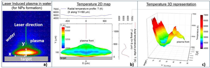
For example, Figure 6 shows the temperature map of an induced plasma on a water-immersed aluminum target. By examining the figure, it can be seen that the plasma kernel has a temperature higher than the dense temperature of most metals. However, due to the high pressure of the plasma under water-enclosed conditions, it allows the density at higher temperatures compared to standard conditions [30, 31]. These observations show that nanoparticles can be formed at the plasma boundary (the area between plasma and liquid) and in the significant part of the plasma. In this case, particles of different sizes and shapes are predicted to be produced at the plasma boundary because the plasma boundary is out of equilibrium. In contrast, the particles formed in the major part of the plasma, in which the processes of growth and evaporation are in thermodynamic equilibrium, are obtained with a spherical form of beam size distribution [23].
Figure 6.
(a) Image of laser-induced plasma on an Al target immersed in water taken with ICCD camera, (b) 2D map of calculated plasma temperature (laser energy = 270 MJ, laser beam diameter = 1 ± 0.2 mm, laser wavelength = 532 nm, laser pulse time = 6 ns, laser frequency = 10 Hz), and (c) 3D display of plasma temperature on the 2D map of part (b) [23].
The mechanism of growth in the plasma phase during the nanosecond (ns)-PLAL laser ablation process has been studied with a theoretical model in which the competition between evaporation and electrostatic growth is examined [31]. These observations show that the electrons are connected as soon as small clusters form due to the high concentration efficiency at the early ablation stage. As a result, the particles are negative and absorb the plasma ions. With the reaction of the ions with a negative charge, particles are formed (electrostatic growth), and particles begin to grow until the growth rate is balanced by the evaporation process due to the high temperature in the plasma (4000–6000 K). In this case, the equilibrium between electrostatic growth and thermodynamic evaporation regulates the dimensions of nanoparticles, and a spherical morphology is achieved. It has recently been shown that when the particle exits the plasma, it still has extra electrons. This additional negative charge can prevent the accumulation of nanoparticles due to repulsion [23, 32].
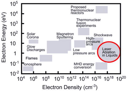
Electron density is one of the essential parameters to determine plasma properties. The electron density equals the number of free electrons in the plasma. Given the quasi-neutral state, the number of free electrons equals the number of charged heavy particles (ions) (n = n+ = ne, where n represents plasma density), in which case the number of charged heavy particles can be estimated. These charged particles may significantly affect the dynamics of chemical reactions through plasma chemistry. A general classification of PLAL-induced plasma compared to other plasmas such as solar corona, ionosphere, magnetron sputtering, glow discharge, flames, thermonuclear fusion, low-pressure arcs, high-pressure arcs, thermonuclear reactors, and magnetohydrodynamic energy conversion is shown in Figure 7. This figure shows that the plasma density in the PLAL is significantly higher than other plasma [33].
Figure 7.
The classification of PLAL-induced plasma compared to other methods [33].
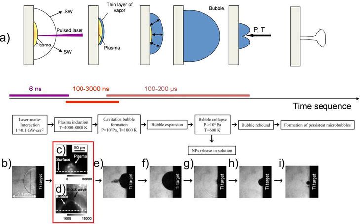
3.2 Dynamic behavior of bubbles caused by cavitation
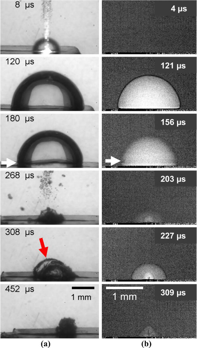
According to Figure 8, the development of bubbles caused by cavitation can be classified into three stages: bubble expansion caused by cavitation, contraction, and collapse (Figure 8(a)). The schematic shows the time sequence of the main events during the PLAL. Figure 8(b) and (e)-(i) show images of the time evolution of the bubbles caused by cavitation after the interaction of a laser pulse with the aim of water-immersed titanium. Figure 8(c) and (d) show images of PLAL of a copper target in the water where a thin layer of vapor is formed around the plasma boundary [33].
Figure 8.
Dynamics time evolution of the bubble caused by cavitation from the growth of the first bubble to microbubbles created after the collapse, (a) the schematic of the time sequence of the main events that occurred during the PLAL, (b) and (e)-(i) titanium-immersed in the water target, (c) and (d) images after the PLAL, a copper in water target [33].
In stroboscopy and X-ray radiography images, Figure 9 shows that the bubble caused by cavitation expands and, after reaching a fixed point with the maximum size and shape of the pseudo-hemisphere, begins to shrink and eventually collapse. For each collapse, part of the mechanical energy of the bubble caused by cavitation is released through the release of a new shock wave [33].
Figure 9.
(a) Stroboscopic images and (b) X-ray radiographic imaging of the PLAL, silver-immersed in the water target (the white arrow indicates the formation of the bubble wall with a pseudo-hemisphere and the red arrow shows the depression and deformation of the bubble) [33].
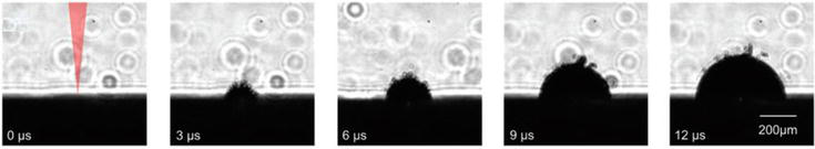
According to Figure 10, during the expansion of bubbles caused by cavitation, the formation of tiny microbubbles around the growing bubble is observed. Using single-pulse experiments, Tanabe et al. [34] concluded that these microbubbles are related to pre-existing nanoparticles. These nanoparticles act as targets by absorbing the laser beam and forming microbubbles.
Figure 10.
The formation of satellite microbubbles around the bubble caused by cavitation after single-laser pulse radiation (2 ps) to a gold target [33].
Shih et al. [35] showed that tiny satellite bubbles around the bubbles caused by cavitation were also created with PS laser pulses when applying single pulses in liquids where the PLAL process has not been performed. They suggested that larger, faster-formed nanoparticles are placed at the bubble phase boundary caused by cavitation and are responsible for forming satellite microbubbles. However, explaining exactly how the presence of large nanoparticles at the bubble boundary due to cavitation causes the microbubbles still requires further studies [33].
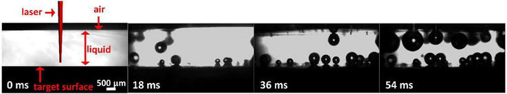
Figure 11 shows that the collapse of bubbles caused by cavitation can lead to the formation of microbubbles that have a much longer life (in ms to s) and are about a few millimeters. The time of growth and the stay of these microbubbles depend on the viscosity of the liquid. The lifespan and size of the bubble caused by cavitation depend on laser parameters such as laser density, pulse length, and fluid properties [33].
Figure 11.
The formation of sustainable microbubbles of a few ms after the PLAL, a gold-in-water target [33].
Laser density is critical because the increase in laser density increases the bubble size caused by cavitation and the amount of ablated mass. Based on the relation (1), the maximum bubble radius at high energy density is obtained. In this relation, Rmax, ρ, tcollapse, Pin, and Pv are the maximum radius of the bubble, the fluid density, collapse time, the liquid pressure, and the vapor saturation pressure inside the bubble, respectively [28].
Rmax=tcollapse0.915ρ/Pinf−PvE1
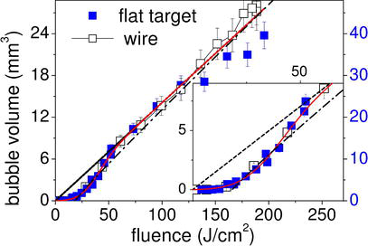
Reich et al. [36] found a linear relation between bubble size caused by cavitation and higher ablation efficiency than the laser density threshold (39 J.cm−2) for the ns-PLAL, which showed almost the entire laser energy on the laser density threshold (39 J.cm−2) is used for the formation of bubbles (Figure 12).
Figure 12.
The bubble volume produced by the PLAL, a flat and wired silver target, in terms of laser density [36].
The direct study of laser ablation in fluid is very challenging for the following reasons:
Most structural study techniques do not have the time and space resolution required for the highly rapid dynamic monitoring of the PLAL process.
The amount of material required to study is in the range of 10−1–10−3 μg per pulse because the sensitivity of most of the techniques is structural study.
The light emission of the plasma plume and the laser pulse interfere with most optical absorption spectroscopy techniques.
To measure the size of the nanoparticles in the liquid, such as the analysis of Dynamic Light Scattering (DLS), which operates based on volume weight signals. This means that a micrometer material separated from the target produces more signals than thousands of nanometer nanoparticles, thus not measuring nanometer particles.
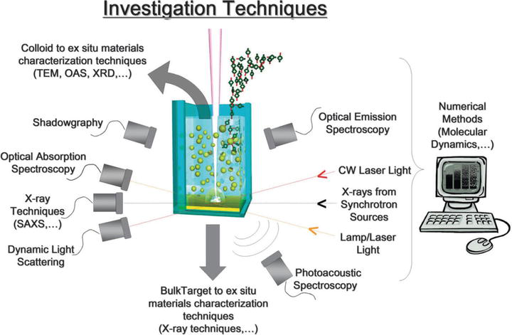
So far, many methods have been used to investigate the process of laser ablation and nanoparticles at various moments, presented in Figure 13. Figure 14 also includes different characterization methods for different PLAL stages with time scales. The following is an explanation of characterization methods.
The optical emission spectroscopy (OES) is performed with a time separation for plasma detection with a timely resolution below the ns and a sub-millimeter spatial resolution with the charged coupled device (CCD) camera.
Ns shadowgraph imaging provides information about the time evolution of the plasma target, shock waves, and cavitation bubbles with micrometer spatial resolution.
Photoacoustic spectroscopy can be performed at a temporal evolution of 10−6 seconds, which provides information about the time evolution of shock waves.
Ultraviolet-visible spectroscopy (UV-VIS) can be performed with a CCD camera with a millimeter space resolution.
DLS provides information about the presence of objects of a few nanometers or larger with temporal evolution of minute. This technique does not distinguish between bubbles and nanoparticles; larger objects heavily influence signals.
Recently, X-ray techniques have been used to investigate the real formation time of nanoparticles during laser ablation. However, these methods require high-temporal resolution X-ray sources such as synchrotron radiation to obtain detectable signals with ns or ms temporal resolution. Different X-ray techniques, such as X-ray diffraction (XRD), small-angle X-ray scattering (SAXS), and X-ray absorption spectroscopy (XAS), have synchronous sources. Among these methods, they are mainly focused on SAXS. This is probably because it gives relatively intense signals and provides information about the size of the nanoparticles rather than their composition.
Figure 13.
The schematic of the methods used to study the PLAL method [27].
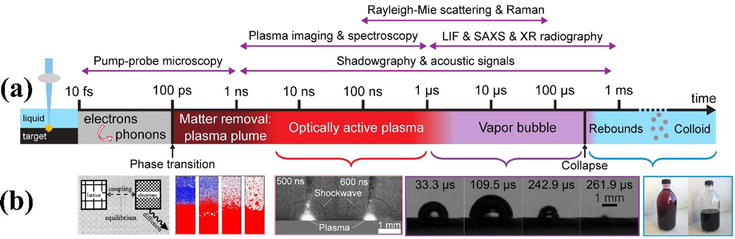
Figure 14.
(a) The different steps in the PLAL method for the ultrashort pulses and the characterization methods at different times and (b) from left to right, respectively: The schematic of electron-phonon, phase transition transmission simulation using molecular dynamics modeling, plasma and released shock wave, bubble dynamics, and colloid produced [2].
In general, to remove existing limitations, theoretical and numerical models can ideally overcome most of the limitations of empirical research methods. Laser ablation and nanoparticle formation can be modeled by molecular dynamics with an atomic spatial and almost unlimited temporal resolution. Molecular dynamics calculations consider the non-thermodynamic equilibrium of laser ablation, and they usually examine electron-ion collisions using Monte Carlo’s method. One of the drawbacks of molecular dynamics measures is that they do not consider ultrafast nonlinear light phenomena, such as photoionization or multi-photon absorption, for charge separation fields from space and Coulomb explosions [27].
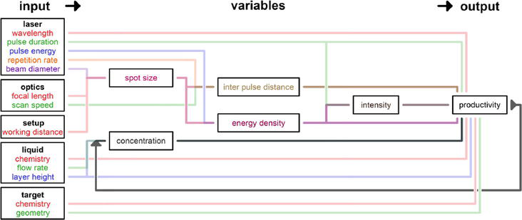
5. Effect of the PLAL process parameters on the production of nanoparticles
The PLAL process parameters are adjustable and related to the process components, including the type of laser, the laser optical adjustment components, the ablation chamber, the target piece, and the liquid environment. A process begins with the input of the primary parameters and creates an output that includes the results of the process. Variables can be defined between these parameters and the process results. The dependency graph in Figure 15 shows a combination of PLAL process parameters. In addition, the dependence of the parameters on each other in the process variables and, finally, the result of the process (product) is also shown in the figure. For example, the laser pulse width parameter directly affects the output efficiency and indirectly affects the laser current intensity variable. Pulse width colors (green) and laser fluence (purple) reach a dark brown, indicating an existing intensity variable. It should be noted that product characteristics such as particle size distribution also represent an output of the process but are not considered in detail here [23].
Figure 15.
The dependency graph of the influential and interdependent process parameters in the PLAL process [23].
5.1 Effect of laser wavelength
Laser wavelengths affect the amount of ablation in the PLAL. Almost all studies show higher efficiency using IR laser light than UV or VIS. However, in the PC process, nanoparticle protection due to Rayleigh scattering [37] can also cause differences in efficiency.
In addition, for almost all metals, higher absorption cross sections are presented for nanoparticles at UV or VIS wavelengths compared to IR wavelengths [38].
The short laser wavelength increases the metal target’s absorption energy and the efficiency of the nanoparticles produced. However, this effect is insignificant due to its open absorption effect that increases ablation efficiency, especially in noble metal materials such as silver, gold, and platinum. This is due to the exacerbation properties of the plasmon in the UV-Vis area. Due to this property of synthesized nanoparticles in the liquid environment, they will be able to re-absorb the input laser pulse, which has adverse effects, reduces the ablation rate, and expands the size distribution of particles. The produced plasma plume can also re-absorb the laser with a shorter input wavelength. However, a near-infrared laser wavelength can eliminate this open absorption effect. The distinct absorption of target matter at different wavelengths results in the production of nanoparticles at different concentrations that affect the size of the particles. However, it should be noted that nanoparticle morphology does not change [39].
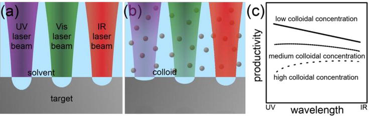
It can be concluded that higher ablation rates (higher efficiency) for metals generally occur throughout the UV laser wavelength due to inter-band absorption. However, the efficiency in VIS and IR wavelengths can be comparable to UV wavelengths. There is also severe dependence on the metal target, laser fluence, and laser pulse duration. In the case of high colloidal concentrations, the absorption of shorter wavelengths by particles can reduce ablation. These relationships are summarized in Figure 16. It is worth noting that for semiconductor and dielectric materials, the initial ablation rate in the absence of a light-extinction colloid can be higher at red and near-IR wavelengths under certain conditions due to the difference in the ablation mechanism compared to metals [23, 40, 41].
Figure 16.
The schematic of laser ablation of metals in liquids in equal laser fluence in different wavelengths, (a) in the absence of colloidal particles, (b) in the presence of colloidal particles, and (c) the effect of increased colloidal concentration on nanoscale efficiency in the PLAL process in different wavelengths [23].
Also, when laser ablation is performed in a liquid environment, the effect of wavelength is very noticeable because, as mentioned earlier, the particles are placed in the path of subsequent laser pulses. On the other hand, the nanoparticles produced depend on the composition of the target matter and have different absorption at different wavelengths. For example, the silver nanoparticles absorb 0.1, 0.28, and 0.52% of the 1064, 532, and 352 nm wavelengths, respectively. Therefore, the self-absorption effects of colloids are more significant at shorter wavelengths. Sending a wavelength at which the absorption of nanoparticles is high causes thermal stimulation, and as a result, the particles are crushed. It should be noted that when self-absorption is small, the size of the particles is determined by the penetration depth of the laser light in the target [42].
Solati et al. [43] reported that radiation of laser pulses at 1064 nm reduced the size of Ag colloids because of the self-absorption of laser pulses by colloidal particles. Tsuji et al. [44] reported that the increasing radiation of colloids by laser after laser ablation results in smaller particles. This phenomenon was also observed in the case of gold colloids prepared using 1064- and 532-nm lasers. Amendola and Meneghetti [45] also examined the laser ablation of gold nanoparticles at 532 and 355 nm. They reported that these wavelengths led to the expansion of the size distribution and a decrease in the average size of nanoparticles. In addition, laser-induced nanoparticles can increase the reaction to other species in the solution and destruction of adjacent organic molecules.
These values have shown that the size of the colloids can be controlled by changing the total number of laser pulses. Also, the size of nanoparticles can be altered by changing the energy of the laser photon (wavelength). Still, it should be noted that an increase in the wavelength will lead to increased particle size [46].
5.2 Effect of laser beam radiation time
The observed absorption features of the UV-Vis spectrum showed that the size of the synthesized nanoparticles is related to laser ablation time (LAT) and laser fluence (LF). A set of experiments determined the effect of parameters on nanoparticle size. Increasing ablation time increases nanoparticle density and decreases liquid molecules [47].
The number of radiation pulses is also adequate in the production of nanoparticles. At the same time, the laser wavelength is 1064 nm, and the total number of pulses is low; the production of nanoparticles is increased linearly with the increase in pulses, but when the laser wavelength is 532 nm. The total number of laser radiation pulses is very high; production is much lower than the pulse number. Initially, the production of nanoparticles increases linearly with the increase in the number of pulses. However, when it reaches saturation, it is due to nanoparticle absorption of laser light [42]. To prevent saturation, the liquid must enter the container from one side and exit from the other, in which case the concentration of nanoparticles in the colloids increases for many laser pulses [27]. An example is the synthesis of copper nanoparticles in a laser fluence of 151 J.cm−2 and LAT of 10 to 50 minutes by Desarkar et al. [48]. Mie theory was used to investigate the results and extract data.
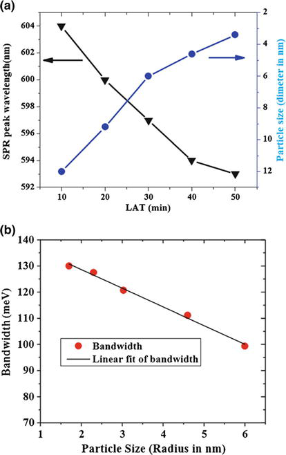
Figure 17(a) shows changes to SPR wavelength and particle size (diameter in nanometer) regarding LAT. This figure shows the TEM measurements and SPR changes with LAT.
Figure 17.
(a) Changes of the SPR wavelength and the average particle size (diameter in nanometer) obtained from the analysis of TEM images with the LAT (min) and (b) line width change (meV) with particle size (radius in nanometer) [48].
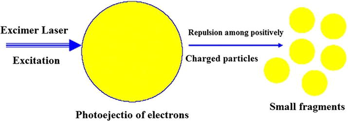
As shown in Figure 17(b), it is observed that the width of the surface plasmon resonance line (bandwidth) is inversely proportional to particle size. In the mentioned figure, a Lorentzian proportion was used to determine the width of the Lorentzian line (according to meV) of the SPR peaks. As the ablation time increases, the volume of nanoparticle production is initially increased and then fixed. The target nanoparticles protect the piece from laser radiation, which is why, after reaching the critical time, no new nanoparticles are produced, and the extra radiation of laser pulses on the colloid through the laser ablation of the liquid phase reduces the size of the nanoparticles which this mechanism (fragmentation of nanoparticles due to the self-absorption phenomenon) is as shown in Figure 18 [49].
Figure 18.
The process of nanoparticle fragmentation due to excess laser radiation [49].
5.3 Effect of laser pulse time changes on the size of nanoparticles
The pulse time (pulse width) is an essential parameter in the synthesis of nanoparticles. Laser pulse width is effective in the production and size of nanoparticles produced by the laser ablation method. There are two theories based on the size of nanoparticles and the efficiency of nanoparticle production at different pulse widths. In the following, these two theories will be explained along with their analysis.
The first theory reported that the size and efficiency of the nanoparticles increase during the longer pulse time (ns) over the shorter pulse time (fs and ps). The efficiency of nanoparticle production and size is believed to increase with the depth of the molten layer on the target surface. The cavity produced by the fs laser pulses is much shallower than the cavity made by nanocrystal laser pulses, indicating lower efficiency of nanoparticle production. For this reason, the efficiency of ablation and size of nanoparticles produced with ns laser pulses is greater than that of fs laser pulses. Also, in ns laser ablation in the liquid environment, plasma is created after the laser pulse; therefore, the plasma’s life is very low. For this reason, the amount of nanoparticle production decreases. Reports show that plasma pressure plays a vital role in the amount of material being slowed down from the target surface in laser ablation.
In the second theory, it is believed that by changing the pulse time from ps and fs to ns, the size of the particle will be reduced, and efficiency will decrease. It is suggested that by changing the pulse time from the ns to the ps and the fs, the ablation mechanism changes from melting and thermal evaporation to phase explosion. The shorter pulse time leads to a more efficient ablation process, resulting in instant evaporation and a minimum heat-affected area. In ps, ablation is faster than ns due to the lower threshold limit for metals [50]. It has also been reported that the energy absorbed by the target for ultra-short laser pulses remains very low in the target piece. As a result, extra-short laser pulse time (ps and fs) is beneficial because the heat loss is low, and the ablation efficiency increases [51]. The thermal nature of the laser ablation is intensified over a longer pulse. Heat losses become more prevalent in this case, so the ablation rate decreases. In addition, the life of the plasma-induced with laser and pulse time on a comparable time scale (more than ten or one hundred ns) for laser pulses intensifies self-induced plasma protection from laser pulses.
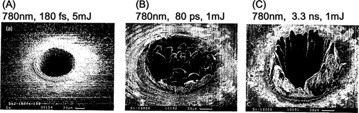
Figure 19 shows the remaining action point by colliding a laser pulse with an fs, ps, and ns laser. The remaining point of action by the laser pulse on the target clearly shows the occurrence of thermal ablation processes in the gas and liquid phases. In the fs state shown in Figure 19(a), the remaining point of action has sharp borders and corresponds to the laser point of action (diameter size of the laser beam), an example of ultra-fast local heating. In this case, the process is mainly influenced by direct photoionization. In the ps pulses (Figure 19(b)), the action point is less than the fs state (Figure 19(a)). It has sharp borders, an example of the simultaneous direct photoionization and thermal ablation processes (melting and evaporation) that have been driven out due to the high pressure created by the material [27].
Figure 19.
The remaining effect of the action on metal targets after laser ablation with pulses (a) fs, (b) ps, and (c) ns [52].
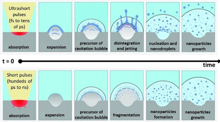
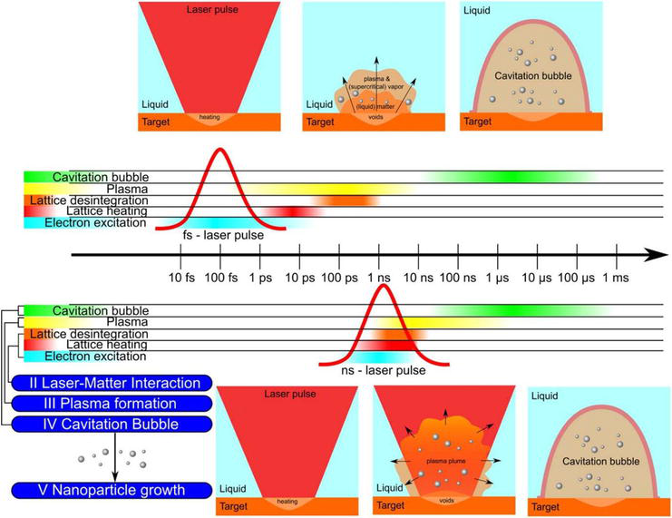
With ns pulses, ablated materials and laser pulses will be used together due to the plasma protective effect for a relatively long period. This time is sufficient to transfer part of the laser energy to the plasma plume and thus increase its temperature and pressure. In such conditions, as shown in Figure 20 as a schematic, the melted droplets from the target into the plume have a better chance of maximizing evaporation [2]. This mechanism also prevails in the ns state for the shrinking nanoparticles. Researchers have reported this mechanism for gold laser ablation, and the size of nanoparticles obtained in fs and ps pulses is larger than in ns pulses [45, 53, 54]. In addition, it has often been observed that the size distribution of nanoparticles obtained with ns pulses is thinner than with ps and fs pulses. This may be another effect of improved homogeneity due to the overlap of the plasma plume with the laser pulse [27, 45].
Figure 20.
The schematic of the PLAL process with lasers with ultra-short pulses (fs to dozens of ps) and short (hundreds of ps to ns) [2].
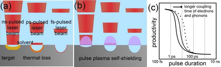
As shown in Figure 21, in the fs laser pulse (10−15), the laser energy is released to the electron in the metal target faster than the thermalization process of the electron-phonon target. In the ps laser (10−12) and ns laser (10−9), the thermal relaxation process is more remarkable, which leads to energy being released thermally to the liquid environment before the end of the pulse. In a few tenths of ps of the laser radiation, plasma is produced and takes tens of ns after ablation. Therefore, no time overlap between the ablated materials and the laser pulse does not occur in the ps and fs laser. However, in the ns laser pulse, there is an overlap in ablated materials due to heat conductivity [28]. The long laser pulse time leads to the absorption of the input laser energy in the plasma plume and increases the plasma temperature and pressure. The plasma then atomizes the materials in the plume. This process homogeneously makes the ablated materials. The energy absorbed by the metal target is reduced because the plasma plume creates an optical protector around the metal target [45].
Figure 21.
An image of laser ablation of metals in liquids during different pulse times for (a) heat losses, (b) self-protection by laser plasma during different pulse times, and (c) effect of further coupling of electrons and phonons on nanoparticle efficiency produced in the PLAL process during different pulse time [23].
In μs and ms pulses, the mechanisms of thermal ablation are largely dominant because the material is separated by melting and evaporation, while the formation of a plasma plume is no longer required for laser ablation. The nanoparticles and atoms evaporated from the target are thrown into the liquid in which three types of reactions include: (a) the reactions of the target vapor phase and liquid solution at temperatures and pressure less than the plasma plume, (b) the reactions of the target liquid phase and the liquid solution, and (c) the reactions of the solid phase from the target and the liquid solution, may occur [27].
Figure 22 also offers a schematic of the laser ablation process at different time scales for fs and ns laser pulses. Laser energy transfer to the target, target phase transfer, plasma life, and bubble life occur on the order of multiple ps, above 100 ps, several μs, and several hundred μs, respectively [33].
Figure 22.
The schematic of the laser ablation process at different time scales [33].
5.4 Effect of the repetition rate of laser pulses (RRLP)
The distance between the two continuous laser pulses determines the quantity called the pulse repetition rate (frequency); therefore, increasing the pulse repetition rate will mean reducing the time between pulses. The pulse repetition rate can change the average size of nanoparticles, which is usually a nonlinear dynamic process in laser ablation. Initially, nanoparticles are produced by a laser pulse. During the subsequent pulse collision, the temperature drops sharply, and this time is an excellent opportunity for particles to cool down and stick to other particles. This process increases the size of the particles, and as the next pulse arrives, the particles are crushed by the absorption of this pulse and converted to smaller sizes. Therefore, in the process of laser ablation, targeting, and, on the other hand, fragmentation of the particles occurs by the above two mechanisms. Lumping and fragmentation interact with each other during ablation [23].
The longer the two-consecutive pulse distance, the more particles can bond with other particles and increase in size. As a result, when we use higher repetition rates, we expect the size of the particles to be smaller. On the other hand, the higher repetition rate (KHz) increases effective ablation due to the greater heat density it creates on the target [42]. Valverde et al. [55] have analyzed the effect of RRLP at 1 to 10 Hz on the synthesis of silver nanoparticles (Ag-NPS) by laser ablation in ethanol. The results showed that the efficiency of silver nanoparticles was decreased by reducing the RRLP from 10 to 1 Hz.
However, linear efficiency increases only at repetition rates exceeding the bubble’s life caused by cavitation (10−3–10−4 s). This is related to repetition rates below 103–104 Hz. The bubble caused by cavitation is characterized by the failure of the refractive coefficient at the liquid/gas interface, which disperses the laser light and reduces the laser energy to reach the target. In addition, during the expansion of bubbles caused by cavitation, target laser ablation is performed in a hot phase with low density, similar to laser ablation in gas. These effects are limiting factors for laser ablation at high laser pulse repetition rates on the target, which is the easiest solution to improve the efficiency of the laser ablation process. However, it has not yet been determined what other possible changes in the mechanism of nanoparticle formation when overlapping laser pulses with bubbles caused by cavitation can be.
Increasing the repetition rate also increases the local concentration of nanoparticles in the target/liquid interface because enough time to distribute nanoparticles in the liquid is reduced away from the ablation area in the liquid. This effect can lead to nanoparticle agglomeration. It also results in the dispersal of input laser pulses and thus reduces ablation efficiency [27]. Therefore, to achieve the distribution and size of smaller particles, it is necessary to optimize the RRLP because, at low RRLP, the particles can be intertwined due to enough time. At high RRLP, due to insufficient time to distribute in the liquid, the particles can be agglomerated.
5.5 Laser fluence effect
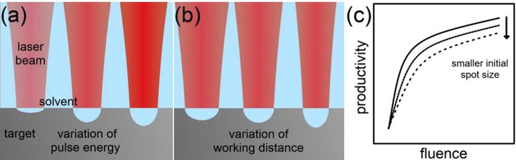
Laser fluence describes the pulse energy penetrating the effective area on the ablation target surface. In other words, the laser fluence is the energy of each pulse per unit area. The laser fluence threshold helps ablation efficiency. The ablation energy threshold refers to the minimum density (optical energy in each pulse area) needed to separate materials from the area under radiation in the target metal and the production of plasma [28]. Laser ablation in the liquid can synthesize nanoparticles in the different laser fluences. Pulse energy or working distance changes may realize laser fluence change. Different results by both methods can occur due to changes in the release of beams in the fluid phase.
By changing the intensity of the laser light, several parameters, such as target absorption and nanoparticle absorption, change simultaneously. Increasing laser fluence on the target surface increases the temperature and improves ablation. The laser’s interaction with matter depends on the laser fluence. When the laser fluence increases, the maximum metal temperature will increase during the warming process. Therefore, despite the high density, the possibility of atoms arousing and ablation rises [56] and increases the efficiency of nanoparticle production [57]. The volumetric ablation rate increases logarithmic with laser fluence. The progress of the logarithmic function depends most on the mechanism of ablation, the surrounding liquid, and the target matter. If the efficiency in PLAL is more related to the laser pulse energy, the optimal laser fluence can be obtained in this case [23]. Figure 23 shows the dependence of nanoparticle production efficiency on laser fluence and beam diameter.
Figure 23.
An image of metal laser ablation in liquids in (a) different pulse energy (fixed beam diameter), (b) different working distances, and (c) indicating laser fluence changes in different sizes of beam diameter on nanoparticle production efficiency (Gaussian beam profile is considered) [23].
When the working distance and diameter of the beam are constant, the amount of ablation and production of nanoparticles increases with increased laser power. However, in the case that power is considered consistent and the diameter of the beam is changed, as the beam diameter increases, the efficiency increases with the smaller beam diameter.
Higher pulse energy separates the material from the metal and increases the target metal concentration in the plume; therefore, with less energy, it is possible to distribute the thinner size of the nanoparticles in laser ablation. In addition, the higher laser fluence also extends the bubble’s lifespan, leading to the bubble reaching a maximum radius [28].
An example is laser ablation of an aluminum target with laser Nd: YAG and a wavelength of 1064 nm for synthesizing Al2O3 nanoparticles. After the process of laser ablation, the liquid becomes opaque. As the laser fluence increases, the particle size varies from 27 to 49 nm. However, as the laser fluence increases, the self-absorption of nanoparticles will prevent the piece target ablation and lead to the crushing of the previous nanoparticles [58]. Mafune et al. [59] also studied gold laser ablation with ns laser and wavelength of 1064 nm in the energy range of 10–100 MJ. They observed that with the increase in pulse energy, the size distribution and the average size of nanoparticles increase.
On the other hand, the concentration of materials increases with increased energy. In other words, with increasing pulse energy, multiple mechanisms of separation of materials, such as fragmentation, phase explosion, and evaporation, co-occur. For example, high-energy gold laser ablation has distributed 2D size due to the simultaneous ablation mechanisms. In other studies, the distribution of single-dimensional size is obtained in low energy [54, 60]. Researchers have reported similar laser ablation results for platinum nanoparticles [61].
The theory of nucleation and growth can be used to explain the difference in particle size. Low laser fluence has less nucleation, leading to a smaller nucleus and particle size. In contrast, in high laser fluence, more nucleation results in large amounts of the nucleus and larger particle size [62].
5.6 Effect of laser focal length on nanoparticles produced
Concentration conditions (target position with focal point) are critical to forming a beam size distribution of nanoparticles. By changing the distance between the lens and the target, the diameter of the laser beam changes. The focal length affects the distribution and size of nanoparticles [63]. It has been observed that the result of ablation is highly dependent on local conditions with laser beam diameter on the target surface. The minimum size of the laser beam’s diameter depends on the focal length, which changes the laser’s fluence. As a result, nanoparticles are created with multiple shapes and sizes. At the same time, if the diameter of the laser beam is small, the size of the nanoparticles becomes uniform, and its diameter is about 2 to 5 nm. If the diameter of the laser beam is too large, the size of the nanoparticles is enlarged, and the average size is about 20 nm [42].
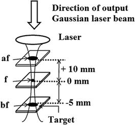
Nath et al. [64] changed the position of the target lens under different focal conditions (up, down, and at the focal point) in Figure 24. They showed that focal condition is an important parameter for synthesizing small-size nanoparticles with beam distribution. This factor changes the laser fluence and the degree of ionization of the liquid environment containing colloids.
Figure 24.
Changing the diameter of the laser beam to above the focal point (af), the focal point (f), and below the focal point (bf) [64].
A regular plasma plume can be detected during the laser radiation. Severe plasma will be created if the target is below the focal point. At low laser energy, the target temperature increases, but its evaporation rate decreases due to the liquid’s function as a cooling agent on its surface, eliminating the formation of plumes. At medium laser beam energy, the plume is formed slowly. While at high energy, target laser ablation in the liquid environment results in the production of a plasma plume, which is visible to the naked eye near the target surface and creates a significant sound that can be due to the breaking bubble caused by cavitation from the evaporation of the fluid layer that is in close contact with the plasma. The release of the plasma and related sounds near the focal point are much greater [23].
According to Figure 25, a plasma plume can be seen during the radiation. When the laser is performed in a liquid environment, the fluid-induced enclosure on the solid target produces a shock wave in the plasma plume. As shown in Figure 25, evaporated species are classified as highly aroused ionic particles that are inappropriately abandoned in their primary quantum modes and emit electromagnetic radiation. It has been shown that the most severe plasma is produced by placing the target at a point a little before the geometric focal point [65].
Figure 25.
The design shows the propagation of sound waves in the enclosed liquid (liquid and shock waves on the solid target) [65].
5.7 Effect of scan speed
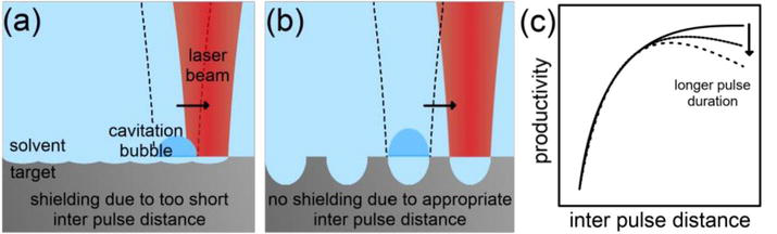
The interaction of laser pulses with plasma, bubbles, and laser-caused particles limits the efficiency of nanoparticles in the PLAL. It is only possible to reduce the protective effects caused by self-induced plasma by using shorter pulse times and changing the scan speed to minimize the effect caused by previous laser pulses. The spatial separation strategy of consecutive laser pulses is controlled by adjusting the interaction of the laser point size, the scan speed (the relative movement between the laser beam and the target), and the pulse repetition rate to prevent previous pulse protection [23].
Sattari et al. [66] found that the Al2O3 target PLAL efficiency can be increased by reducing the spatial overlap of consecutive laser pulses by increasing the scan speed in the IR laser with the ns pulse. The researchers reached the maximum efficiency by completely separating the pulses from each other. However, the linear decrease in efficiency was achieved at much higher distances due to the less target heat heating. Wagener et al. [67] studied the distance between the optical pulses in the laser ablation with the VIS laser with a 7 ps pulse of zinc metal target in tetrahydrofuran. However, reducing efficiency at distances between pulses was not more noticeable without overlapping. This was probably due to these researchers’ lower pulse time (ps pulse). Streubel et al. [18] showed no efficiency decreases between longer pulse distances during 3 ps pulse time.
The thermal heating of the ablation target during the longer pulses will affect the distance between the optimal pulses because the heating mechanism is more prevalent in longer pulses. In short, the PLAL efficiency can be optimized by performing a complete pulse separation. For longer pulse time, the efficiency between the optimum pulses is maximized. Figure 26 shows the effects of the distance between the appropriate pulses for the production of nanoparticles by the PLAL method.
Figure 26.
(a) The distance image between the very short pulse, (b) the distance between the appropriate pulse in the metal laser ablation in liquids, and (c) the graph of the relative effects of the laser pulse time on the dependence of the efficiency on the distance between the pulses [23].
6. Effect of ablation environment on nanoparticles produced
6.1 Effect of liquid environment on the size of nanoparticles
The size of nanoparticles in the liquid after ablation in the liquid phase can be controlled with parameters such as laser fluence, pulse width and pulse repetition rate, wavelength, focal length, and ablation environment. In addition, the average size of colloidal nanoparticles is inversely related to fluid depth [57]. Increased liquid temperature changes the morphology of the nanoparticles from spherical to long [68].
Control of nanoparticles is an essential aspect of colloidal synthesis because the physical and chemical properties of the metal nanoparticles depend very much on their size. The physical and chemical properties of liquids and stabilizers in the liquid environment affect the synthesized nanoparticles. The viscosity, density, and fluid surface tension affect the bubble caused by cavitation and the enclosure of the plasma plume. Increasing viscosity in the liquid environment increases ablation efficiency by improving the plasma plume enclosure. Also, reducing the accumulation causes nanoparticle stability in the liquid [27].
Stabilizers change viscosity, density, and liquid surface tension, which affect the bubble dynamics caused by cavitation and enclosure of the plasma plume on the target. An example of this is the silver nanoparticle colloids prepared in deionized water (DW), ethanol, and polyvinylpyrrolidone (PVP) via laser ablation with a wavelength of 1064 nm to determine the effect of the liquid environment on the average size of nanoparticles. The nanoparticle distribution results and ablation efficiency results are presented in Table 1. Changing the fluid environment in the DW, ethanol, and PVP leads to changes in the average size of nanoparticles [57].
Liquid environment
The average size of nanoparticles
Ablation efficiency
Distribution of nanoparticle size
PVP
16
High
Narrow
DW
23
Low
Wide
Ethanol
26
Very low
Narrow
Table 1.
Average size, ablation efficiency, and colloidal silver nanoparticle size distribution in three different PVP, DW, and ethanol environments [57].
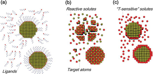
Also, the effect of changing the physical-chemical properties of liquid is associated with the interactions of nanoparticles and stabilizers. Table 2, for example, shows the physical properties of DW and acetone as two commonly used liquid environments in the PLAL process [69, 70]. Figure 27 shows the interaction between stabilizers and ablated materials in different degrees. In studies on the effect of SDS, researchers concluded that SDS anions have electrostatic interactions with nanoparticles and form a molecular layer that restricts the accumulation and growth of nanoparticles by absorbing free atoms [71]. For example, the size of the gold nanoparticles from 20 nm in pure water is reduced to about 10 nm in a 10 mM SDS aqueous solution [59]. As shown in Figure 27(a), reducing the size of nanoparticles during growth is only effective if the concentration of ligands is large enough to overlap the nanoparticles and prevent the accumulation and deposition of nanoparticles. Similar effects are also observed with stabilizers such as cyclodextrins, biopolymers, PVP, or cetyltrimethylammonium bromide (CTAB) [27]. As shown in Figure 27(b), some molecules change the composition of nanoparticles at the highest level of interaction between stabilizers and ablated materials. For example, gold laser ablation in NaCl aqueous solution causes Au-Cl chemical bonds. In the case of NaCl, creating a high zeta potential increases surface repulsion among nanoparticles and creates smaller particles than pure water [45]. Similar results have been reported for silver laser ablation with NIR pulses [72]. In addition, as shown in Figure 27(c), if spontaneous or temperature chemical reactions activate the solution, they can act as nanoparticle nucleation sites. For example, noble metal salts on the metal nanoparticles are reduced during the laser ablation process to create core-shell structures [27, 73].
Parameters
Acetone
DW
Dielectric constant
21.01
80.10
Boiling point (°C)
56
100
Density (g.ml−1)
0.7845
0.9970
Special heat capacity on 25°C (J.g.K−1)
2.175
4.180
Surface tension in 25°C (mN.m−1)
22.72
71.99
Thermal conductive on 25°C (Wm.K−1)
0.161
0.6062
Dipole moment (D)
2.88
1.8546
Viscosity on 25°C (mPa.S)
0.306
0.890
Reduction potential (V)
−1.2
−0.83
The refractive index in wavelength of 1064 nm
1.36135
1.32604
The passage of light (thickness of 1 cm)
≈1
0.54559
Table 2.
Physical and chemical properties of distilled water and acetone [69, 70].
Figure 27.
Effect of stabilizers on nanoparticles (a) preventing growth, accumulation, and deposition of nanoparticles with physical or chemical interactions of ligands on the nanoparticle surface, (b) combining stabilizers with ablated materials and formation of new composition, and (c) the nucleation place of nanoparticles on stabilizers [27].
Applying the magnetic field during the laser ablation process also increases the concentration and size of the nanoparticles, thereby increasing the absorption and efficiency. Using the external electric field decreases the size of the resulting nanoparticles, and tin and gold samples created under different electric fields can be mentioned [74].
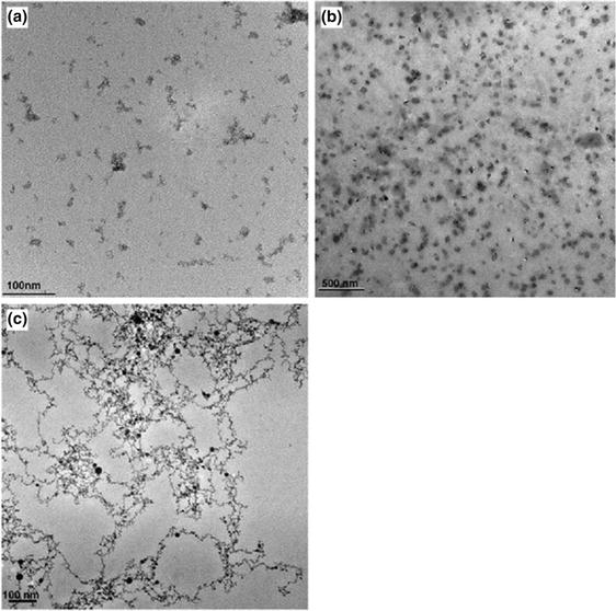
6.2 Effect of the fluid environment on the morphology of the nanoparticles
It has also been found that liquids such as ethanol, DW, and acetone affect the morphology of the synthesized nanoparticles in the laser ablation method. The liquid controls the morphology, size, and distribution of nanoparticles and their composition. Its effect can be seen in Figure 28 on the average size, distribution, and morphology of the tin produced by the PLAL [75]. The dipole moment in liquid is essential for producing smaller nanoparticles. The dipole moment is much higher for the acetone environment than DW and ethanol. For this reason, acetone’s significant dipole moment leads to the smaller size of the tin nanoparticles because it prevents the cluster from growing in the plume. Therefore, the likelihood of spherical nanoparticles also increases [75].
Figure 28.
TEM images of nanoparticles synthesized in the laser fluence 2.3 J.cm−2 in (a) ethanol, (b) DW, and (c) acetone [75].
Laser ablation for the production of nanoparticles relies on two mechanisms: (1) direct nucleation of atoms in the dense plume and (2) the act of nanoparticles as a growth center for the production of new nanoparticles. The effect of these mechanisms results in the distribution of the wide size of the nanoparticles. Compact and strong bonds are absorbed onto the surface by very polar molecules. The electrostatic repulsion force of the dual electrical layers on the overlaps of the nuclei and the clusters in the plume prevents more growth, accumulation, or deposition. The average size, size distribution, and shape of particles obtained in different liquid environments are presented in Table 3.
Liquid
Dipole moment (D)
Average size of particles (nm)
Size distribution (nm)
Morphology
Ethanol
1.69
—
—
Filamentous state
DW
1.85
37
±10
Stretched state
Acetone
2.89
2
±1
Spherical state
Table 3.
Nanoparticle size, size distribution, and morphology were observed for tin nanoparticles prepared by laser ablation in different liquid environments [75].
Azawi et al. [57] synthesized metal nanoparticles with spherical morphology by controlling the process parameters in water, ethanol, and PVP aqueous solutions at a concentration of 20 mM. Aye et al. [76] also concluded that synthesized nanoparticles’ morphology and physical and chemical properties can be regulated by controlling laser ablation parameters in the liquid environment. For example, when using copper as a target, immersing it in acetone instead of water at low laser fluence results in smaller nanoparticles and non-oxidation [23]. In addition, it is said that laser ablation in a stirring liquid environment is more effective than in a static liquid environment and increases ablation efficiency (up to 30%). The quality of colloidal properties is achieved [77].
7. The composition of the target matter in the process of ablation
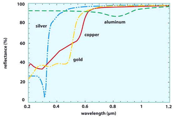
All quantities that affect laser ablation can be somehow related. The characteristics of the target exposed to radiation significantly influence the ablation process. This material has a distinct melting temperature, and the melting process is initiated only when the laser energy is capable of raising the surface temperature of the material to this threshold. For example, the melting temperature of the copper is about 1358 K. If the laser with a power of 102 W.cm−2 is used, the surface temperature can reach 1500 K, which is not too much to produce a significant amount of molten layer on the surface. However, a laser with a power of 108 W.cm−2 reaches a surface temperature of 8000 K and converts about 1.8 μm of the target surface to a liquid phase. It is undoubtedly different in other cases. Other quantities, such as boiling temperature, special heat capacity, and heat conductivity of the target matter, are important in the ablation process. On the other hand, nanoparticles produced with the composition of the target matter have the maximum absorption at a specific wavelength, and the composition of nanoparticles can be found even by examining the nanoparticle absorption spectrum [42]. For example, Table 4 shows the physical properties of copper and silver [69, 70, 78, 79, 80]. In this table, the values of the refractive index (n) and the extinction coefficient (k) indicate that the more k> > n in metals show more shine (higher reflection). On the other hand, if the values of k ≈ n ≈ 3 are divided into gray metals. For example for silver in the wavelength of 500 nm is n = 0.13 and K = 2.92, known as shiny metal. However, tungsten in this wavelength (500 nm) is n = 3.4 and k = 2.69, known as gray metal. Figure 29 shows the reflection of some shiny metals (copper, silver, gold, and aluminum) in the wavelengths of 200 to 1200 nm [80].
In this study, different parameters of the PLAL process for the preparation of metal nanoparticles were examined. Higher laser fluence results in an increase in nanoparticle production efficiency. However, further increased laser fluence reduces metal target ablation due to its effect of self-absorption of nanoparticles. Laser pulse time, by increasing the pulse width from the ps and fs laser to the ns laser, increases efficiency due to the larger cavity in the target and the size of the smaller nanoparticles due to the maximum evaporation of the nanoparticles in the plasma plume. As the repetition rate (frequency) increases of laser pulses, due to the effect of higher heat density, the efficiency of the target increases, and the size of the nanoparticles is reduced due to the crushing of nanoparticles due to successive pulses. Also, with increased wavelengths from UV to NIR, the efficiency and size of nanoparticles will increase due to the self-absorption of nanoparticles. It should be noted that due to the interactions of the laser parameters and the liquid environment of the synthesis, it is difficult to have a conclusion about the efficiency and size of the nanoparticles, and this is a general result according to the results of most researchers, which are mentioned in the sources. An essential key to the synthesis of nanoparticles by the PLAL method is to optimize the process parameters.
1.Zhang D, Gokce B, Barcikowski S. Laser synthesis and processing of colloids: Fundamentals and applications. Chemical Reviews. 2017;117(5):3990-4103
2.Amendola V et al. Room-temperature laser synthesis in liquid of oxide, metal-oxide core-shells, and doped oxide nanoparticles. Chemistry–A European Journal. 2020;26(42):9206-9242
3.Amans D, Cai W, Barcikowski S. Status and Demand of Research to Bring Laser Generation of Nanoparticles in Liquids to Maturity. Vol. 488. Netherlands: Elsevier; 2019. pp. 445-454
4.Ziefuß AR et al. Laser fragmentation of colloidal gold nanoparticles with high-intensity nanosecond pulses is driven by a single-step fragmentation mechanism with a defined educt particle-size threshold. The Journal of Physical Chemistry C. 2018;122(38):22125-22136
5.Meader VK, John MG, Frias Batista LM, Ahsan S, Tibbetts KM. Radical chemistry in a femtosecond laser plasma: Photochemical reduction of Ag+ in liquid ammonia solution. Molecules. 2018;23(3):532
6.Hunter BM, Blakemore JD, Deimund M, Gray HB, Winkler JR, Muller AM. Highly active mixed-metal nanosheet water oxidation catalysts made by pulsed-laser ablation in liquids. Journal of the American Chemical Society. 2014;136(38):13118-13121
7.Marzun G, Levish A, Mackert V, Kallio T, Barcikowski S, Wagener P. Laser synthesis, structure and chemical properties of colloidal nickel-molybdenum nanoparticles for the substitution of noble metals in heterogeneous catalysis. Journal of Colloid and Interface Science. 2017;489:57-67
8.Ma C, Yan J, Huang Y, Yang G. Directional scattering in a germanium nanosphere in the visible light region. Advanced Optical Materials. 2017;5(24):1700761
9.Niyuki R et al. Double threshold behavior in a resonance-controlled ZnO random laser. APL Photonics. 2017;2(3):036101
10.Petersen S, Barcikowski S. In situ bioconjugation: Single step approach to tailored nanoparticle-bioconjugates by ultrashort pulsed laser ablation. Advanced Functional Materials. 2009;19(8):1167-1172
11.Hess C et al. Dose-dependent surface endothelialization and biocompatibility of polyurethane noble metal nanocomposites. Journal of Biomedical Materials Research Part A. 2014;102(6):1909-1920
12.Zhang D, Gökce B. Perspective of laser-prototyping nanoparticle-polymer composites. Applied Surface Science. 2017;392:991-1003
13.Maurer E, Barcikowski S, Gökce B. Process chain for the fabrication of nanoparticle polymer composites by laser ablation synthesis. Chemical Engineering & Technology. 2017;40(9):1535-1543
14.Compagnini G, Scalisi AA, Puglisi O. Ablation of noble metals in liquids: A method to obtain nanoparticles in a thin polymeric film. Physical Chemistry Chemical Physics. 2002;4(12):2787-2791
15.Jonušauskas L et al. Plasmon assisted 3D microstructuring of gold nanoparticle-doped polymers. Nanotechnology. 2016;27(15):154001
16.Doñate-Buendía C et al. Oxide dispersion-strengthened alloys generated by laser metal deposition of laser-generated nanoparticle-metal powder composites. Materials & Design. 2018;154:360-369
17.Hupfeld T et al. A new approach to coat PA12 powders with laser-generated nanoparticles for selective laser sintering. Procedia CIRP. 2018;74:244-248
18.Streubel R, Barcikowski S, Gökce B. Continuous multigram nanoparticle synthesis by high-power, high-repetition-rate ultrafast laser ablation in liquids. Optics Letters. 2016;41(7):1486-1489
19.Jendrzej S, Gökce B, Epple M, Barcikowski S. How size determines the value of gold: Economic aspects of wet chemical and laser-based metal colloid synthesis. ChemPhysChem. 2017;18(9):1012-1019
20.Tymoczko A et al. How the crystal structure and phase segregation of Au–Fe alloy nanoparticles are ruled by the molar fraction and size. Nanoscale. 2018;10(35):16434-16437
21.Wagener P et al. Solvent-surface interactions control the phase structure in laser-generated iron-gold core-shell nanoparticles. Scientific Reports. 2016;6(1):1-12
22.Du H, Castaing V, Guo D, Viana B. Rare-earths doped-nanoparticles prepared by pulsed laser ablation in liquids. Ceramics International. 2020;46(16):26299-26308
23.Fazio E et al. Nanoparticles engineering by pulsed laser ablation in liquids: Concepts and applications. Nanomaterials. 2020;10(11):2317
24.Blatchford C-G, Campbell J, Creighton JA. Plasma resonance—Enhanced Raman scattering by absorbates on gold colloids: The effects of aggregation. Surface Science. 1982;120(2):435-455
25.Nano, Nanoparticle Production by Pulse Laser Ablation in Liquid. IMRA. Available from: http://nano.imra.com/ [Accessed: June 13, 2022]
26.Particular, Nanoparticle Production by Pulse Laser Ablation in Liquid. Particular GmbH. Available from: http://particular.eu/start.html [Accessed: June 13, 2022]
27.Amendola V, Meneghetti M. What controls the composition and the structure of nanomaterials generated by laser ablation in liquid solution? Physical Chemistry Chemical Physics. 2013;15(9):3027-3046
28.Mat Isa SZ, Zainon R, Tamal M. State of the art in gold nanoparticle Synthesisation via pulsed laser ablation in liquid and its characterisation for molecular imaging: A review. Materials. 2022;15(3):875
29.De Giacomo A et al. Cavitation dynamics of laser ablation of bulk and wire-shaped metals in water during nanoparticles production. Physical Chemistry Chemical Physics. 2013;15(9):3083-3092
30.Dell’Aglio M, Motto-Ros V, Pelascini F, Gornushkin IB, De Giacomo A. Investigation on the material in the plasma phase by high temporally and spectrally resolved emission imaging during pulsed laser ablation in liquid (PLAL) for NPs production and consequent considerations on NPs formation. Plasma Sources Science and Technology. 2019;28(8):085017
31.Taccogna F, Dell’Aglio M, Rutigliano M, Valenza G, De Giacomo A. On the growth mechanism of nanoparticles in plasma during pulsed laser ablation in liquids. Plasma Sources Science and Technology. 2017;26(4):045002
32.Dell'Aglio M, De Giacomo A. Plasma charging effect on the nanoparticles releasing from the cavitation bubble to the solution during nanosecond pulsed laser ablation in liquid. Applied Surface Science. 2020;515:146031
33.Kanitz A, Kalus M, Gurevich E, Ostendorf A, Barcikowski S, Amans D. Review on experimental and theoretical investigations of the early stage, femtoseconds to microseconds processes during laser ablation in liquid-phase for the synthesis of colloidal nanoparticles. Plasma Sources Science and Technology. 2019;28(10):103001
34.Tanabe R, Nguyen TT, Sugiura T, Ito Y. Bubble dynamics in metal nanoparticle formation by laser ablation in liquid studied through high-speed laser stroboscopic videography. Applied Surface Science. 2015;351:327-331
35.Shih C-Y et al. Two mechanisms of nanoparticle generation in picosecond laser ablation in liquids: The origin of the bimodal size distribution. Nanoscale. 2018;10(15):6900-6910
36.Reich S et al. Fluence threshold behaviour on ablation and bubble formation in pulsed laser ablation in liquids. ChemPhysChem. 2017;18(9):1084-1090
37.Strutt JW. LVIII. On the scattering of light by small particles. The London, Edinburgh, and Dublin Philosophical Magazine and Journal of Science. 1871;41(275):447-454
38.Creighton JA, Eadon DG. Ultraviolet–visible absorption spectra of the colloidal metallic elements. Journal of the Chemical Society, Faraday Transactions. 1991;87(24):3881-3891
39.Chewchinda P, Tsuge T, Funakubo H, Odawara O, Wada H. Laser wavelength effect on size and morphology of silicon nanoparticles prepared by laser ablation in liquid. Japanese Journal of Applied Physics. 2013;52(2R):025001
40.Nedialkov NN, Atanasov P, Sawczak M, Sliwinski G. Ablation of ceramics with ultraviolet, visible, and infrared nanosecond laser pulses. In: XIV International Symposium on Gas Flow, Chemical Lasers, and High-Power Lasers. Vol. 5120. USA: SPIE; 2003. pp. 703-708
41.Sikora A, Grojo D, Sentis M. Wavelength scaling of silicon laser ablation in picosecond regime. Journal of Applied Physics. 2017;122(4):045702
42.Bogaerts A, Chen Zh. Effect of laser parameters on laser ablation and laser-induced plasma formation: A numerical modeling investigation. Spectrochimica Acta Part B: Atomic Spectroscopy. 2005;60(9-10):1280-1307
43.Solati E, Mashayekh M, Dorranian D. Effects of laser pulse wavelength and laser fluence on the characteristics of silver nanoparticle generated by laser ablation. Applied Physics A. 2013;112(3):689-694
44.Tsuji T, Okazaki Y, Tsuji M. Photo-induced morphological conversions of silver nanoparticles prepared using laser ablation in water—Enhanced morphological conversions using halogen etching. Journal of Photochemistry and Photobiology A: Chemistry. 2008;194(2–3, 253):247
45.Amendola V, Meneghetti M. Laser ablation synthesis in solution and size manipulation of noble metal nanoparticles. Physical Chemistry Chemical Physics. 2009;11(20):3805-3821
46.Patra N et al. Parametric investigations on the influence of nano-second Nd3+: YAG laser wavelength and fluence in synthesizing NiTi nano-particles using liquid assisted laser ablation technique. Applied Surface Science. 2016;366:104-111
47.Chaturvedi A, Joshi M, Mondal P, Sinha A, Srivastava A. Growth of anatase and rutile phase TiO2 nanoparticles using pulsed laser ablation in liquid: Influence of surfactant addition and ablation time variation. Applied Surface Science. 2017;396:303-309
48.Desarkar HS, Kumbhakar P, Mitra A. Optical properties of tin oxide nanoparticles prepared by laser ablation in water: Influence of laser ablation time duration and laser fluence. Materials Characterization. 2012;73:158-165
49.Badr Y, Mahmoud M. Excimer laser photofragmentation of metallic nanoparticles. Physics Letters A. 2007;370(2):158-161
50.Giorgetti E et al. TiO2 nanoparticles obtained by laser ablation in water: Influence of pulse energy and duration on the crystalline phase. Journal of Alloys and Compounds. 2015;643:S75-S79
51.Schwenke A, Wagener P, Nolte S, Barcikowski S. Influence of processing time on nanoparticle generation during picosecond-pulsed fundamental and second harmonic laser ablation of metals in tetrahydrofuran. Applied Physics A. 2011;104(1):77-82
52.Momma C et al. Short-pulse laser ablation of solid targets. Optics Communications. 1996;129(1-2, 142):134
53.Sylvestre J-P, Poulin S, Kabashin AV, Sacher E, Meunier M, Luong JH. Surface chemistry of gold nanoparticles produced by laser ablation in aqueous media. The Journal of Physical Chemistry B. 2004;108(43):16864-16869
54.Sylvestre J-P, Kabashin AV, Sacher E, Meunier M. Femtosecond laser ablation of gold in water: Influence of the laser-produced plasma on the nanoparticle size distribution. Applied Physics A. 2005;80(4):753-758
55.Valverde-Alva M et al. Laser ablation efficiency during the production of Ag nanoparticles in ethanol at a low pulse repetition rate (1–10 Hz). Laser Physics Letters. 2016;13(10):106002
56.Tsuji T, Tsuboi Y, Kitamura N, Tsuji M. Microsecond-resolved imaging of laser ablation at solid–liquid interface: Investigation of formation process of nano-size metal colloids. Applied Surface Science. 2004;229(1-4):365-371
57.Al-Azawi MA et al. The effects of the ambient liquid medium on the ablation efficiency, size and stability of silver nanoparticles prepared by pulse laser ablation in liquid technique. Jurnal Teknologi. 2016;78(3):7-11
58.Piriyawong V, Thongpool V, Asanithi P, Limsuwan P. Effect of laser pulse energy on the formation of alumina nanoparticles synthesized by laser ablation in water. Procedia Engineering. 2012;32:1107-1112
59.Mafuné F, Kohno J-y, Takeda Y, Kondow T, Sawabe H. Formation of gold nanoparticles by laser ablation in aqueous solution of surfactant. The Journal of Physical Chemistry B. 2001;105(22):5114-5120
60.Kabashin AV, Meunier M. Synthesis of colloidal nanoparticles during femtosecond laser ablation of gold in water. Journal of Applied Physics. 2003;94(12):7941-7943
61.Nichols WT, Sasaki T, Koshizaki N. Laser ablation of a platinum target in water. II. Ablation rate and nanoparticle size distributions. Journal of Applied Physics. 2006;100(11):114911
62.Chewchinda P et al. Preparation of Si nanoparticles by laser ablation in liquid and their application as photovoltaic material in quantum dot sensitized solar cell. Journal of Physics: Conference Series. 2014;518(1):012023
63.Elsayed KA, Imam H, Ahmed M, Ramadan R. Effect of focusing conditions and laser parameters on the fabrication of gold nanoparticles via laser ablation in liquid. Optics & Laser Technology. 2013;45:495-502
64.Nath A, Laha S, Khare A. Effect of focusing conditions on synthesis of titanium oxide nanoparticles via laser ablation in titanium–water interface. Applied Surface Science. 2011;257(7):3118-3122
65.Messina E. Metal Nanoparticles Produced by Pulsed Laser Ablation in Liquid Environment. Italy: Universita'degli Studi di Catania; 2011
66.Sattari R, Sajti C, Khan S, Barcikowski S. Scale-up of nanoparticle production during laser ablation of ceramics in liquid media. In: International Congress on Applications of Lasers & Electro-Optics. Vol. 2008. USA: Laser Institute of America; 2008. p. N204
67.Wagener P, Schwenke A, Chichkov BN, Barcikowski S. Pulsed laser ablation of zinc in tetrahydrofuran: Bypassing the cavitation bubble. The Journal of Physical Chemistry C. 2010;114(17):7618-7625
68.Al-Dahash G, Obaid NM, Majeed HA. The effect of liquid environment and magnetic field on optical properties of Pt nanoparticles colloidal prepared by pulsed laser ablation. International Journal of ChemTech Research. 2016;9:118-130
69.Haynes WM. CRC Handbook of Chemistry and Physics. USA: CRC Press; 2016
70.Charlot G. Selected Constants Oxydo-Reduction Potentials: Tables of Constants and Numerical Data Affiliated to the International Union of Pure and Applied Chemistry. Vol. 8. Turkiye: Elsevier; 2013
71.Baiee RM. Generation of Ultra-Fine Nanoparticles by Laser Ablation in Liquid. United Kingdom: The University of Manchester; 2019
72.Šišková K, Vlckova B, Turpin P, Fayet C. Ion-specific effects on laser ablation of silver in aqueous electrolyte solutions. The Journal of Physical Chemistry C. 2008;112(12):4435-4443
73.Jimenez E, Abderrafi K, Abargues R, Valdes JL, Martinez-Pastor JP. Laser-ablation-induced synthesis of SiO2-capped noble metal nanoparticles in a single step. Langmuir. 2010;26(10):7458-7463
74.Sapkota D, Li Y, Musaev OR, Wrobel JM, Kruger MB. Effect of electric fields on tin nanoparticles prepared by laser ablation in water. Journal of Laser Applications. 2017;29(1):012002
75.Bajaj G, Soni R. Effect of liquid medium on size and shape of nanoparticles prepared by pulsed laser ablation of tin. Applied Physics A. 2009;97(2):481-487
76.Aye HL, Choopun S, Chairuangsri T. Influence of solvents on characteristics of nanoparticles prepared by pulsed laser ablation on iron target. Chiang Mai University Journal of Natural Sciences. 2014;31:37-42
77.Resano-Garcia A, Battie Y, Koch A, En Naciri A, Chaoui N. Influence of the laser light absorption by the colloid on the properties of silver nanoparticles produced by laser ablation in stirred and stationary liquid. Journal of Applied Physics. 2015;117(11):113103
78.Vitos L, Ruban A, Skriver HL, Kollár J. The surface energy of metals. Surface Science. 1998;411(1–2):186-202
79.Grimvall G. The electron-phonon interaction in normal metals. Physica Scripta. 1976;14(1–2):63
80.Photonics marketplace, refractive index, mirrors: Coating choice makes a difference. Available from: https://www.photonics.com/Articles/Mirrors_Coating_Choice_Makes_a_Difference/a25501 [Accessed: June 14, 2020]
Written By
Hamed Naderi-Samani and Reza Shoja Razavi
Submitted: 09 January 2024Reviewed: 16 January 2024Published: 09 April 2024