Open access peer-reviewed chapter - ONLINE FIRST

Invasive and Non-Invasive Methods of Diagnosing H. pylori Infection: A Review of Current Practice

Written By

Taylor Fontenot and Annabel E. Barber

Submitted: 09 December 2023 Reviewed: 29 December 2023 Published: 10 April 2024

DOI: 10.5772/intechopen.1004779

Towards the Eradication of <em>Helicobacter pylori</em> Infection IntechOpen
Towards the Eradication of Helicobacter pylori Infection Rapid Diagnosis and Precision Treatment Edited by Liang Wang

From the Edited Volume

Towards the Eradication of Helicobacter pylori Infection - Rapid Diagnosis and Precision Treatment [Working Title]

Liang Wang, Alfred Chin Yen Tay and Barry J. Marshall

Chapter metrics overview

14 Chapter Downloads

View Full Metrics

Abstract

H. pylori infection can be diagnosed using both noninvasive and invasive methods. There is no one gold standard test that is used for diagnosis. Invasive methods of diagnosis involve endoscopy with biopsy, histologic examination, culture, and rapid urease testing. There are certain clinical situations that are appropriate for noninvasive testing and invasive testing. Choosing the appropriate method of diagnosis is dependent upon multiple factors including pretest probability of Infection as well as cost-effectiveness and availability. The aim of this chapter is to discuss the current options of diagnosis, when invasive testing is indicated, and the interpretation of the results obtained.

Keywords

  • H. pylori
  • endoscopy
  • invasive testing
  • peptic ulcer disease
  • urease
  • culture
  • histology

1. Introduction

H. pylori infection remains the most common chronic bacterial infection in humans. It is estimated that half of the world’s population is infected with H. pylori, making its clinical and global impact of great significance. It is the leading cause of gastritis and peptic ulcer disease. Additionally, prolonged colonization and infection can progress to development of gastric carcinoma. H. pylori infection is also associated with gastric lymphoma. Protective factors against H. pylori infection include adequate nutritional status, especially enteral consumption of fruits, vegetables, and foods with high vitamin C content. Higher prevalence of H. pylori infection has been linked to poor sanitation practices, low socioeconomic class, and crowded or high-density living conditions, facilitating intrafamilial and institutional transmission [1]. In general, the most plausible mode of transmission is via oral-oral (i.e., emesis or saliva) or fecal-oral route. Oral-oral transmission between persons is supported by higher incidence of infection among institutionalized individuals and clustering of familial H. pylori infections [1]. Fecal contamination of water supply in developing regions is a significant source of infection as well. The above modes of transmission form the basis of detection of H. pylori through the various non-invasive methods of diagnosis. Treatment of H. pylori-related illness, especially ulcers and gastric cancers, contributes significantly to the financial burden of healthcare globally. In some regions with high prevalence rates, screening programs have been developed to target at-risk populations to detect and treat H. pylori infections early, aiming to prevent sequelae of disease. The aim of this chapter will be to discuss the salient points regarding the history of H. pylori and its clinical relevance, indications for testing, methods of diagnosis, and confirmation of bacterial eradication.

Advertisement

2. Historical background of H. pylori

The discovery of Helicobacter pylori stands as a testament to the transformative power of scientific investigation and serves as a model for challenging established beliefs in medicine. Prior to discovery, the pathology of peptic ulcer disease was thought to be a result of excess acid production secondary to endogenous stress from catecholamine induced acid production and exogenous exposure of gastric mucosa to food with high acid content. Drs Marshall and Warren were credited with discovering the link between H. pylori infection and peptic ulcers and gastric cancer in 1984 [2]. They suspected that a bacterium was the underlying cause, given observations of a spiral-spaced bacteria present in gastric biopsy samples from patients suffering from gastritis and peptic ulcers. Many prior studies attempting to prove the link in animal models were unsuccessful in establishing the association between H. pylori colonization and pathologic disease. In their Nobel Prize-winning landmark paper, Marshall conducted the famous self-experiment in which he administered a solution containing a suspension of cultured H. pylori organisms. He documented onset of symptoms within 5 days of ingestion and further confirmed progression of pathology with endoscopic evaluation. Their work revolutionized modern medicine, leading to a paradigm shift in gastroenterology.

2.1 Bacteriology and pathophysiology of H. pylori

The bacteria that is responsible for infection of the stomach was originally noted as an incidental finding on many gastric biopsies. Further speciation of bacteria found that it was related to campylobacter. It was later further defined and speciated and renamed to be Helicobacter pylori.

H. pylori is a spiral shaped, gram-negative bacterium that can be cultured on blood agar or selective media (Skirrow’s) [3]. This bacterium is catalase, oxidase and urease positive. Urease positivity is both vital to the organism’s survival and clinically relevant as it is the basis of several diagnostic assays.

There are several inherent factors that ensure survival in a hostile environment within the gastric mucosa including urease activity, motility, and the ability to adhere to gastric epithelial lining [4].

Bacterial urease hydrolyzes gastric urea to form ammonia, which facilitates invasion of the gastric mucus layer via neutralization of the acidic protective layer of the stomach [5]. The unique morphology of the spiral shaped bacterium and flagellated motility contribute to the ability to adhere to gastric mucosa. H. pylori attaches to gastric epithelium via receptor-mediated adhesion. Susceptibility of the host is multifactorial and dependent on cellular expression of surface receptors, which renders some individuals more susceptible to penetration and colonization leading to pathologic response. In summation, the four steps necessary for successful colonization described by Kao et al. are survival in the acidic environment, movement toward epithelium via flagella motility, attachment to host cells by adhesin-receptor interactions, and tissue damage by bacterial toxin release [6].

Advertisement

3. Indications for testing

The established indications for testing patients for H. pylori include gastric marginal zone lymphoma of mucosa associated lymphoid tissue, active peptic ulcer disease or history of peptic ulcer if the cure of H. pylori infection has not been documented, and early gastric cancer [7].

If any of the following are identified on endoscopic evaluation: actively bleeding peptic ulcer, other endoscopic abnormality requiring gastric biopsy, or a clinical history of proton pumping inhibitor, bismuth, or antibiotic use warrant biopsy for H. pylori testing. Endoscopy is not indicated for the sole purpose of establishing a diagnosis of H. pylori infection. Other indications for H. pylori that are more controversial include uninvestigated dyspepsia and patients less than 60 years of age without alarm features, chronic treatment with non-steroidal anti-inflammatory drugs or aspirin, unexplained iron deficiency [8], and adults with immune thrombocytopenia [9].

Medications that should be discontinued prior to H. pylori testing include proton pump inhibitors as well as bismuth and antibiotics. Proton pump inhibitors within one to 2 weeks can decrease sensitivity of all endoscopic based testing as well as noninvasive testing. Antibiotics and bismuth within 2–4 weeks of testing also decreases sensitivity. Patients should be advised to stop proton pump inhibitors one to 2 weeks prior to testing if feasible testing should be performed at least 4 weeks after completion of any bismuth and antibiotic treatment [7]. Stool antigen testing and urea breath testing are appropriate for people who do not otherwise require endoscopy.

3.1 Methods of noninvasive diagnosis—Urea breath and stool antigen assay, serology

Of the myriad diagnostic tests available, all with various ranges of sensitivity and specificity, there is not one single test that can be considered a gold standard. Typically, a combination of invasive and noninvasive tests can lead to an accurate diagnosis. One of the notable disadvantages of invasive testing is the requirements of tertiary laboratory equipment and staff. Thus, several noninvasive tests have been developed.

3.1.1 Urea breath testing (UBT)

The basis of UBT involves the bacterium urease mechanism of action. Urease enzyme hydrolyzes urea within the gastric lumen to produce carbon dioxide and ammonia. The assay involves ingestion of urea with a carbon labeled isotope (typically, nonradioactive 13C or radioactive 14C). In afflicted individuals, H. pylori releases the labeled CO2, which is detected in breath samples. The test can be performed with relative ease and within 15–20 minutes. The sensitivity and specificity of UBT are greater than 95% [10]. False-positive results are uncommon; however, false negative results are seen in patients with recent PPI and antibiotic use. Additionally special equipment is required for the test which can limit its clinical use in areas of limited resources.

3.1.2 Stool antigen testing

The presence of bacterial antigen within stool samples is indicative of an ongoing age pylori infection. Thus, stool antigen testing can be used to diagnose and confirm H. pylori infection as well as confirm eradication. The sensitivity and specificity of stool antigen testing is comparable to that of urea breath testing at greater than 95%. Additionally, stool antigen testing can be affected by recent PP, bismuth, and antibiotic use. A limitation of stool antigen testing not seen in UBT is a patient reluctance to give stool samples. Of the available noninvasive testing, stool antigen testing is the most cost effective in areas of limited resources as well as intermediate prevalence H. pylori infection [11].

3.1.3 Serology

ELISA test to detect immunoglobulin G antibodies generated against H. pylori is both inexpensive and noninvasive. This method requires reliable laboratory staffing with appropriate controls and can be impractical in routine practice. In areas with low prevalence populations and limited resources and limited resources can lead to inaccurate serologic testing results. While the sensitivity is greater than 95, specificity has been reported as low as 60% [12]. Another limitation of this method is the failure of it to distinguish between active and past infection.

3.2 Invasive methods of diagnosis

Endoscopy is the basis for invasive methods of diagnosing H. pylori. Tissue biopsies are used to perform several assays including urease testing, culture, and histologic examination to determine presence of H. pylori organisms. The ideal location of biopsy has been studied, both antral and fundal biopsies have been determined to yield the most accurate diagnosis. Incisura angularis biopsies are not recommended as they have been shown to have the lowest yield of positive diagnosis [13, 14, 15].

3.2.1 Histology

Historically, histologic evaluation of gastric mucosa was the first method of accurate diagnosis of H. pylori infection. The presence of the unique spiral shaped bacteria along with inflammatory changes within the mucosa on microscopic evaluation is considered a positive diagnosis [16]. A multitude of stains, including Giemsa, Hp silver stain, and immunostaining have been used to detect H. pylori. Hematoxylin and eosin stains help to evaluate the extent of inflammatory changes as well as with the detection of the bacteria [17]. Since its discovery, more complex histologic examination techniques have been employed in the microscopic evaluation of H. pylori. Genta stain, a combination of three commonly available stains (Steiner, hematoxylin-eosin, and alcian blue) is particularly advantageous as it visualizes both inflammatory changes and H. pylori [18]. Typically, the routine use of Giemsa stain is a simple, highly sensitive and cost-effective method of histologic diagnosis of H. pylori infection.

The Sydney System of classification of gastritis was originally described in 1996 and has had multiple revisions since its inception. The Sydney system classifies the severity of gastritis by the following histologic findings: the extent of lymphocyte and neutrophil invasion and the lamina propria, glandular atrophy within the corpus and antrum, intestinal metaplasia of the mucosal epithelium, and the amount of
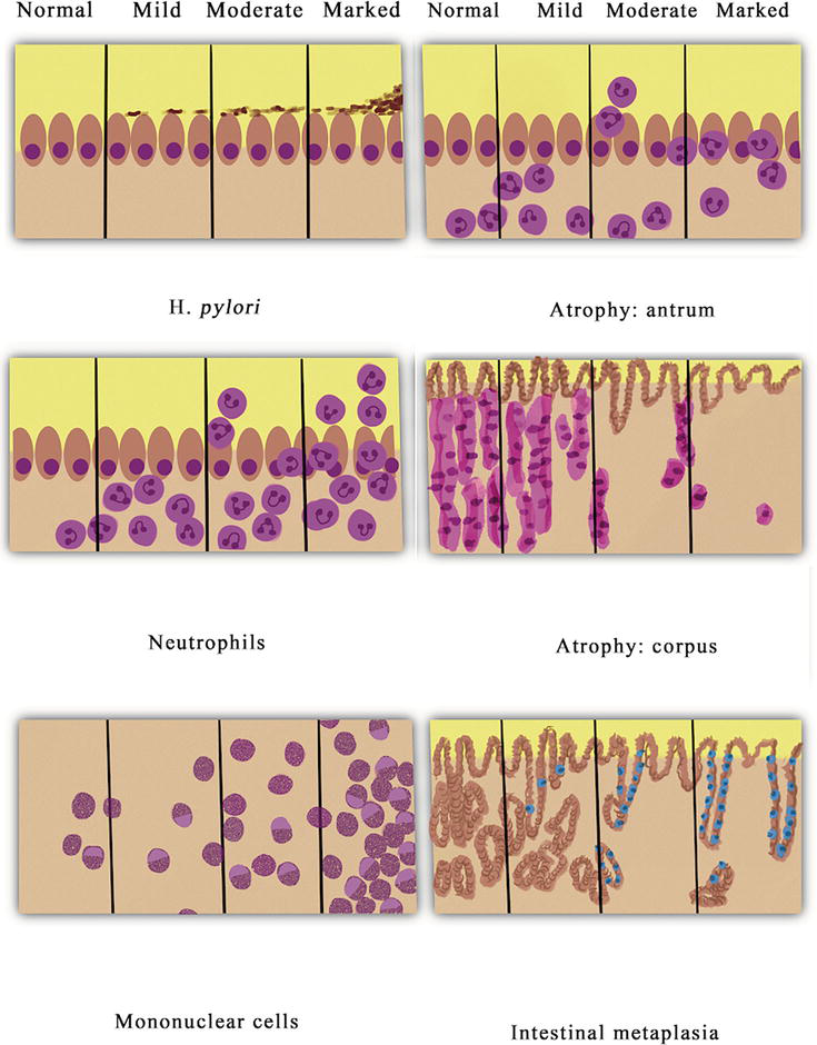
 H. pylori organisms present (Figure 1) [19].

Figure 1.

Grading of gastritis by Sydney system: acute inflammation, chronic inflammation, atrophic gastritis, intestinal metaplasia, and Helicobacter pylori density.

3.2.2 Rapid urease test (RUT)

Rapid Urease test (RUT) is another invasive method of detection of H. pylori infection. The basis of the test revolves around the ability of H. pylori bacteria to convert urea into carbon dioxide and ammonia. Once the biopsy is obtained, it is placed within a test container. Once the H. pylori within the specimen splits the urea to ammonia, the elevation of pH by ammonium hydroxide is detected by pH indicator color change. Tests can be performed either in solution or a solid supporting medium [20]. The Campylobacter-like organism (CLO) test is a commercially available kit that contains solid media, and typically takes up to 24 hours to result. The Pyloritek kit utilizes a reagent strip for the rapid urease test where a positive result can be obtained within 1 hour. Both kits have similar sensitivity and specificity; however, the Pyloritek kits are more convenient for use in endoscopy clinics given rapidity of results [21].

It is notable that while both kits have sensitivity and specificity greater than 90%, there are several factors that influence these measures. The bacterial density of at least 104 organisms within the biopsy specimen is required for a positive result [22]. Additional factors that influence the specificity and sensitivity of the RUT kits include bacterial density post-treatment, patients with bleeding ulcers at the time of biopsy, patients taking H2-receptor antagonists or PPIs [23, 24]. Urease testing does have the advantage over culture and histology given the low cost, user ease, and rapidity of results.

3.2.3 Culture

H. pylori can be isolated by culture from gastric biopsy samples. The microbe requires a hypercapnic, microaerophilic environment and complex media for isolation [3]. Following biopsy, samples must be placed in saline vials and not formalin for successful culture and analysis. Typical media includes Agar base supplemented with blood or serum. Various antibiotics such as vancomycin, bacitracin, polymyxin, are often used in selective media for H. pylori [25]. Isolated bacteria grow best in a humid atmosphere at a temperature of 37C and require five to 7 days of incubation. For optimal growth, 5–10% carbon dioxide can be added to the culture atmosphere [26]. Because of the patchy distribution of H. pylori within the stomach, it is recommended to take biopsies 2 cm proximal to the pylorus and at least two biopsies from the anterior and posterior corpus [27]. Culture is not considered first line testing for establishing a diagnosis of H. pylori because it is time-consuming and often tedious; however, culture data allows for testing of antibiotic sensitivity to choose appropriate antibiotic regimen for eradication.

3.2.4 Polymerase chain reaction (PCR)

PCR is a molecular analysis method that involves rapid production of copies of a specific segment of DNA. It uses short synthetic DNA fragments called primers to select a segment of the genome to be amplified followed by multiple rounds of DNA synthesis to amplify the segment of interest. PCR is useful in providing data involving the presence of certain virulent factors as well as antibiotic resistance [26]. An advantage of PCR is that a variety of clinical samples can be used to extract H. pylori DNA including gastric biopsy, gastric secretions, saliva, dental plaque, and stool samples; therefore, it can be utilized for both invasive and non-invasive diagnosis. When utilizing stool samples for PCR assay, a separate extraction step must be performed to separate the H. pylori DNA from the stool sample, as it typically contains a variety of commensal bacteria and PCR-inhibiting chemicals that may lead to false-negative and false-positive results [28]. PCR stool testing is advantageous as the patient needs to provide only a small sample (usually 50–200 mg) of fresh stool sample, and results can be obtained within hours of sample submission, facilitating treatment decisions in real time [28]. The overall cost of fecal PCR testing is lower than invasive methods, negating the need for the expense of hospital admission and anesthesia.

Antibiotic resistance is one of the leading causes of failure of treatment of H. pylori. Specific gene identification can be used for both bacterial detection and lend information regarding antibiotic resistance, notably, the 23S rRNA gene. Specific point mutations within this gene can lead to clarithromycin resistance [29]. Other genes that have been identified that confer antibiotic resistance include rdxA/frxA with metronidazole resistance [30, 31], 16S ribosomal DNA with tetracycline resistance [32], and gyrA gene in fluoroquinolone resistance [33]. PCR based testing has a significant role in both non-invasive and invasive methods of diagnosis of H. pylori and can guide effective treatment.

Advertisement

4. Conclusions

There is no one gold standard test that is recommended for a diagnosis of H. pylori infection. Of the non-invasive tests discussed, each has their distinct advantages and disadvantages as well as varied sensitivity and specificity. Invasive testing across the board has higher sensitivity and specificity, but requires biopsy, which may not be indicated in certain patient populations for initial screening for H. pylori infection. A combination of one non-invasive and invasive test tends to be the overall recommended gold standard for accurate diagnosis of H. pylori.

References

  1. 1. Brown LM. Helicobacter pylori: Epidemiology and routes of transmission. Epidemiologic Reviews. 2000;22(2):283-297. DOI: 10.1093/oxfordjournals.epirev.a018040
  2. 2. Marshall BJ, Warren JR. Unidentified curved bacilli in the stomach of patients with gastritis and peptic ulceration. Lancet. 1984;1(8390):1311-1315. DOI: 10.1016/s0140-6736(84)91816-6
  3. 3. Goodwin CS, Worsley BW. Microbiology of helicobacter pylori. Gastroenterology Clinics of North America. 1993;22(1):5-19
  4. 4. Amieva MR, El-Omar EM. Host-bacterial interactions in helicobacter pylori infection. Gastroenterology. 2008;134(1):306-323. DOI: 10.1053/j.gastro.2007.11.009
  5. 5. Mobley HL. The role of helicobacter pylori urease in the pathogenesis of gastritis and peptic ulceration. Alimentary Pharmacology & Therapeutics. 1996;10(Suppl. 1):57-64. DOI: 10.1046/j.1365-2036.1996.22164006.x
  6. 6. Kao CY, Sheu BS, Wu JJ. Helicobacter pylori infection: An overview of bacterial virulence factors and pathogenesis. Biomedical Journal. 2016;39(1):14-23
  7. 7. Malfertheiner P, Megraud F, O'Morain CA, Atherton J, Axon AT, Bazzoli F, et al. Management of Helicobacter pylori infection--the Maastricht IV/ Florence consensus report. Gut. 2012;61(5):646-664. DOI: 10.1136/gutjnl-2012-302084
  8. 8. DuBois S, Kearney DJ. Iron-deficiency anemia and helicobacter pylori infection: A review of the evidence. The American Journal of Gastroenterology. 2005;100(2):453-459. DOI: 10.1111/j.1572-0241.2005.30252.x
  9. 9. Rostami N, Keshtkar-Jahromi M, Rahnavardi M, Keshtkar-Jahromi M, Esfahani FS. Effect of eradication of helicobacter pylori on platelet recovery in patients with chronic idiopathic thrombocytopenic purpura: A controlled trial. American Journal of Hematology. 2008;83(5):376-381. DOI: 10.1002/ajh.21125
  10. 10. Howden CW, Hunt RH. Guidelines for the management of helicobacter pylori infection. Ad hoc committee on practice parameters of the American College of Gastroenterology. The American Journal of Gastroenterology. 1998;93(12):2330-2338. DOI: 10.1111/j.1572-0241.1998.00684.x
  11. 11. Vakil N, Rhew D, Soll A, Ofman JJ. The cost-effectiveness of diagnostic testing strategies for helicobacter pylori. The American Journal of Gastroenterology. 2000;95(7):1691-1698. DOI: 10.1111/j.1572-0241.2000.02193.x
  12. 12. Chey WD, Wong BC. Practice parameters Committee of the American College of gastroenterology. American College of Gastroenterology guideline on the management of helicobacter pylori infection. The American Journal of Gastroenterology. 2007;102(8):1808-1825. DOI: 10.1111/j.1572-0241.2007.01393.x. Epub 2007 Jun 29
  13. 13. Patel SK, Pratap CB, Jain AK, Gulati AK, Nath G. Diagnosis of helicobacter pylori: What should be the gold standard? World Journal of Gastroenterology. 2014;20(36):12847-12859. DOI: 10.3748/wjg.v20.i36.12847
  14. 14. Lash JG, Genta RM. Adherence to the Sydney system guidelines increases the detection of helicobacter gastritis and intestinal metaplasia in 400738 sets of gastric biopsies. Alimentary Pharmacology & Therapeutics. 2013;38:424-431. DOI: 10.1111/apt.12383
  15. 15. Lopes AI, Vale FF, Oleastro M. Helicobacter pylori infection—Recent developments in diagnosis. World Journal of Gastroenterology. 2014;20(28):9299-9313. DOI: 10.3748/wjg.v20.i28.9299
  16. 16. Rotimi O, Cairns A, Gray S, Moayyedi P, Dixon MF. Histological identification of helicobacter pylori: Comparison of staining methods. Journal of Clinical Pathology. 2000;53:756-759
  17. 17. Laine L, Lewin DN, Naritoku W, Cohen H. Prospective comparison of H&E, Giemsa, and Genta stains for the diagnosis of helicobacter pylori. Gastrointestinal Endoscopy. 1997;45:463-467
  18. 18. Genta RM, Robason GO, Graham DY. Simultaneous visualization of helicobacter pylori and gastric morphology: A new stain. Human Pathology. 1994;25(3):221-226. DOI: 10.1016/0046-8177(94)90191-0
  19. 19. Stolte M, Meining A. The updated Sydney system: Classification and grading of gastritis as the basis of diagnosis and treatment. Canadian Journal of Gastroenterology. 2001;15(9):591-598. DOI: 10.1155/2001/367832
  20. 20. Kato M, Shimizu Y, Kobayashi T, Komatsu Y, Takeda H, Sugiyama T, et al. Rapid urease test. Nihon Rinsho. 2003;61(1):72-78 Japanese
  21. 21. Mcnicholl AG, Ducons J, et al. Helicobacter pylori study Group of the Asociación Española de Gastroenterología (AEG). Accuracy of the ultra-rapid urease test for diagnosis of helicobacter pylori infection. Gastroenterología y Hepatología. 2017;40(10):651-657. English, Spanish. DOI: 10.1016/j.gastrohep.2017.07.007
  22. 22. Puetz T, Vakil N, Phadnis S, Dunn B, Robinson J. The Pyloritek test and the CLO test: Accuracy and incremental cost analysis. The American Journal of Gastroenterology. 1997;92(2):254-257
  23. 23. Mégraud F. Advantages and disadvantages of current diagnostic tests for the detection of helicobacter pylori. Scandinavian Journal of Gastroenterology. Supplement. 1996;215:57-62. DOI: 10.3109/00365529609094536
  24. 24. Lerang F, Moum B, Mowinckel P, Haug JB, Ragnhildstveit E, Berge T, et al. Accuracy of seven different tests for the diagnosis of helicobacter pylori infection and the impact of H2-receptor antagonists on test results. Scandinavian Journal of Gastroenterology. 1998;33(4):364-369. DOI: 10.1080/00365529850170982
  25. 25. Mégraud F, Lehours P. Helicobacter pylori detection and antimicrobial susceptibility testing. Clinical Microbiology Reviews. 2007;20:280-322
  26. 26. Saez J, Belda S, Santibáñez M, Rodríguez JC, Sola-Vera J, Galiana A, et al. Real-time PCR for diagnosing helicobacter pylori infection in patients with upper gastrointestinal bleeding: Comparison with other classical diagnostic methods. Journal of Clinical Microbiology. 2012;50:3233-3323
  27. 27. Cover TL. Perspectives on methodology for in vitro culture of helicobacter pylori. Methods in Molecular Biology. 2012;921:11-15. DOI: 10.1007/978-1-62703-005-2_3
  28. 28. Rimbara E, Sasatsu M, Graham DY. PCR detection of helicobacter pylori in clinical samples. Methods in Molecular Biology. 2013;943:279-287. DOI: 10.1007/978-1-60327-353-4_19
  29. 29. Gong RJ, Xu CX, Li H, Liu XM. Polymerase chain reaction-based tests for detecting helicobacter pylori clarithromycin resistance in stool samples: A meta-analysis. World Journal of Clinical Cases. 2021;9(1):133-147. DOI: 10.12998/wjcc.v9.i1.133
  30. 30. Kwon DH, Kato M, El-Zaatari FA, Osato MS, Graham DY. Frame-shift mutations in NAD(P)H flavin oxidoreductase encoding gene (frxA) from metronidazole resistant helicobacter pylori ATCC43504 and its involvement in metronidazole resistance. FEMS Microbiology Letters. 2000;188:197-202
  31. 31. Jenks PJ, Ferrero RL, Labigne A. The role of the rdxA gene in the evolution of metronidazole resistance in helicobacter pylori. The Journal of Antimicrobial Chemotherapy. 1999;43:753-758
  32. 32. Dailidiene D, Bertoli MT, Miciuleviciene J, Mukhopadhyay AK, Dailide G, Pascasio MA, et al. Emergence of tetracycline resistance in helicobacter pylori: Multiple mutational changes in 16S ribosomal DNA and other genetic loci. Antimicrobial Agents and Chemotherapy. 2002;46:3940-3946
  33. 33. Moore RA, Beckthold B, Wong S, Kureishi A, Bryan LE. Nucleotide sequence of the gyrA gene and characterization of ciprofloxacin-resistant mutants of helicobacter pylori. Antimicrobial Agents and Chemotherapy. 1995;39:107-111

Written By

Taylor Fontenot and Annabel E. Barber

Submitted: 09 December 2023 Reviewed: 29 December 2023 Published: 10 April 2024