
Abstract
The purpose of this work is to highlight the significance of applying scanning electron microscopy coupled with X-ray energy dispersive (SEM–EDX) as a method of analyzing the surface of kidney stones, to assess the antilithiatic activity of extracts from medicinal plants, such as Saussurea costus (Falc) Lipsch. This chapter aims to showcase the substantial application and use of electron microscopy in the field of medical research, with particular emphasis on urinary lithiasis. Initially, we will present the pathology of urinary lithiasis and kidney stones. We will then examine the evaluation of kidney stones and the importance of characterizing them using various methods, including electron microscopy. Subsequently, we will provide an overview of scanning electron microscopy coupled to X-ray energy dispersive (SEM–EDX) and its implementation in antilithiatic activity using the stone dissolution test built upon our previous study.
Keywords
- antilithiatic activity
- kidney stones
- dissolution test
- scanning electron microscopy
- SEM–EDX
1. Introduction
The electron microscope, a revolutionary type of microscope [1], was developed by German physicists Ernst Ruska and Max Knoll at the University of Berlin in 1931 [2]. In 1986, Ernst Ruska was awarded the Nobel Prize in Physics for his invention and development of the transmission electron microscope [3]. Electron microscope use electron beams [4] to produce images with significantly higher precision and resolution than those obtained with ordinary optical microscopes [4]. Electron microscope are limited by the wavelength of the light, while the resolution of an optical microscope is limited by the phenomenon of diffraction, with the size of the details observed becoming comparable to the wavelength of the light used, resulting in the mixing of information and blurring of fine details [5]. Electron microscopes, on the other hand, have been specially designed to overcome this resolution constraint. Instead of using visible light, they use electron beams, that is, fast electrons, as a source of radiation. One of the main benefits of electron radiation is its short wavelength, allowing for high resolution. This means that finer details can be distinguished [5], such as transmission electron microscopy, which uses fast electrons, usually around 100 to 200 keV, approximately equivalent to half the speed of light [5]. Electron microscopy offers an innovative approach thanks to various components such as an electron gun, responsible for producing the electron beam, a set of magnetic lenses, a scanning coil and an electron detection system [6].
The main types of electron microscopy include scanning electron microscopy (SEM), which has made great strides in terms of resolution and flexibility, enabling us to observe the surface of samples with nanometric resolution [7]; transmission electron microscopy (TEM), to examine the internal structure and composition of samples at atomic resolution [8]; scanning transmission electron microscopy (STEM), which combines the advantages of both techniques to provide high-resolution images of the internal structure and surface of samples [9] and finally, scanning tunnel electron microscopy (STEM), which enables the topography and surface properties of materials to be studied at the atomic scale, allowing detailed exploration of surface structure and characteristics [10]. All these different types of electron microscopy can be coupled with energy dispersive X-ray (EDX) microanalysis, which provides information on the structure and chemical composition at the atomic level of the sample being analyzed. This makes it possible to explore the composition of samples in greater detail. Electron microscopes can be used in many fields, including biology and biotechnology [11], nanotechnology in the ability to manipulate and observe the nanoscale world [12], materials chemistry, particularly for energy devices such as rechargeable batteries and fuel cells [13], the geology of mineral deposits [14] and medical research, in particular for accurate diagnostic assessment of kidney lesions in native medical kidney biopsies [15]; they are also highly accurate in the field of urinary lithiasis, making it possible to accurately determine the composition of kidney stones [16]. As medical scientific research is our field of interest, we will focus on the scientific study of urinary lithiasis pathology and its relationships using electron microscopy.
This chapter focuses on presenting the application and use of electron microscopy in the context of medical research related to urinary lithiasis, and highlights the importance of accurately determining the composition of kidney stones, as it is crucial to correctly determine the type of stone to prescribe the appropriate preventive treatment. First, we will identify and examine the pathology associated with urinary lithiasis and kidney stones. Next, we will explore the assessment of kidney stones, highlighting the importance of characterizing them using a variety of methods, including electron microscopy. We will then give an overview of the scanning electron microscope coupled to X-ray energy dispersive (SEM–EDX) and its application in the field of urinary lithiasis, focusing on the stone dissolution test. Finally, we will look at future developments in this field.
2. Pathology of urinary lithiasis and kidney stones
2.1 Pathophysiology of urinary lithiasis
The global prevalence of kidney disease is increasing, primarily due to factors that contribute to its occurrence, such as urinary stones, which have become more common in the general population in recent decades [17]. It is crucial to first understand the pathology of urinary lithiasis in this chapter, which corresponds to the formation of kidney stones in the kidneys or urinary tract, often accompanied by severe pain in most cases of lithiasis. Urolithiasis is a complex disease involving multiple factors [18]. Factors that increase the likelihood of stone formation and development include urinary pH abnormalities, such as pH >6.5, which favors alkaline precipitation and the presence of struvite [19]; nutritional factors, such as increased dietary intake of oxalate [20]; environmental factors, such as climate, which are involved in the epidemiology of urinary lithiasis [21]; age and sex in general, with a higher incidence of urolithiasis in men over 50 and women over 40 [22] and genetic factors, with urolithiasis induced by hereditary diseases being relatively rare [23].
This pathology occurs when crystals saturate the urine due to their high concentration and begin to accumulate, crystallize and agglomerate in the renal parenchyma, moving to other parts of the urinary tract where they can become trapped in the urethra or bladder, forming kidney stones [24]. These stones follow a formation process called lithogenesis, which takes place in several stages [25].
2.2 Urinary stones formation steps
2.2.1 Lithogenesis
The pathology of urinary lithiasis is the result of a urinary biochemical imbalance between inhibitors and promoters of the stone-forming process known as lithogenesis [26]. Lithogenesis is the set of processes that lead to the development and formation of a calculus in the urinary tract [27]. It comprises various stages that are represented in succession, and are eventually divided into seven distinct stages: urine supersaturation, crystal growth, crystal aggregation, crystal agglomeration, retention of crystallized particles and growth of calculus [28].
Urine supersaturation: Supersaturation occurs when the concentration of one or more molecules or atoms dissolved in urine exceeds the maximum limit, beyond which any new fraction of the added substance remains insoluble due to physiological changes and physicochemical conditions.
Crystal growth: Happens when the level of urinary supersaturation level is high enough for undissociated dissolved molecules to accumulate to form crystalline nuclei.
Crystal aggregation: This stage ensures that crystals are enlarged by the mixing of new ions or new molecules, thus converting primitive crystals of a few tens of nanometers into micrometer-sized crystals.
Crystalline agglomeration: This process result in the rapid formation of large particles, driven by electrostatic attraction forces and interactions between crystalline nuclei and urinary macromolecules. Urinary macromolecules have the ability to bind new crystals and form agglomerates.
Retention of crystallized particles: The crystalline particles formed during the various phases of crystallogenesis are likely to be retained in the kidneys or urinary tract, where they may grow to form a calculus.
Calculus growth: In the urinary tract, calculus development depends on several factors, such as the increase in promoter concentration, which allows new crystals to be attached, with highly variable calculus growth rates.
2.2.2 Promoters and inhibitors of lithogenesis
There is generally a balance between promoters and inhibitors of crystallization in urine [29]. This equilibrium can be disturbed either by an excess of promoters or by a deficit of inhibitors. The ions involved in the constitution of insoluble species are called crystallization promoters, such as calcium, urate and cystine. Numbering approximately ten, they very often combine in twos or threes to form a crystallizable substance, which can occur in several crystalline species. Lithogenesis inhibitors are molecules that slow crystal growth. These inhibitors fall into two categories: urinary ionic molecules, such as Zn2+, Fe3+ and Mg2+, act by forming a soluble complex with crystallizable substances, thus reducing supersaturation. High molecular weight inhibitors such as uropontine and bikunine act directly on the crystals by blocking the growth sites on their surface [30].
2.3 Types of urinary calculi
The classification of urinary stones is important for medical diagnosis, as it helps identify the primary causes of their formation to be determined, thus facilitating the treatment of the disease [31]. For this, a morpho-constitutional analysis must be carried out to determine the chemical composition and crystalline form of the stones, thus revealing the specific cause of the pathology [32]. There are several types of urinary calculi, the most common of which are oxalocalcic calculi, which account for up to 80% of cases and can be divided into two forms: calcium oxalate dihydrate (weddellite) and calcium oxalate monohydrate (whewellite) [33]. In addition, phosphate calculi can be divided into two forms: calcium phosphates, which have a macroscopic chalky appearance, as in the case of brushite, and ammonium-magnesium phosphates, of which struvite represents 20%, which are included in the category of infectious lithiasis, because the presence of struvite necessarily attests to the intervention of a ureolytic germ (Proteus or Klebsiella), which can cause urinary alkalinity to be sufficiently high to provoke the simultaneous precipitation of ammonium and magnesium phosphates [19]. In addition, there are rare types of stones such as cystine stones, which account for 1% and are present as smooth, light-yellow and waxy-looking stones. They are caused by a genetic abnormality of the renal tubules, which leads to reduced reabsorption of cystine by the proximal tubules, resulting in increased cystine concentrations in the urine [34]. Another rare type is drug-induced calculi, which can occur in patients on long-term, high-dosage treatments, such as atazanavir [35].
All these types of stones need to be identified and defined to control patient treatment using urinary stone analysis methods. So, what are these methods?
3. Urinary stone analysis method
Characterizing urinary calculi requires a thorough understanding of their chemical composition and origin, facilitating the optimal selection of treatment and prevention strategies tailored to each individual patient. To achieve this goal, it is crucial to analyze the structure in order to comprehend the pathological conditions that lead to their formation. This assessment of urinary calculi involves evaluating both their qualitative and quantitative composition [36]. As far as qualitative characterization is concerned, several techniques are used to determine the physicochemical characteristics of urinary calculi. These include chemical laboratory approaches such as binocular loupe and scanning electron microscopy. The latter allows us to describe the topology and surface of stones, examining both superficial and internal characteristics. Various methods are used to quantitatively characterize the composition of urinary calculi, each based on a specific principle. Infrared spectrophotometry, for example, identifies the chemical composition of stones by analyzing the chemical bonds between atoms, allowing for the determination of stones types. X-ray diffraction, on the other hand, provides a chemical composition analysis that focuses on atomic proportions [37].
3.1 Quantitative composition
3.1.1 Binocular stereomicroscope
Binocular stereomicroscope utilizes fiber optics and offers 40x magnification. It is employed for morphological analysis of urinary calculi and to identify their morphological type of calculus according to Daudon’s classification [34]. Its purpose is to inspect the stone optically, noting its surface characteristics. He examines the surface, including aspects such as color, texture, Randall’s spots and the possible presence of umbilical cords. Then, after making a cross-section of the calculi using a scalpel, he highlights the presence of the nucleus, deep layers, middle layers and peripheral layers of the calculus. The use of this stereomicroscope is often linked to the use of the Fourier transform infrared microscope, a quantitative technique [38]. This shows that combining quantitative and qualitative techniques in the characterization of stones improves the reliability and accuracy of the results obtained.
3.1.2 Scanning electron microscopy
Scanning electron microscopy (SEM) is a powerful technique for high-resolution imaging and for characterizing texture and local chemical composition, in other words determining the predominant elements on the surface of massive materials, as is the case when examining the surface structure of calculi at nanometric resolution. In this context, the imaging of pathological calcifications plays a crucial role, in whether they manifest themselves as mesoscopic-sized calculi [38]. The capabilities of scanning electron microscopy go far beyond the precise visualization of the topology of these calcifications on a submicron scale. Indeed, scanning electron microscopy accurately visualizes not only the topology of these submicron calcifications but also maps them chemically, thanks to coupling with energy-dispersive X-ray spectroscopy (EDS) [39].
3.2 Qualitative composition
3.2.1 Fourier-transform infrared spectroscopy
The spectrophotometer is a physical technique for molecular analysis. It has undergone a major evolution with the advent of Fourier transform spectrophotometers (FTIR). This method is frequently utilized in nephrology departments to identify the composition of stones by analyzing the distinct bands that indicate the bonding types between the atoms forming the molecules of the calculus. This information can be used to guide the patient’s diet or treatment. The operating principle is based on the use of an infrared beam that induces a specific vibration for each molecule exposed. These vibrations produce a spectrum whose absorption bands correspond to the specific presence of one or more molecular bonds [40].
3.2.2 X-ray diffraction
X-ray diffraction (XRD) is a technique for characterizing crystallized materials and is used to determine the phase composition of all urinary calculi. This technique makes it possible to identify the crystals, in other words, just the minerals present in the stones which is essential for classifying the type of stone [16]. X-ray diffraction measurements are a simple means of phase recognition. Once the diagram has been obtained, the positions and intensities of the observed peaks are compared with those in the database. This enables rapid verification of a synthesis result (correct crystal phase, presence of impurities, etc.) or confirmation that a new compound has been obtained. This technique then provides a product identity card by comparison with a database. X-ray diffraction can also be utilized to determine the chemical composition, as long as the phase is crystallized, and it also indicates the size of the crystal [41].
4. Overview of MEB-EDX
4.1 Principle of scanning electron microscopy
Scanning electron microscopy (SEM) has marked a significant advance in scientific research, proving to be a powerful tool for both textural and local chemical characterization of massive materials. Based on the interaction between electrons and matter, the term “electron” reflects the use of electrons as probe particles. Similarly, the use of the term “scanning” recalls that the image of the sample surface is formed as the electron beam moves along its surface, and this image is then displayed on a viewing screen [42]. SEM merges high-resolution imaging with an extended depth of field, taking advantage of electrons’ short wavelengths and their ability to be focused by electrostatic and electromagnetic lenses [2].
Scanning electron microscopy is based on the principles of electron-sample interaction. Samples are observed by bombarding their surface with an electron beam generated in an electron gun. In this gun, electrons are emitted from a Joule-heated lanthanum hexaboride filament. They are then accelerated by a potential difference created between a cathode and an anode positioned at the center of the gun, allowing the beam to pass through. The beam thus formed passes through several elements of the optical column. Multiple holes are present throughout this column, and electromagnetic lenses (condensers) enable the beam size to be adjusted and modified. By controlling the current (in a range from 1011 to 1017 electrons per second), it becomes possible to regulate the beam size. The lower the current is, the finer the beam size. The interaction between the electrons in the beam and the sample occurs at a precise point, usually less than 100 nm away. This means that certain areas of the sample can only be analyzed by scanning with the beam. To this end, two scanning lenses (one for each plane direction) are positioned close to a condensing lens. These scanning lenses guide the beam in both directions, allowing for the sample’s surface to be scanned line by line. When this scan is combined with a CRT screen, it produces an image of the sample surface [3, 43].
4.2 Energy-dispersive X-ray spectroscopy
X-rays are photons with energies ranging from 10 ev to 100 Kev, and the control of these X-rays depends on the precision of a target with a medium-energy electron. The energy-dispersive X-ray detector was developed in the 1960s, primarily for nuclear purposes, but its adaptation to SEM analysis around 1970 was extremely effective and has since become an essential tool in various applications, from production environments to cutting-edge research laboratories. Energy- and wavelength-dispersive X-ray spectroscopy involves the emission of X-rays and their interaction with the sample, to analyze the chemical elemental properties of the sample, whether organic or inorganic, as a function of the energy and wavelength of the dispersed X-rays [44, 45].
Scanning electron microscopy (SEM) was used to examine the surface of the stone and energy dispersive X-ray (EDX) spectroscopy was used to identify all the sample atomic elements. However, one approach is to merge these two methods to characterize urinary calculi, particularly in the context of lithiasis research. This approach focuses on studying the anti-lithiasis properties of medicinal plants. Scanning electron microscopy (SEM) coupled with energy dispersive X-ray spectroscopy (EDS) was used to study compositional mapping. This means they examined the distribution of chemical elements in kidney stone samples. This technique makes it possible to visualize the distribution of different elements in stones, which can help to identify the type of stone based on its chemical composition. The combination of scanning electron microscopy (SEM) and energy dispersive X-ray spectroscopy (EDS) appears to be a relevant method for exploring the field of urinary lithiasis [16].
So, how is the SEM–EDS technique used in the context of anti-lithiasis activity?
5. Anti-lithiasis activity by dissolution test and use of SEM-EDS
5.1 General description
As mentioned earlier in this chapter, urinary lithiasis is a disease resulting from the formation of stones in the kidneys or urinary tract, caused by a variety of factors, including anatomical, genetic and metabolic factors. Conventional treatment of urinary lithiasis, involving a range of techniques and medications, can be complex and have significant side effects. It is in this context that the use of medicinal plants, or phytotherapy, has emerged as a promising alternative [33].
The world’s plant diversity offers a vast heritage, including medicinal plants that have long been used in the traditional treatment of various pathologies, including urinary lithiasis. These include
In this context, other work involves assessing the anti-lithiasis activity of medicinal plant extracts, such as the urinary stone dissolution test, which consists of monitoring the variation in stone mass (of all types) and structural modifications before and after treatment with plants, by observing the stones using an energy-disappearing X-ray scanning electron microscope (SEM–EDS).
5.2 Dissolution test
5.2.1 Anti-Lithiasis exploration
Once the medicinal plant has been selected according to a logical scientific approach based on ethnobotanical surveys, and extracted using extraction techniques such as maceration, sohxhlet and decoction, depending on the solvent involved (water, ethanol, hexane, apolar affinity, etc.), the active ingredients are chemically characterized using techniques such as gas chromatography coupled with mass spectrometry (GC–MS) and high-performance liquid chromatography coupled with mass spectrometry (HPLC-MS). Once the active ingredients have been chemically characterized, the transition to
5.2.2 Evaluation of the effect of plants on urinary calculi
The aim of the stone dissolution test is to evaluate the effect of herbal extracts on the loss of urinary stone mass over a given period, under optimized physiological conditions. To start the test, the initial step involves gathering urinary stones. Then, the chemical composition of these stones was determined using Fourier transform infrared spectroscopy (FTIR) as a qualitative method. These findings were subsequently validated by scanning electron microscopy coupled with energy dispersive spectrometry (SEM-EDS) [33].
5.2.3 Protocols for dissolving urinary calculi
Setting up a dissolution protocol depends on the choice of plants and urinary stones. For example, in our recent study, we chose aqueous and ethanolic extracts of the plant
NaCl solution was used as a negative control to monitor variations in stone mass and structure, while potassium citrate was used as a positive control. The stones were then immersed in Erlenmeyer flasks, each containing one extract, and maintained at a temperature of 37°C. Each sample was subjected to constant magnetic agitation at 130 rpm for 6 weeks. Throughout the experiment, the pH of the solution was measured every 7 days using a pH meter. In addition, the loss of mass of the kidney stones was assessed by measuring their weight after drying in an oven at 40°C for 18 hours. For further analysis, the surface of the stones was examined before and after the experiment using scanning electron microscopy (SEM) in conjunction with energy dispersive spectrometry (EDX). This made it possible to characterize the chemical elements present in the stones and corroborate the results previously obtained by infrared spectroscopy, thus confirming the composition of these cystine stones [33].
5.2.4 Dissolution test results
The results obtained from this protocol involve the use of scanning electron microscopy coupled to energy dispersive spectrometry (SEM–EDS), and are of great importance in the context of this chapter. The results of our recent study significantly highlight the effect of the plant extracts studied on the dissolution of cystine kidney stones. To confirm the interactions between plant extracts and cystine stones, changes in crystallite morphology were monitored (Figure 1).
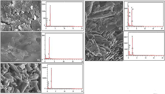
Figure 1.
A) Crystal surface visualized by SEM coupled to EDX before any treatment, B) crystal surface visualized by SEM coupled to EDX after treatment with citrate, C) crystal surface visualized by SEM coupled to EDX after treatment with aqueous NaCl solution, D) the surface of crystals visualized by SEM that is coupled to EDX after treatment with the solution containing the ethanolic extract of
Data from this study revealed that, prior to treatment, the composition of cystine stones consisted mainly of carbon (C), nitrogen (N), oxygen (O) and sulfur (S), an analysis corroborated by infrared spectra. However, after treatment with plant extracts (aqueous and ethanolic), a new composition emerged, including elements such as oxygen (O), nitrogen (N), sulfur (S) and chlorine (Cl), in addition to those initially present. Similar changes were observed in
The application of scanning electron microscopy in conjunction with energy dispersive spectrometry analysis fully confirmed the changes in the ultrastructural characteristics and elemental composition of the cystine stones before and after treatment. These results suggest the possibility of a curative effect induced by the plant in question, probably thanks to the presence of various compounds acting as active principles in the plant extracts studied, although we did not observe any significant change in mass levels in comparison with the other plant extracts used in Hannache’s experiment [42], which showed a high dissolution percentage for the cystine stones, indicating that our plant does indeed have a slight effect for this type of stone, but is probably effective for another type.
6. Conclusion
In conclusion, this chapter demonstrates the significance of electron microscopy in scientific research within the medical field. It allows for the exploration of the effects of
Furthermore, scanning electron microscopy coupled with X-ray energy dispersive spectrometry (SEM–EDS), as a qualitative and quantitative technique, has a high potential for studying and detecting surface changes, as well as for determining the chemical elements present in the structure of stone crystals. This technique confirms the results of Fourier transform infrared spectroscopy as a quantitative approach. In brief, this technology has the potential to equal or even exceed infrared technology in the chemical analysis of patient stones.
References
- 1.
Sandberg PA, Hay WW. Study of Microfossils by Means of the Scanning Electron Microscope. Tulsa, Oklahoma: SEPM Society for Sedimentary Geology; 2023 - 2.
Freundlich MM. Origin of the electron microscope. Science. 1963; 142 (3589):185-188. DOI: 10.1126/science.142.3589.185 - 3.
Mestres Ventura PJ. The electron microscope on the eve of its first centenary. EJA. 2023; 27 :111-127. DOI: 10.1126/science.142.3589.185. Available from:https://eurjanat.com/articles/the-electron-microscope-on-the-eve-of-its-first-centenary - 4.
Masters BR. History of the Electron Microscope in Cell Biology. 1st ed. John Wiley & Sons, Ltd; 2009 [cited 2023 Sep 4]. Available from: https://onlinelibrary.wiley.com/doi/10.1002/9780470015902.a0021539 - 5.
Kociak M. Introduction à la microscopie électronique. JDN. 2007; 12 :61-74. Available from:http://www.neutron-sciences.org/10.1051/sfn:2007006 - 6.
López De La Rosa F, Sánchez-Reolid R, Gómez-Sirvent JL, Morales R, Fernández-Caballero A. A review on machine and deep learning for semiconductor defect classification in scanning electron microscope images. Applied Sciences. 2021; 11 (20):9508. Available from:https://www.mdpi.com/2076-3417/11/20/9508 - 7.
Roussel LY, Stokes DJ, Gestmann I, Darus M, Young RJ. In: Postek MT, Newbury DE, Platek SF, Joy DC, editors. Extreme High Resolution Scanning electron Microscopy (XHR SEM) and beyond. Monterey, CA; 2009. p. 73780W. Available from: http://proceedings.spiedigitallibrary.org/proceeding.aspx?doi=10.1117/12.821826 - 8.
Ross FM. Opportunities and challenges in liquid cell electron microscopy. Sciences. 2015; 350 (6267):aaa9886. Available from:https://www.science.org/doi/10.1126/science.aaa9886 - 9.
Ge B. Scanning transmission electron microscopy (STEM). In: Wang R, Wang C, Zhang H, Tao J, Bai X, editors. Progress in Nanoscale Characterization and Manipulation. Singapore: Springer; 2018. pp. 205-254. (Springer Tracts in Modern Physics; vol. 272). Available from: http://link.springer.com/10.1007/978-981-13-0454-5_4 - 10.
Binnig G, Rohrer H. Scanning tunneling microscopy. Surface Science (North-Holland Publishing Company). 1983; 126 :236-244 - 11.
Smith D, Starborg T. Serial block face scanning electron microscopy in cell biology: Applications and technology. Tissue and Cell. 2019; 57 :111-122. Available from:https://linkinghub.elsevier.com/retrieve/pii/S004081661830212X - 12.
Schaming D, Remita H. Nanotechnology: From the ancient time to nowadays. Foundational Chemistry. 2015; 17 (3):187-205. Available from:http://link.springer.com/10.1007/s10698-015-9235-y - 13.
Fan Z, Zhang L, Baumann D, Mei L, Yao Y, Duan X, et al. In situ transmission electron microscopy for energy materials and devices. Advanced Materials. 2019; 31 (33):1900608. Available from:https://onlinelibrary.wiley.com/doi/10.1002/adma.201900608 - 14.
Frelinger SN, Ledvina MD, Kyle JR, Zhao D. Scanning electron microscopy cathodoluminescence of quartz: Principles, techniques and applications in ore geology. Ore Geology Reviews. 2015; 65 :840-852. Available from:https://linkinghub.elsevier.com/retrieve/pii/S0169136814002595 - 15.
Yamashita M, Lin MY, Hou J, Ren KYM, Haas M. The continuing need for electron microscopy in examination of medical renal biopsies: Examples in practice. Glomerular Diseases. 2021; 1 (3):145-159. Available from:https://www.karger.com/Article/FullText/516831 - 16.
(Porikli) Durdaği S, AHH A-J, Yalçin P, Bozkurt AS, Salcan S. Morphological characterization and phase determination of kidney stones using X-ray diffractometer and scanning electron microscopy. Chinese Journal of Physics. 2023; 83 :379-388. Available from:https://linkinghub.elsevier.com/retrieve/pii/S0577907322002258 - 17.
Siener R. Nutrition and kidney stone disease. Nutrients. 2021; 13 (6):1917. Available from:https://www.mdpi.com/2072-6643/13/6/1917 - 18.
Grases F, Costa-Bauza A, Prieto RM. Renal lithiasis and nutrition. Nutrition Journal. 2006; 5 (1):23. Available from:http://nutritionj.biomedcentral.com/articles/10.1186/1475-2891-5-23 - 19.
Mammate N, El Oumari FE, Imtara H, Belchkar S, Benjelloun Touimi G, Al-Zharani M, et al. Anti-struvite, antimicrobial, and anti-inflammatory activities of aqueous and ethanolic extracts of Saussurea costus (Falc) Lipsch Asteraceae. Molecules. 2023;28 (2):6673. Available from:https://www.mdpi.com/1420-3049/28/2/667 - 20.
Dako E, Retta N, Desse G. Comparison of Three Sweet Potato ( Ipomoea batatas (L.) Lam) Varieties on Nutritional and Anti-Nutritional Factors. USA; 2016 - 21.
Wróbel G, Kuder T. The role of selected environmental factors and the type of work performed on the development of urolithiasis – A review paper. International Journal of Occupational Medical and Environmental Health 2019 32(6):761-775. Available from: http://www.journalssystem.com/ijomeh/The-role-of-selected-environmental-factors-and-the-type-of-work-performed-on-the ,111685,0,2.html - 22.
Temporal trends in the incidence of kidney stone disease. Kidney International. 2013; 83 (1):146-152. Available from:https://www.sciencedirect.com/science/article/pii/S008525385556949 - 23.
Dalibon P. La lithiase urinaire, une affection sous surveillance. Actualités Pharmaceutiques. 2015; 54 (542):23-29. Available from:https://linkinghub.elsevier.com/retrieve/pii/S0515370014004303 - 24.
Thakore P, Liang TH. Urolithiasis. StatPearls. Treasure Island, FL; 2023. Available from: https://www.ncbi.nlm.nih.govbooksNBK559101.pdf . - 25.
Srivastava A, Swain KK, Chahar V, Bhardwaj S, Ajith N, Mete U, et al. Role of diet and trace elements in lithogenesis of renal calculi. Journal of Radioanalytical Nuclear Chemistry. 2019; 319 (1):271-278. Available from:http://link.springer.com/10.1007/s10967-018-6335-x - 26.
Singh P, Harris PC, Sas DJ, Lieske JC. The genetics of kidney stone disease and nephrocalcinosis. Nature Reviews Nephrology. 2022; 18 (4):224-240. DOI: 10.1038/s41581-021-00513-4 - 27.
Wang M, Chiu Y, Flahaut D, Jones IP, Zhang Z. Secondary phase area fraction determination using SEM-EDS quantitative mapping. Materials Characterization. 2020; 167 :110506. Available from:https://linkinghub.elsevier.com/retrieve/pii/S104458032031977X - 28.
Miller NL, Evan AP, Lingeman JE. Pathogenesis of renal calculi. Urologic Clinics of North America. 2007; 34 (3):295-313. Available from:https://linkinghub.elsevier.com/retrieve/pii/S0094014307000468 - 29.
Cochat P, Bacchetta J, Sabot JF, Bertholet-Thomas A, Demède D. Lithiase urinaire de l’enfant. Journal de Pédiatrie et de Puériculture. 2012; 25 (5):255-268. Available from:https://linkinghub.elsevier.com/retrieve/pii/S0987798312001065 - 30.
Daudon M, Traxer O, Lechevallier E, Saussine C. La lithogenèse. Progrès en Urologie. 2008; 18 (12):815-827. Available from:https://linkinghub.elsevier.com/retrieve/pii/S1166708708003904 - 31.
Bazin D, Daudon M, Chevallier P, Rouziere S, Elkaim E, Thiaudiere D, et al. Les techniques de rayonnement synchrotron au service de la caractérisation d’objets biologiques : un exemple d’application, les calculs rénaux. Annales de Biologie Clinique. 2006; 2006 :64 - 32.
El Habbani R, Chaqroune A, Sqalli Houssaini T, Arrayhani M, El Ammari J, Dami F, et al. Étude épidémiologique sur les calculs urinaires dans la région de Fès et sur le risque de récidive. Progrès en Urologie. 2016; 26 (5):287-294. Available from:https://linkinghub.elsevier.com/retrieve/pii/S1166708716000294 - 33.
Mammate N, El Oumari FE, Imtara H, Belchkar S, Lahrichi A, Alqahtani AS, et al. Antioxidant and anti-urolithiatic activity of aqueous and ethanolic extracts from Saussurea costus (Falc) Lispich using scanning electron microscopy. Life. 2022; 12 (7):1026. Available from:https://www.mdpi.com/2075-1729/12/7/1026 - 34.
Daudon M, Jungers P, Traxer O. Lithiase Urinaire. Paris: Lavoisier; 2012 - 35.
Plawecki M, Bistoquet M, Grillet PE, Abdo N, Souweine JS, Cristol JP. Drug-induced urinary stone of atazanavir incidentally found in an asymptomatic patient: A case report. Case Reports in Urology. 2023; 2023 :4. Article ID 4890711. Available from:https://www.hindawi.com/journals/criu/2023/4890711/ - 36.
Singh VK, Rai PK. Kidney stone analysis techniques and the role of major and trace elements on their pathogenesis: A review. Biophysical Reviews. 2014; 6 (3-4):291-310. DOI: 10.1007/s12551-014-0144-4 - 37.
Tcheka C, Moubarik A, Outzourhit A, Mbarki M, Benguellah BL, Mbadcam JK, et al. Epidemiological exploration of urinary stones. Physical and Chemical News. 2011; 61 :120-127 - 38.
Hannache B. La lithiase urinaire : épidémiologie, rôle des éléments traces et des plantes médicinales [Internet] [thesis]. Paris 11; 2014. Available from: http://www.theses.fr/2014PA114804 - 39.
Bougouma M, Keraghel F, Sawadogo J, Adama K, Buess-Herman C, Doneux T. Caractérisation physico-chimique et électrochimique de monocristaux de Mo0,75W0,25Se2 obtenus par croissance cristalline. 2020 - 40.
Paramita C, Arup C, Mukherjee AK. Phase composition and morphological characterization of human kidney stones using IR spectroscopy, scanning electron microscopy and X-ray Rietveld analysis. Spectrochimica Acta Part A: Molecular and Biomolecular Spectroscopy. 2018; 200 :33-42. Available from:https://www.sciencedirect.com/science/article/pii/S1386142518303044 - 41.
Guinebretière R. La diffraction des rayons X sur des échantillons polycristallins- l’actualité chimique - juil.-août-sept.-oct. 2014 - n° 387-388-389 - 42.
Hannache B. La lithiase urinaire : épidémiologie, rôle des éléments traces et des plantes médicinales [These de doctorat]. Paris 11; 2014 [cited 2023]. Available from: https://www.theses.fr/2014PA114804 - 43.
Doumalin P. Microextensométrie locale par corrélation d’images numériques. Application aux études micromécaniques par microscopie électronique à balayage. Thèse de doctorat en Sciences de l’ingénieur [physique]. Ecole Polytechnique X, 2000. En français. Numéro de thèse: 2000EPXX0027. Identifiant pastel: pastel-00001162 - 44.
Bell DC, Garratt-Reed AJ. Energy dispersive X-ray analysis in the electron microscope. Garland Science. 2003; 2003 :163 - 45.
Technique (ANRT) AN de la R. Electron-probe microanalysis: x-ray spectroscopy. Association Nationale de la Recherche Technique (ANRT); 1987 [cited 2023 Sep 10]. Report No.: FRNC-R--274. Available from: http://inis.iaea.org/Search/search.aspx?orig_q=RN:21052160 - 46.
Taïbi K, Aït Abderrahim L, Boussaid M, Taibi F, Achir M, Souana K, et al. Unraveling the ethnopharmacological potential of medicinal plants used in Algerian traditional medicine for urinary diseases. European Journal of Integrative Medicine [Internet]. 2021; 44 :101339. Available from:https://linkinghub.elsevier.com/retrieve/pii/S1876382021000573 - 47.
Idmhand E, Msanda F, Cherifi K. Étude ethnobotanique des plantes médicinales utilisées dans le traitement de la lithiase urinaire dans la province de Tarfaya (Maroc). International Journal of Innovation and Applied Studies. Jun 2019; 26 (3):711-719. ISSN: 2028-9324