
Abstract
Trace evidence analysis is essential in criminal investigations as it provides vital information for establishing connections between suspects and scenes. Minute or complicated trace evidence is sometimes difficult for traditional microscopic techniques to handle. At micro- and nanoscale, electron microscopy (EM) shows great promise as a potent technique for characterization and visualization. Scanning electron microscopy (SEM) and transmission electron microscopy (TEM) offer valuable insights into morphology, chemical composition, and crystalline structure of trace evidence, enabling the identification and differentiation of similar materials. TEM allows high-resolution examination of paint components, dirt particles, gunshot residues (GSR), fibers, hair structures, glass shards, nano-particles, explosive materials, etc. In forensic investigations, SEM is a crucial instrument, especially when it comes to GSR analysis, which uses SEM to correlate bullets to firearms more successfully than visual approaches. Additionally, SEM plays a major role in the examination of gemstones and jewelry by identifying manufactured and natural gems, analyzing surface imperfections, and determining elemental compositions. SEM also improves forensic inspection in non-conductive material analysis, paint and fiber analysis, filament bulb investigations, handwriting analysis, and counterfeit detection. The adoption of EM in forensic trace evidence analysis has potential to revolutionize the field, offering valuable insights that were previously unattainable.
Keywords
- forensic trace evidence
- criminal investigations
- electron microscopy (EM)
- transmission electron microscopy (TEM)
- scanning electron microscopy (SEM)
1. Introduction
Throughout human history, crime has been a persistent aspect of society, harming people, animals, and entire communities. It is interesting to note that great minds focused on this mystery in the late nineteenth century, which is when the scientific study of criminal phenomena started to gain traction. This led to the creation of the field of forensic science, an applied discipline that is seamlessly incorporated into a society’s legal framework and serves the dual purposes of providing justice to victims of crime and understanding the motivations behind the actions of those who commit it to discourage such evil deeds within the community. In the wake of the renowned forensic scientist Sir Edmond Locard, who famously declared that “every contact leaves a trace,” a crucial advancement in forensic science arose: the identification and application of trace evidence. This type of evidence plays a crucial role in forensic analysis because it provides important information about the presence or absence of people and things at crime scenes, illuminating the complex network of relationships that connects the criminal act to its surroundings.
Every contact contains trace evidence, and the crucial question is how much our scientific knowledge can identify and track these minute remnants. What is just as important is the competence of the people using the technology; they need to be able to predict the kinds of traces that could be found.
Scientific progress, combined with the use of a wide range of instrumental methods, has greatly improved the ability to perform in-depth analyses of traces of evidence. Notably, in this dynamic environment, electron microscopy technology arises as a revolutionary instrument, providing hitherto unseen accuracy and understanding into the micro-scale realm of trace evidence.
The first electron microscope was invented in 1931 by Ernst Ruska and Max Knoll, two University of Berlin physicists and electrical engineers, respectively. This prototype was the first to demonstrate what could be done with electron microscopy, producing a 400-power magnification [1].
Ten years later, Ruska developed a comparable but distinct method that delivered information about the topography and composition of a sample by scanning its surface in a rectangular pattern with a focused electron beam. In contrast to TEM, the new scanning electron microscope’s (SEM) image was produced after the instrument gathered and tallied the dispersed electrons.
Many believe that the introduction of the digital microscope by Japanese scientists in 1986 transformed the field of microscopy. They devised a method for moving the image from the microscope to a computer so that it could be examined right away. These days, high-resolution monitors are integrated into microscopes, so viewing images does not require an extra computer [2].
Fibers, hairs, glass shards, paint chips, soil particles, plants, insects, gunshot residue, and more can be included in this category. By connecting these items to certain sources or people, trace evidence analysis seeks to provide important details for criminal investigations [3]. With its unmatched resolution and extraordinarily high magnification powers, electron microscopy is a technological marvel when it comes to trace evidence examination. This sophisticated technique, in contrast to traditional optical microscopy, reduces material distortion commonly associated with sample preparation, maintaining the integrity and fidelity of the materials under examination. Furthermore, the use of electron microscopy makes it possible to examine specimens at different strata and to investigate a wider depth of field. This function plays a crucial role in revealing minute details and nuanced aspects within the trace evidence, offering priceless insights for forensic analyses and contributing to a thorough comprehension of the evidentiary landscape [4].
The minute number and exquisite structure of these evidential elements present a major difficulty for laboratory analysis and research employing trace evidence. It is now essential to use electron microscopes to address this problem. Because of the incredibly small size and complex structure of these materials, conventional methods may not be able to reach the precise visualization and analysis that this sophisticated equipment enables when examining trace evidence at the micro- and nanoscales.
The study of trace evidence presents difficulties because the evidence is complex and does not lend itself to standard microscopic procedures. These difficulties include low resolution, complicated evidence (e.g., nanoparticles or intricate fiber structures), and the incapacity to identify minute characteristics that are essential for differentiating between materials and connecting evidence to crime scenes. Nevertheless, electron microscopy (EM), particularly scanning electron microscopy (SEM) and transmission electron microscopy (TEM), skillfully handles these difficulties. Excellent high-resolution imaging capabilities provided by EM methods allow the viewing of minuscule details, even at the nanoscale. Furthermore, EM enables thorough characterization of traces, supporting material analysis, identification, and differentiation according to elemental composition, morphology, and structure. By facilitating the identification of minute details and minute variations between materials, the improved discriminating power of EM considerably advances forensic investigation by offering crucial insights into the nature and provenance of trace evidence.
In this chapter, we examine a number of situations in which the application of electron microscopes to the examination of trace evidence provides not only improved structural analysis but also special chances to examine characteristics that are not accessible through conventional microscopy techniques. The utilization of electron microscopes facilitates a comprehensive analysis at the micro- and nanoscales, offering unmatched clarity and permitting the discovery of minuscule structural subtleties and more minute details that were previously unobservable by traditional microscopic methods.
The first section of the chapter covers the definitions of electron microscopes (EM) and the essential structural elements that are present in transmission electron microscopes (TEM) and scanning electron microscopes (SEM). From the second section, a methodical investigation into the various domains in which TEM finds application is then undertaken. The second section carefully explains the use of TEM in paint analysis, soil analysis, gunshot residue (GSR) characterization, fiber and hair structure clarification, glass analysis, and nanoparticle scrutiny. After that, in the third section, the story shifts to a detailed examination of SEM applications. In particular, thorough research is conducted into the critical role that SEM plays in GSR analysis, firearm identification, gemstone investigation, paint and fiber analysis, and filament bulb scrutiny. The final and last section of the chapters deals with a through summery of the key learning.
This chapter delves into the complex and subtle uses of TEM and SEM in several fields of trace evidence analysis using a thorough scientific methodology.
1.1 Electron microscope
Currently, one of the most important tools for evaluating the surface or internal structure is electron microscopy (EM), which has a greater resolution than light microscopy (LM) since it creates images using an accelerated electron beam rather than photons. Electron beams can identify tiny structures because their wavelengths are smaller than those of visible light. To reduce electron beam scatter, conventional electron microscopy operates at high vacuum (10−5–10−7 Pa).
Two primary types of electron microscopy techniques are employed: scanning electron microscopy (SEM) and transmitted electron microscopy (TEM). SEM and TEM have different principles and resolutions, and the information obtained by both methodologies complements each other [5].
1.2 Scanning electron microscope (SEM)
Using a thermal electron source, the scanning electron microscope (SEM) (Figure 1) projects a high-energy electron beam (100–30,000 eV) onto an object. Through the use of lenses, the spot size is reduced to less than 10 nm to accomplish fine imaging. Electrons generate signals for picture creation by penetrating the specimen up to 1 μm. Using scan coil movements that change depending on the necessary magnification, the SEM creates an image point by point. Signals generated by electrons are detected by electron detectors; backscattered electrons (BSE) and secondary electrons (SE) are employed in imaging. The degree of surface or inner sample features is determined by the electron voltage mode; greater voltages penetrate deeper and reveal more surface information. The SEM offers a three-dimensional picture that highlights the topography of the sample, which is influenced by the surface’s degree of inclination [7].
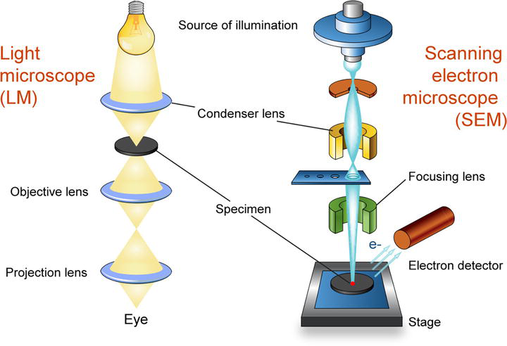
Figure 1.
Parts of scanning electron microscope [
1.3 Transmission electron microscope (TEM)
In materials research, transmission electron microscopy (TEM) (Figure 2) is a useful instrument. It employs thermionic or field emission to release electrons from an electron source, such as a tungsten filament or lanthanum hexaboride (LaB6), that is linked to a high-voltage source. Upper TEM lenses shape the released electrons into an electron probe, and they can be manipulated by applying magnetic and electrostatic fields. Condenser lenses, objective lenses, and projector lenses make up TEM optics, which allow for variations in magnification through the manipulation of current flow. To display images, one uses an imaging device, such as a phosphor screen. TEM works in a low-pressure setting to improve the clarity of the electron path. Sample grids are supported by specimen stages, while electron beams are produced and managed by electron cannons. Electromagnetic coils are used in electron lenses for electron path correction and focus. Electron beams are selectively filtered by apertures to enhance imaging quality. The methods used for sample preparation—which include grid deposition and fixation for biological samples—depend on the materials and data needed [9].
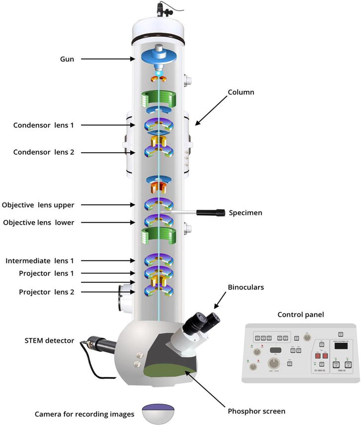
Figure 2.
Parts of transmission electron microscope [
2. Sample preparations
2.1 Transmission electron microscope (TEM)
2.1.1 General preparation
Preparing the specimen for TEM visualization: In order to facilitate visualization and produce a crisp image, the specimen to be examined under the TEM needs to go through a certain preparation process.
On solid materials, electrons are readily absorbed and dispersed, resulting in poor visibility for thick specimens. For accurate and clear vision that also forms a crisp image, very thin specimens are used. The specimen should have a diameter of 0.025–0.1 nm and a thickness of roughly 20–100 nm, or as small as a bacterial cell. Thin specimens are able to preserve their inherent structure while interacting with electrons in a vacuum.
The specimen is initially fixed on a plastic surface using glutaraldehyde or osmium tetraoxide in order to obtain thin-slice specimens. These chemical substances preserve the cell’s uniqueness while stabilizing its structure. When an organic solvent, such as alcohol, is added, since ethanol will totally dry out the cell, it is necessary to embed the specimen in the plastics.
After that, the specimen is permeated with unpolymerized liquid epoxy plastic, which solidifies it into a block. This is the area where thin sections are cut with a glass knife and an ultramicrotome, a piece of specialized equipment.
Subsequently, the material is suitably dyed (using the suitable stain) to ensure consistent electron dispersion. After that, the thin sections are immersed in heavy metallic substances such as uranyl acetate and lead citrate, which enable the lean and aluminum ions to bond to the arrangements of cells. To improve contrast, this creates an opaque coating to block the electrons on the cell structures.
After staining, the thin pieces are put on copper grids so they can be seen. Negative staining combined with a strong metallic element coating is the main staining approach used for TEM viewing. The study of bacterial and viral cell morphologies and structures is facilitated by the metallic coating’s ability to scatter electrons, which show up on the photographic film in uncoated portions.
2.1.2 Freeze-itching treatment
Unlike chemical fixation, dehydration, and embedding, freeze-itching is specifically employed for the treatment of microbial cells in order to minimize the potential risks of artifacts.
Microbial cell organelles receive a unique treatment called freeze-itching, where the specimens are prepped with liquid nitrogen and subsequently heated at −100°C in a vacuum chamber. This is the process when the majority of specimens become contaminated.
The parts are then cut in liquid nitrogen at −196°C using a knife that has been precooled. The sectioned specimen can be covered with platinum and carbon layer-forming replicas after being heated in a high vacuum for roughly 2 minutes.
These can then be examined under a TEM, which shows the cell’s inside architecture in greater detail in three dimensions. This phase of liquid nitrogen therapy is referred to as “freeze-itching.”
2.2 Scanning electron microscope
2.2.1 Scrubbing the specimen’s surface
It is critical to thoroughly clean the sample’s surface because it may include a range of undesirable deposits, such as silt, dust, detritus, media elements, or other pollutants, contingent upon the origin of the biological material and any experiment that might have been carried out before the fabrication of the SEM specimen.
2.2.2 Maintaining the specimen’s stability
Fixatives are usually used for stabilization. Fixation can be accomplished through perfusion, for instance, by employing a variety of fixatives, such as aldehydes, osmium tetroxide, tannic acid, or thiocarbohydrazide,
Rinsing the specimen: Following the fixation phase, samples need to be rinsed to get rid of extra fixative.
2.2.3 Dehydration the specimen
It is imperative that a biological sample be dehydrated with extreme caution. Either a graded series of acetone or ethanol is usually used for it.
2.2.4 Mounting the specimen
The samples need to be mounted on a holder that can be placed inside the scanning electron microscope once they have been cleaned, fixed, washed, dehydrated, and dried according to the proper technique. Usually, the double-stick tape is used to mount samples on metallic (aluminum) stubs. Prior to attaching the specimen, the investigator must determine the optimal orientation for it on the mounting stub. Reorienting the sample proves to be challenging and may cause serious harm.
2.2.5 Coating the specimen
By transporting the charge to the ground, the coating increases the specimen’s conductivity under the scanning electron microscope and inhibits the accumulation of high-voltage charges on it. Usually, specimens have a thin layer of conductive metal (such as platinum, gold–palladium, or gold) covering them, ranging from 20 to 30 nm. Since they are all conductive, metals can be used without any prior treatment. A small layer of conductive material must be applied to the sample in order to make all non-metals conductive. The tool used to accomplish this is known as a “sputter coater.” Argon gas and an electric field are used by the sputter coater. The sample is put in a tiny vacuum-shielded chamber.
The atoms become positively charged when argon gas and an electric field remove one of the atoms’ electrons.
A negatively charged gold foil then attracts the argon ions. Gold atoms are removed from the gold foil’s surface by the argon ions. A thin layer of gold is created on the sample’s surface as a result of these gold atoms falling and settling there.
3. Applications of electron microscopy for trace evidence analysis
3.1 Application of transmission electron microscope
The investigation of trace evidence is greatly aided by transmission electron microscopy (TEM), which provides in-depth information at the nanoscale, leading to recognize and describe minuscule particles such as paint bits, fibers, and gunshot residue. The great resolution of TEM makes it possible to determine the chemical and elemental makeup of materials, which facilitates source identification. When it comes to examining fibers, this technique is quite helpful as it provides crucial information that helps connect items to crime scenes. Furthermore, TEM offers tiny details that conventional techniques would overlook, greatly assisting forensic investigations in the evaluation of biological trace evidence, tool marks, and counterfeit materials.
3.1.1 Paint
Forensic investigations in particular heavily rely on transmission electron microscopy (TEM) for paint chip examination. With the ability to give high-resolution images of tiny paint fragments and fine-grained compositional data, TEM can help identify pigments, binders, and layer structures, as shown in Figure 3, as well as facilitate material characterization. This analytical method is essential for investigating trace evidence discovered at crime scenes because it provides a way to identify the paint materials’ inorganic components with remarkable precision and clarity.
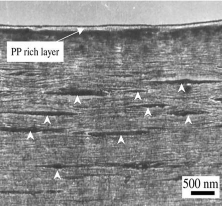
Figure 3.
Three layers of paint [
The adhesive factor of paints is an important aspect for its application on any surface, especially automobile surfaces, which are exposed to a lot of environmental factors. In such cases, thermoplastic polyolefins (TPO) are mostly used as an adhesive, but they have poor adhesive properties; to compensate for this, we use chlorinated polyolefins, an extra layer over the TPO layer, and through observation in injection-molded TPO samples, a rich layer of polypropylene (PP layer) is present [10], which can be seen in Figure 4.
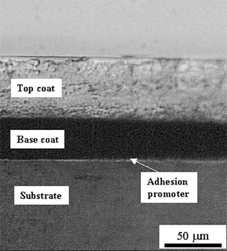
Figure 4.
Polypropylene homo-polymer seen in paint layer under TEM [
A study examining paintings from the fourteenth century serves as an example of the usefulness of TEM in analyzing the authenticity of antique paintings. In this instance, the original layer arrangement was preserved, while TEM was used to examine the makeup and structure of paint and base layers. Meticulously manufactured thin micro-samples showed a vivid red paint coat and underlying layers. Major and minor components within these layers were identified by examining individual particles using electron diffraction. The findings showed that gypsum made up the majority of the basal layer, with small amounts of calcite and dolomite. Notably, it was discovered that the paint layer was made entirely of vermilion [11]. But we encounter paint samples mostly in case of car accidents. A lot of work has gone into identifying special characteristics, especially for primers and fillers. TEM is crucial for the assessment of their micromorphology because of its remarkable capacity to reveal minute features. Furthermore, pearl luster pigments such as Iriodin and Afflair, which are mostly found in cars manufactured in the United States and Europe, can also be examined using TEM in paint analysis [12].
Additionally, when paint samples are generated with focused ion beam technology, TEM provides useful capabilities for imaging and identifying nanometer-sized particles scattered throughout the surface zone of the samples. This cutting-edge method makes it possible to see and identify particular components, such as lead-rich particles, on a 0.5-micrometer surface area. For tasks that conventional scanning electron microscopy (SEM) and electron probe microanalysis (EPMA) cannot do, TEM’s high resolution and specificity make it an invaluable tool [13].
3.1.2 Soil
Soil, being a complex and dynamic material, is an essential trace evidence in criminal investigations. Most soil particles are of a variety of sizes, ranging from several nanometers to several centimeters. Soil mostly acts as associative evidence in criminal investigation helping us in determining the place from where our person of interest has arrived or where he went. Transmission electron microscopy (TEM) is an indispensable tool for the accurate characterization of tiny soil particles, particularly those with a size between a few nanometers and a few micrometers.
The investigation of crystallographic alterations in terrestrial particles under physicochemical and environmental factors is made easier by TEM. It is essential to comprehend how living things, such as fungi and bacteria, alter the makeup of soil, including the transformation of its elemental chemical forms. Current studies highlight the usefulness of TEM in examining micrometer-scale micro aggregates, offering insights into the impact of microbes on soil function. Soil near the mining area contains a characteristic amount of the mineral present in the soil as nanoparticles, and even in other soil profiles, trace minerals can be found in the soil, which can generate a unique profile for the soil and point us toward the probable source.
TEM makes it possible to qualitatively evaluate the interactions that take place between bacteria and fungus in oregano-mineral connections, which include biogenic structures such as bulk soil, the soil-root interface, and casts. This information takes into account the inherent diversity of soil and aids in assessing the state of these microorganisms at the time of sample collection [14, 15], as shown in Figure 5.
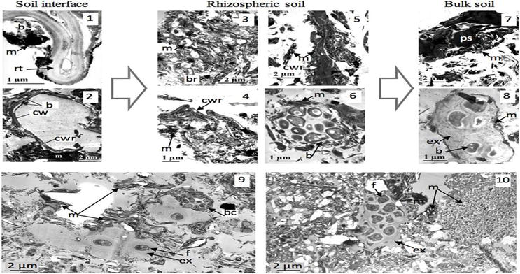
Figure 5.
Soil-microorganism interactions at hotspots of biological activity. TEM views (1–8) (soil 4): Micro-aggregation from maize root-soil interface (1, 2) to rhizospheric soil (3–6) and to bulk soil (7, 8) [
For a thorough analysis of soil particles magnified more than 100,000 times, transmission electron microscopy (TEM) and scanning electron microscopy (SEM) are also used. TEM and SEM make it possible to use methods like energy-dispersive X-ray spectroscopy to investigate the morphology and chemical composition of particles. These high-resolution techniques are very helpful for analysis and discrimination, greatly advancing our knowledge of pollen spores, minerals found in soil, and fossils—all of which are pertinent to forensic soil study [17].
3.1.3 Gun shot residue (GSR)
Particles created when a firearm is discharged are referred to as gunshot residue, cartridge discharge residue (CDR), or firearms discharge residue. The primer and propellant combustion products will be emitted simultaneously when a cartridge round is shot in a weapon [18]. Gunshot residues are made up of partially and unburned materials, smoke, propellant powder, bullet primer fragments, metals, grease, and lubricants from the cartridge in addition to the actual weapon [19].
According to a scientific study, depending on the type of ammunition, gunshots release sub-micrometer nanoparticles into the environment. About 80 to 94% of the samples were in the nanometer size range, as shown in Figure 6.
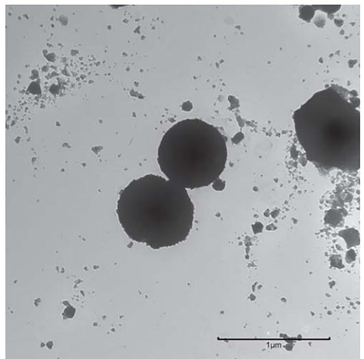
Figure 6.
TEM image of particles collected on the filter during the discharge of one round of S&B ammunition. Scale bar = 1 μm. [
These tiny particles—particularly gunshot residue (GSR) nanoparticles—could linger in suspension in the atmosphere for as long as 12 hours. GSR nanoparticles are especially significant in forensic investigations because they are more common and persistent than bigger particles. The elemental analysis and high-resolution imaging capabilities of transmission electron microscopy with energy-dispersive X-ray spectroscopy (TEM-EDS) make it an invaluable tool for the analysis of GSR nanoparticles. Future forensic analyses depend on the detection of GSR nanoparticles [21].
TEM can recognize distinctive components that are particular to producers or geographical areas. Gunshot residue (GSR) examination using ammunition manufactured in Brazil revealed that TEM provided a better comprehension of nanoscale particles than SEM [22].
3.1.4 Fiber
Another type of trace evidence is fiber, which includes compounds derived from synthetic materials such as rayon and nylon as well as natural sources such as cotton and silk. These fibers often have a major impact on assault and burglary cases. They are useful associative evidence, and it is frequently necessary to examine their distinctive architecture to determine their origins. Although scanning electron microscopy (SEM) is the most commonly used technique for fiber analysis, transmission electron microscopy (TEM) has also shown value in some situations.
It has been found useful where it was found that analytical TEM can be of great application for finding mineral fibers and residues in the lung tissue of the dead, which can be found due to occupational hazards and death due to working in poor health-sanctioned paces [23].
Asbestos fiber has caused some major health issues over the decades, namely asbestosis, carcinoma of the lung (LC), pleural plaques, and malignant mesothelioma (MM). TEM has been useful in identifying it in tissue samples from lungs, which can be useful in case of death due to this reason [23]; it is useful in the study of the orientation of microfibrils present in wooden fiber and other forms of fibers, as shown in Figure 7.
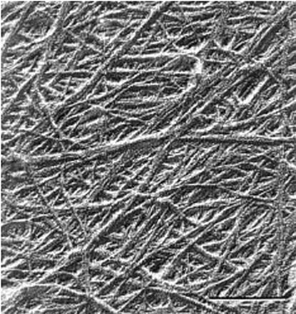
Figure 7.
TEM image of a spruce Kraft pulp fiber surface replica showing the microfibril orientation [
3.1.5 Hair
In forensic science, hair follicles are mostly used to get DNA samples, which allows for individualization. Still, these follicles conceal a more profound mystery concerning the coordinated actions of neighboring cells and their complex interaction with our nervous system. The application of the transmission electron microscope’s (TEM) potent imaging capabilities becomes essential to revealing this hidden knowledge. Morioka used TEM images in a noteworthy work to interpret the complex cellular structure around hair follicles. This study adds to our understanding of this intricate biological system by illuminating the previously unknown relationships and dynamics within this milieu [25].
With scanning electron microscopy, the hair shaft is easily visible (SEM). But hair coloring and hair treatments have become common in modern society, which means that these procedures could be sources of personal traits. Such chemical treatments leave the hair structure with characteristic damage patterns and markings. Transmission electron microscopy (TEM) can be a useful tool for studying and characterizing these effects [26]. This bleaching and coloring of hairs are very particular to a trend in certain communities, creating a mass effect and, hence, sometimes can also be used for racial identification, as shown in Figure 8.
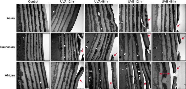
Figure 8.
Conventional transmission electron microscopy findings for all hair samples. Ultraviolet B (UVB) irradiation resulted in more damage than UVA irradiation (red arrow). All three groups exhibited similar patterns of damage. However, the African hair exhibited weaker resistance to UV irradiation than the other groups, and the African hair also exhibited a decreased number of cuticle layers than the other groups (red two-way arrow) [
As bleaching became more severe, leached proteins showed progressive oxidation. Bleached fibers exhibited significant damage to both cuticle layers and the cortex. Even mild bleach treatments led to extensive melanin granule degradation. Protein oxidation primarily affected cortical intermediate filaments, rich in sulfur-containing amino acids, notably the conversion of cystine disulfide bonds to cysteic acid. This data reveals the detrimental impact of bleach on hair structure and proteins, particularly in the cortex [28].
3.1.6 Glass
Glass is an amorphous solid used in various places of our day-to-day life, which includes including building materials from windows to table tops, used in scientific laboratories to our car windshields, and lastly, in our smartphones, glass has become a very common form of trace evidence found on a crime scene.
Glass examination by a forensic scientist may be requested to reconstruct events (e.g., to ascertain whether an exterior or interior window break occurred) or to link an individual or item to the crime scene or a victim. The type of glass involved, whether it is uncommon or common, how accurately it is described, and the background information and comparisons utilized all affect how strong the forensic scientist’s view is [29].
Glass samples’ interior microstructure, including imperfections, inclusions, and cracks, can be seen
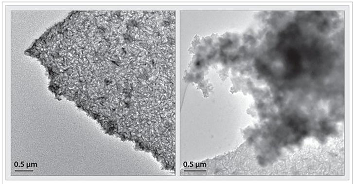
Figure 9.
TEM images of delamination particles filtered from the same vial [
3.1.7 Nanoparticles
The use of nanoparticles as trace evidence in forensic sciences has grown in recent years as a result of improvements in analytical methods and nanotechnology. In forensic science, nanoparticles are used as trace evidence in the following ways:
Characterization of nanoparticles: Other nanoparticles discovered at crime scenes can be identified and characterized using reference materials. Nanoparticles are inspected and categorized according to size, shape, and composition using methods such as transmission electron microscopy (TEM) [31], as shown with silver and gold nanoparticles in Figure 10.
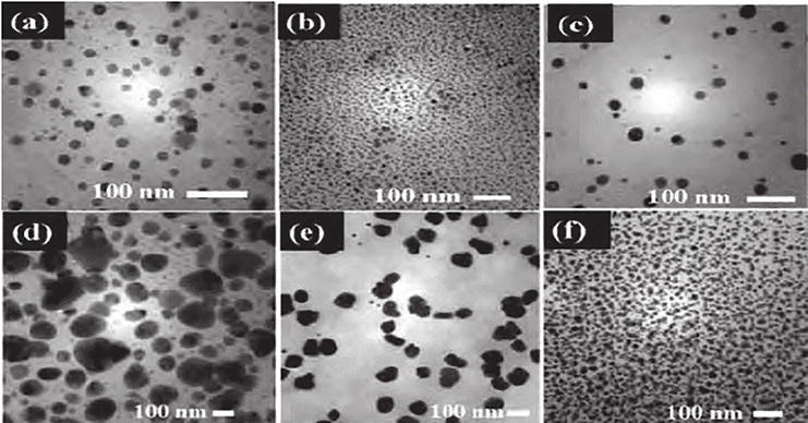
Figure 10.
Transmission electron microscopy (TEM) image of silver nanoparticles [
The manufacturing and utilization of nanoparticles are both steadily rising. Thus, the characterization of these particles is crucial for investigating their influence on trace evidence analysis, creating a particle database with TEM that offers a reference atlas of the different kinds of particles that might be found during trace evidence investigations in the future. The database, which is continuously updated, includes an image, a selected area electron diffraction (SAED) pattern, and an EDS of several particles from different origins. TEM is a useful tool in forensic science trace evidence studies since it may provide morphological, elemental, and internal structural information on very minute particles that may be missed or difficult to analyze with other microscopical techniques. This information can be used to supplement other analytical investigations [33].
3.1.8 Explosives
Explosives are a broad category that includes both high and low explosives. They have become powerful weapons of mass devastation that are frequently used to cover up illicit activity by feigning unintentional explosions. Pre- and post-blast analysis is a two-pronged strategy used by the field of forensic investigation in handling explosion-related situations. Because samples that are thought to be explosives are readily available, pre-blast analysis can be done rather easily. This makes it possible to apply different chemical and analytical tests to ascertain their properties and makeup. On the other hand, post-blast analysis poses a unique set of difficulties. In this case, forensic specialists have the difficult duty of looking through explosion leftovers and wreckage.
Small amounts of the explosive material, frequently in the nanoscale range, are usually present in these residues. Because these traces are so small and scattered throughout the debris, it is a challenging task to analyze them. In a post-blast analysis, the use of TEM-EDX has been found successful in which shot from the air after the blast was studied, and the type of explosive used was identified [34].
Transmission electron microscopy (TEM) has become an important tool to overcome these obstacles. TEM has the extraordinary capacity to examine a material’s crystalline structure at the nanoscale. This characteristic is essential for analyzing the post-blast residue because it allows for a thorough analysis of the structural characteristics of the explosive, which makes it easier to identify and connect it to the explosion. It also helps in the study of quantum dots, which are nowadays used to identify some common nitro base explosives [35].
3.2 Application of scanning electron microscope
The scanning electron microscope (SEM), which provides high-resolution, three-dimensional surface imaging, is an essential instrument in the investigation of trace evidence. SEM is utilized in forensic applications to examine the topography and morphology of trace materials. By offering fine-grained surface pictures, it facilitates the detection of various particles such as paint fragments, dirt, and gunshot residue. SEM is very useful for fiber analysis since it may be used to differentiate between various fiber types depending on surface features. Highlighting minute details on surfaces and connecting tools to particular markings helps with toolmark analysis. SEM is also used for surface imaging of different materials, paint study, hair morphology research, and tire tread analysis, all of which provide crucial information for forensic investigations.
3.2.1 GSR
Gunshot residue (GSR) or firearm discharge residue (FDR) comprises particulate matter generated during the act of discharging a firearm. These residues are commonly linked to components such as the primer, primer cup, propellant, projectile (slug/shot), projectile jacket and lubricant, cartridge case, and firearm barrel.
Anand et al. [36] showed gunshot residue (GSR) constitutes a heterogeneous assemblage of vapors emanating from the explosive components housed within the cartridge. When a firearm is involved in a criminal incident, the dissemination of gunshot residue (GSR) particles occurs not only through the muzzle but also through the cylindrical apertures, ejection ports, and other orifices within the firearm structure. These particles serve as crucial forensic evidence, prompting the need for their meticulous examination. Utilizing scanning electron microscopy coupled with energy-dispersive spectroscopy (SEM/EDX) is a conventional method for the comprehensive imaging and elemental analysis of gunshot residue (GSR) particles extracted from the specimens. Regrettably, a limited scholarly inquiry has explored the detection of GSR particles at extended distances employing SEM. Therefore, this current research introduces a novel methodology designed to identify GSR particles resulting from long-range firing. The approach involves an assessment of lead (Pb), barium (Ba), antimony (Sb), and other trace elements on the target material
Brożek-Mucha et al. [37] state that gunshot residues stemming from six distinct pistol ammunitions were the subject of investigation. Six individuals with no habitual firearm exposure discharged three rounds, each using a different pistol. Samples of the resulting gunshot residues were obtained from the shooters’ hands employing aluminum stubs affixed with black carbon adhesive tabs. These samples were subsequently subjected to morphological and elemental analyses through an automated process utilizing a scanning electron microscope integrated with an energy-dispersive X-ray spectrometer.
In the comparative analysis, focus was solely placed on the examination of primer residues. The percentage of particles representing specific chemical categories relative to the total count of detected particles was computed to determine their occurrence frequency. Varied relationships were observed between the frequencies of particular chemical residue occurrences for most of the studied ammunition types, as discerned through both non-statistical and non-parametric statistical methodologies (R-Spearman and τ-Kendall correlation coefficients). These statistical techniques facilitated the differentiation of one ammunition type from each of the other examined types. The analyses conducted unveiled discernible dissimilarities in the occurrence frequency defines that how many times the same component appeared in a samples under the study. Here it is define that which chemical component appeared to be present in various primer under study hence their occurrence frequency was determined.
Reyes et al. [38] showed that upon discharge of a firearm, a rapid release of a gaseous cloud containing distinct particles of gunshot residue (GSR) occurs, disseminating both forward and backward. These particles are deposited on various surfaces, including clothing, hands, face, and hair. Furthermore, they are also inhaled, leading to retention within the nostrils of the individual who discharged the weapon. GSRs, characterized by specific sizes and morphologies, comprise a composite of lead, antimony, barium, and other elemental components. Although scanning electron microscopy with energy-dispersive X-ray spectroscopy (SEM-EDS) represents a widely accepted technique for GSR analysis, limited research has addressed samples obtained from the suspect’s nostrils. While the analysis of residues found on the hands has been more commonplace, this study introduces a non-invasive collection device (Nasal Stub) designed for the retrieval of GSR particles from the nostrils. Additionally, a corresponding platform compatible with SEM-EDS has been developed. The efficacy of the Nasal Stub in collecting GSR particles from nasal hairs was evaluated using four different-caliber firearms. Nasal samples were collected at specific intervals (0, 2, 4, 6, 8, and 20 hours) post-firing. The findings indicate that the Nasal Stub successfully retrieved GSR particles from nasal hairs associated with all utilized firearms, even after a duration exceeding 20 hours in some instances. Consequently, it was determined that the proposed Nasal Stub Figure 11, in conjunction with the methodology for the analysis of nasal GSR from nose hairs
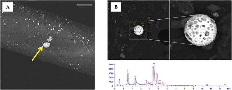
Figure 11.
GSR in nasal swab [
Kara et al. [39] mention that an investigation was focused on examining variations in the quantities of gunshot residues (GSR) collected from the shooter’s right hand using the “swab” method. Scanning electron microscope/energy-dispersive spectroscopy (SEM-EDS) was utilized to explore the potential correlation between these amounts and diverse conditions, including the shooter’s skin color, various physical attributes, hand hair density, hand size, and the presence of sweat or oiliness on the hands.
Analysis of the data revealed no significant disparity in the GSR quantities based on the shooter’s skin color. However, discernible fluctuations in the GSR levels were observed in relation to the shooter’s physical characteristics. These findings are anticipated to offer practical insights for experts engaged in GSR analysis using the swab technique, facilitating a more comprehensive assessment of the occurrence.
Toal et al. [40], Red X Defense has introduced an automated field presumptive lead test, utilizing a sampling pad designed to function as a red-light/green-light indicator. This pad can subsequently undergo processing within a scanning electron microscope (SEM) for the purpose of confirming the presence of gunshot residue (GSR). The XCAT’s sampling card is specifically employed to collect samples from a suspect’s hands at the crime scene, enabling investigators to swiftly ascertain the potential presence of lead, likely originating from primer residue. Notably, positive outcomes can be obtained after the discharge of as few as one shot.
Furthermore, the same sampling card can be forwarded to a crime laboratory for SEM-based GSR analysis, aligning with the procedures outlined in the ASTM E-1588-10 Standard Guide for gunshot residue analysis by scanning electron microscopy/energy-dispersive X-ray spectrometry. This processing method is akin to the current utilization of tape lifts within the field. Notably, the system can detect GSR-characteristic particles, including fused lead, barium, and antimony, as small as 0.8 microns with a resolution of 0.5 microns. This detection is achieved through the utilization of a JEOL JSM-6480LV SEM, equipped with an Oxford Instruments INCA EDS system featuring a 50mm2 SDD detector. The system operates at 350X magnification, with analyses conducted in both low-vacuum and high-vacuum modes, following carbon coating
The GSR particles demonstrate stability on the sampling pad for at least 2 months subsequent to chemical exposure, with ongoing assessments exploring their long-term stability. The immediate actionable intelligence provided by the XCAT’s presumptive result empowers law enforcement in expediting their investigations without compromising the necessity of a confirmatory test crucial for bolstering legal and investigative proceedings.
3.2.2 Firearms
Scanning electron microscopy (SEM) serves as a valuable instrument for assessing and comparing impressed or striated toolmarks, especially in cases concerning rimfire and centerfire cartridge cases, as well as striated marks on bullets. Notably, SEM provides a significantly superior depth of field compared to a light microscope, eliminating the need for side lighting to visualize the object’s surface topography [41].
With the advances in the EM, the field of study has increased which enables to examine the bullets and that bullets shot from the same firearm leave unique marks related to the gun’s barrel shape. Matching these distinct marks on bullets can help determine if they were fired from the same gun. Additionally, the firing pin on the bullet’s cartridge case can create similar markings, providing another way to link a bullet to a specific firearm. While optical techniques can sometimes be used to match bullets, they may lack the ability to reveal fine details at higher magnifications and lack the necessary depth of focus for clear visibility [42].
SEM series possesses three key features that offer significant advantages in comparing bullet markings: With a backscattered electron detector, the contrast of markings can be amplified while irrelevant information like dust contamination is suppressed. Carl Zeiss semiconductor manufacturing technology (SMT) provides a specially designed sub-stage for comparing bullets. This sub-stage can securely hold bullets or cartridge cases, allowing each bullet to be rotated independently for precise matching. The capability to cut and paste images, or segments of images, facilitates the alignment of various images to assist in the matching process.
Korda et al. [43] stated that various distinguishing marks can be identified on cartridge cases, including impressions left by the firing pin and imprints created by the breechblock, extractor, and ejector of the firearm. Given that intricate details in such markings are typically situated at the bottom of impressions, the notable depth of focus offered by the SEM presents a clear advantage over other microscopic examination techniques. Hence, the scanning electron microscope (SEM) can be effectively utilized to directly investigate the surface topography of forensic evidence. Its significant depth of focus and high resolutions in the emissive mode enables the detection of crucial microstructural details that are often inaccessible using other microscopic methods. Although not employed in this specific study, the luminescent and conductive modes of the SEM might also hold value in the field of criminalistic investigations.
Similarly, Bíró et al. [44] stated that in the field of forensic medicine, the use of scanning electron microscopy with energy-dispersive X-ray microanalysis (EDX) offers crucial insights into the morphology of injuries and the instruments causing them. The research conducted aims to validate the efficacy of employing scanning electron microscopy in conjunction with X-ray microanalyzer EDX to assess the interaction of a bullet passing through human tissue. The study involved the SEM and EDX analysis of skin tissue samples obtained from bullet wound sites and brain tissue from the depths of bullet wounds in cases of firearm-related injuries to the head. The research team successfully identified the presence of light particles on the surface of the samples and within the bullet wound canal. Analysis of these particles revealed the presence of various elements, including Mg, Al, Si, P, S, Cl, Ca, Cu, and Zn, in both the skin and brain tissue samples. The abundance of micro-particles was notably higher at the bullet wound site, while their presence was minimal in the deeper regions of the wound canal. The study concludes that the utilization of scanning electron microscopy in combination with EDX analysis represents a suitable approach for forensic investigations of firearm-related wounds. This integrated technique facilitates the determination of projectile parameters and allows for an approximate estimation of the distance between the firearm and the target.
Hopkins et al. [45] stated that with 45 recorded instances of confirmed shootings of hen harriers in the UK since the commencement of records (data unpublished, Royal Society for the Protection of Birds), this study presents the results of a pathological examination conducted on a hen harrier, wherein suspected ballistic fragments were identified
3.2.3 Gemstones and jewelry
Scanning electron microscopes (SEMs) have become fundamental instruments within the domain of gemstone and jewelry examination, enabling thorough analyses of diverse materials and structures. In the context of gemstone identification, SEMs facilitate the scrutiny of both internal and external features of gemstones at amplified magnifications, aiding gemologists in their evaluation of genuineness and caliber. These instruments facilitate the identification of surface irregularities, inclusions, and structural imperfections that may signify origins, whether natural or synthetic, thereby augmenting the capability to differentiate between natural gemstones and their synthetic counterparts. Moreover, SEMs yield significant insights into the surface morphology, elemental composition, and crystalline arrangement of gemstones, thereby supporting the assessment of their geological provenance and potential treatment history. In the field of jewelry forensics, SEMs assume a critical role in examining surface coatings, plating thickness, and material constitution, thereby contributing to the validation of precious metals and the discernment of counterfeit jewelry. Through their capacity to generate images of exceptional resolution and conduct elemental analyses, scanning electron microscopes persist in innovating the exploration and evaluation of gemstones and jewelry, safeguarding the integrity and worth of these treasured artifacts.
Cartier et al. [46] showed that by utilizing scanning electron microscopy, the cross-sectional view of pearl nacre exposes the distinct aragonite tablets, as shown in Figure 12. It is believed that DNA is present within the organic substance situated amid these individual tablets.
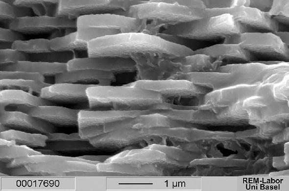
Figure 12.
Aragonite tablets [
According to Tiwari et al. [47], a report was filed at the police station in Raipur City, Chhattisgarh state, pertaining to a case of fraudulent activity. The victim had been deceived into purchasing gold beads amounting to nearly 16 lakhs. Owing to the nature of the case falling under the purview of forensic physics, involving the analysis and comparison of physical properties, it was duly registered at the State Forensic Science Laboratory in Raipur. This study aimed to apply fundamental principles of physics in resolving unresolved cases and foster awareness regarding the purchase of precious ornaments. In this specific instance, a strand of numerous spherical golden beads was submitted for examination to assess the gold content under the assumption that the gold possessed a lower purity level. To this end, scanning electron microscope (SEM) examination and energy-dispersive X-ray analysis (EDAX) were performed in the laboratory. The findings indicated a notable absence of gold (Au) in all the gold beads within the strand. Instead, the analysis revealed the presence of copper (Cu) and zinc (Zn) in a ratio of Cu:Zn::2:1.
Troalen et al. [48] stated that the National Museums Scotland (NMS) houses approximately 6000 artifacts originating from Ancient Egypt and Sudan, which notably encompass gold and electrum jewelry that has previously received limited attention in terms of scientific scrutiny. The assemblage represents various periods ranging from the Predynastic era to the Roman Empire, with a substantial portion of these items originating from reputable archeological excavations conducted during the nineteenth and twentieth centuries. In an effort to conduct an initial exploration into the diverse alloys and production methodologies employed in Ancient Egypt, the study focused on the examination of the Qurneh jewelry alongside a small selection of other gold items dating between the nineteenth and thirteenth centuries BC. As such, for the examination of gold PGE (platinum group element) inclusions, ranging from a few to a few hundred microns in diameter, as shown in Figure 13, this is a characteristic analysis for golden jewelry, and different concentrations may reveal that the source for the golden metal was different. The examination of these objects took place at the NMS, utilizing a stereo-microscope (Olympus SZX12 × 7–90) integrated with a digital camera (Olympus DP70), as well as CamScan scanning electron microscopy in secondary electron mode (SEI). Furthermore, their structural composition was investigated by employing a 300 kV Pantak X-radiography system. Evident signs of wear were observed across all the jewelries, notably prominent in the Qurneh girdle and one of the Amarna finger-rings (A.1883.49.2). Notably, observations revealed loose decorative elements in the girdle, along with deformations on the edges of the beads and within the holes of the wallet spacers, particularly where they make contact with the beads.
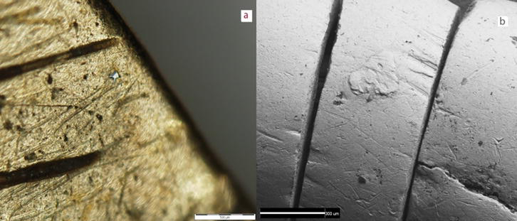
Figure 13.
PGE inclusions in pendant (A.1914.1081) from el-Harāgeh by OM [
Burat et al. [49], the analysis of a representative sample was conducted using scanning electron microscopy alongside energy-dispersive X-ray spectroscopy (SEM-EDS). This study outlines the process of concentrating and extracting Au and Ag from the waste generated during floor-sweeping activities at jewelry workshops. The concentration and recovery procedures involved both physical methods, such as shaking table, multi-gravity separator (MGS), Knelson, and Falcon concentrators, as well as physicochemical methods like froth flotation. Experimental findings highlighted the effectiveness of gravity-based beneficiation techniques in the removal of a significant portion of the waste matrix. Notably, the employment of a shaking table separator yielded a heavy fraction with a content of 701 and 6017 g/t Ag, obtained from a feed assaying 183 Au and 1835 g/t Ag. To further enhance the beneficiation process, the middlings from tabling were subjected to centrifugal separation, resulting in an increase in the Ag grade from 848 to 7812 g/t. Additionally, the tailings from gravity and centrifugal separations, containing the discharged Au and Ag fractions, were effectively concentrated using froth flotation, ultimately leading to an overall recovery rate of approximately 92% for both Au and Ag.
3.2.4 Paints and fiber
The application of scanning electron microscopes (SEMs) has significantly advanced forensic analysis, specifically in the scrutiny of paint and fiber specimens. In the context of paint analysis, SEMs facilitate an intricate exploration of the layers of paint and their constituent components, aiding in the discernment of diverse pigments and supplementary substances. These instruments enable the visualization of the layer arrangement and the identification of distinctive morphological attributes, thereby contributing to the differentiation of paint samples obtained from varying sources. Furthermore, SEMs play a pivotal role in the analysis of fibers, allowing for the examination of fiber structure, surface properties, and elemental constitution. They facilitate the identification of fiber categories, encompassing both natural and synthetic varieties, while assisting in the detection of any modifications or contamination. By offering capabilities in high-resolution imaging and elemental analysis, scanning electron microscopes continue to serve as indispensable tools in the comprehensive scrutiny and verification of paint and fiber evidence within the domain of forensic investigations.
Schreiner et al. [50], the utilization of scanning electron microscopy (SEM), particularly in conjunction with energy-dispersive X-ray microanalysis (SEM/EDX), has been significantly prevalent in the comprehensive examination of materials found in artifacts of both artistic and archeological significances. A series of case studies illustrate the benefits and limitations of SEM/EDX. These include the analysis of pigments within cross-sectional views of paint layers, the quantitative assessment of archeological glass from the Roman era excavated in Ephesos, Turkey, and investigations into glasses with compositions characteristic of the medieval period, focusing on their susceptibility to weathering and degradation phenomena. The integration of scanning electron microscopy, along with energy-dispersive X-ray microanalysis, has played a pivotal role in the extensive exploration of items comprising our cultural heritage. Moreover, it has served as an indispensable tool in unraveling the processes of deterioration affecting ancient materials as well as contemporary materials utilized in art and archaeology. Recent advancements in techniques, such as environmental SEM (ESEM) or low-voltage SEM (LV-SEM), have broadened the scope of material analysis, eliminating the prerequisites of electrical conductivity and resistance to high-vacuum conditions in samples. However, the findings underscore the necessity of conducting scientific investigations through the amalgamation of multiple analytical methods, wherein conventional microscopy and micro-chemical spot tests are implemented alongside sophisticated techniques utilizing synchrotron radiation.
Mahltig and Grethe [51] provide a comprehensive examination of various high-performance fibers like polyester filament fiber, as shown in Figure 14, and functional fiber materials, offering insights into their properties and applications. Microscopic imagery obtained through scanning electron microscopy (SEM) is introduced for fiber materials and fabrics, complemented by electron-dispersive spectroscopy (EDS) analyses performed on the fiber materials, with an overview of the resulting EDS spectra. The distinctive attributes of SEM images and EDS spectra are thoroughly discussed, primarily aimed at aiding professionals engaged in fiber analytics. To furnish a comprehensive understanding of both analytical techniques, SEM and EDS, the review also delineates the challenges and common errors encountered during SEM measurements on textiles. Collectively, the review presents a valuable survey of advanced high-tech fiber materials and their examination using the analytical methods of SEM and EDS, enabling the elucidation and discussion of material properties and composition. It also delves into the analysis and discussion of the composition of industrial fiber materials, as well as the treatments applied to fibers to achieve specific functional properties. Ultimately, the review strives to serve as a practical resource for professionals engaged in fiber and textile analytics and identification.
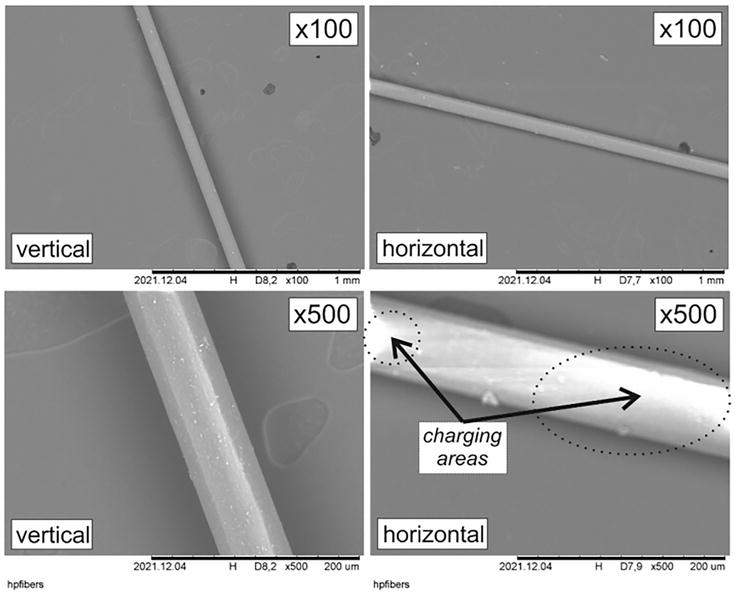
Figure 14.
SEM images of a polyester filament fiber. The SEM images are recorded with two different magnifications (×100 or ×500) and with two different fiber arrangements on the sample holder [
Jaques et al. [52] stated that scanning electron microscopy (SEM) represents a prevalent technique employed for the analysis of micro-samples derived from paintings. Notably, this method’s heightened resolution allows for meticulous surface analysis and can be augmented with an energy-dispersive spectrometer to gather elemental composition data. In the context of light microscopy and SEM analysis, painting micro-samples are commonly prepared as cross sections, where the micro-sample is embedded in resin and subsequently polished to reveal the sequential layers. In instances pertinent to cultural heritage, the cross section’s polished surface is typically coated with a conductive layer, although in this specific domain, measurements are predominantly conducted using a low-vacuum SEM (LV-SEM). Despite the reduced occurrence of the charging effect in LV-SEM, this issue may still arise and remains challenging to mitigate, even with the application of carbon tape or paint. This research introduces two distinct methods for preparing conductive cross sections from non-conductive samples, thereby mitigating charging effects while preserving the integrity of the samples.
News-Medical.net [53] states that the market for fibers and fiber materials, including natural options like hemp fibers, remains extensive. Hemp fibers have served various purposes over the years, finding application in the production of papers, ropes, clothing, and canvas. More recently, these fibers have been integrated into the automotive industry to enhance paint strength. However, hemp phloem contains wax, pectin, lignin, hemicelluloses, and other impurities, typically removed during the degumming process. To assess the impact of enzymatic degumming on the quality of hemp fibers, a combination of scanning electron microscopy (SEM) and fluorescent microscopy was employed to examine untreated and treated hemp fibers. The primary aim of this investigation was to ascertain the protein content and quality of hemp fibers, evaluating the efficacy of the enzymatic degumming procedure. The findings suggest that the biochemical degumming process under scrutiny offers a viable solution with favorable environmental and economic implications, thereby presenting itself as a feasible candidate for industrial-scale implementation.
According to (Paint Analysis, n.d.) [54], methods such as scanning electron microscopy (SEM) offer the capability to investigate both contemporary and historical paint and polychrome samples. Energy-dispersive X-ray analysis enables the identification and examination of inorganic fillers, pigments, and organic components within distinct paint layers. This information facilitates the comprehensive characterization of each layer, including insights into its formulation and thickness.
Park et al. [55], in electron microscopy (EM), the issue of non-conductive biological samples experiencing charging from focused electron beams has hindered the achievement of high-resolution imaging. Commonly employed solutions, such as the application of gold or platinum coatings, have proven effective in preventing sample charging. However, these coatings pose limitations on the feasibility of conducting further quantitative and qualitative chemical analyses, including energy-dispersive spectroscopy (EDS). This study presents findings indicating that the implementation of graphene-coating on biological samples allows for non-destructive high-resolution imaging through EM, alongside facilitating chemical analysis through EDS. The utilization of graphene is supported by its transparency to electron beams and high conductivity, as shown in Figure 15 on biological samples as well as its remarkable mechanical strength and flexibility. It is believed that the introduction of graphene-coated imaging and analytical methods will provide a novel avenue for exploring various biological phenomena previously unseen due to limitations in sample preparation and image resolution, thereby enriching our comprehension of the life mechanisms inherent in diverse living organisms.
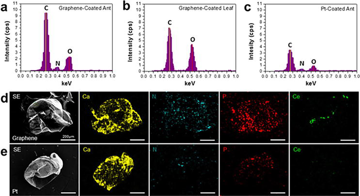
Figure 15.
SEM and EDS analyses of biological samples coated with graphene [
In a study, a compact, untextured multifilament nylon fabric with a plain woven structure was specifically chosen for the experimental evaluation. The generation of damaged samples involved the use of a precise carving knife, a finely honed pair of dressmaker’s shears, and an Elmendorf tear tester. Written guidelines were distributed to 14 panelists, instructing them on the examination of fiber end impairment through comparisons with theoretical models and scanning electron microscopy (SEM) micrographs featuring distinct fiber end appearances resulting from knife cuts, scissor cuts, and impacts causing tears and ductile fractures. Findings indicated a limited probability of accurately identifying the underlying cause of damage, as the fiber ends resulting from various damage mechanisms exhibited overlapping characteristics. Given these outcomes, the study advocates for further exploration into fiber damage, cautioning researchers and practitioners in the forensic domain against relying solely on SEM analyses to ascertain the root cause of fabric damage in forensic investigations [56].
Examining paint chips or flakes is a routine procedure aimed at gathering significant insights. These paint samples are instrumental in deducing essential particulars such as the hues, brands, and models of any vehicle implicated in a crime scene or an incident. Automobile producers typically apply multiple coats of primer, color, and clear finishes to achieve the intended appearance. Unveiling the composition of these paint layers is made feasible through energy-dispersive spectroscopy (EDS), which enables the elemental makeup of each layer to be revealed and cross-referenced against a database of known layer compositions. This analysis can also be utilized for direct comparison with a suspect’s vehicle.
3.2.5 Investigation of filament bulb in traffic accidents
Scanning electron microscopy (SEM) is a helpful way to look closely at filament bulbs that were in car crashes. It can take detailed pictures of the inside of the bulbs, helping investigators understand what happened during the crash. By studying how the surface of the broken parts looks and what they are made of, SEM can find important clues about why the bulb stopped working during the accident. This careful study can find signs of pressure, how the materials changed shape, or if there were any problems with the materials that might explain what happened. Also, SEM can find any other things that should not be there or things that might have affected how the bulb worked. Using SEM to look at the filament bulbs helps investigators learn important things about what caused the car accident.
Thorsen et al. [57] assessed the lighting conditions of a vehicle during an accident as a crucial task in forensic investigations, achieved through the examination of lamps and their remnants. Optical and scanning electron microscopy are two valuable techniques used for this purpose, with the latter gaining prominence due to its high resolution, extensive depth of focus, and the option for EDS microanalysis. To enhance the reliability of light bulb analysis as an investigative tool, it is essential to fully comprehend bulb design, the materials involved, and the functionality of the bulb, particularly its response to impact. This review aims to consolidate the existing but dispersed literature pertaining to these aspects, aiming to facilitate a more systematic and comprehensive approach to light bulb analysis in forensic investigations.
Horvat et al. [58] introduced a novel technical and technological approach for examining automotive light bulbs to detect trace particles of shattered glass in cases involving minimal inertia forces resulting from a traffic accident. Given the limited prior investigations into light bulbs in such accidents, the study focuses on analyzing the filaments of light bulbs used in cars for illuminating the road and signaling, incorporating a new technical procedure utilizing the SEM/EDX method. The research significantly enhances the investigative process for analyzing automotive light bulbs, aiding in the determination of whether the vehicle’s regulatory lights were in operation at the time of the accident. Such findings can be critical in establishing the liability of the parties involved in causing the traffic incident.
Grafiati [59] carried out research in order to introduce the benefits of using surveying techniques in traffic accident investigations and show their impacts on evidence documentation and scene clearance. This is done by focusing on the advantages and the disadvantages of each method based on the relevant works of literature and comparing them. Although comparison result shows that the traditional method (coordinate method) is simpler and cheaper than other methods, surveying technique methods are safer and faster in clearing the accident scene, fewer investigators are needed, the scale can be provided directly, high accuracy measurements can be obtained, and three dimensions’ models can be produced. So, it is worth using the surveying equipment in car accident investigations.
3.2.6 Handwriting and print examination
Scanning electron microscopy (SEM) has emerged as a versatile tool in the realm of handwriting and print examination, with particular relevance in cases involving forgery. Through the utilization of SEM, forensic professionals can conduct a meticulous analysis of the minute features of ink, paper, and other writing materials utilized in various documents. This thorough examination enables the discernment of distinctive handwriting attributes, encompassing aspects such as pen pressure, ink dispersion, and the unique traits of individual pen strokes. SEM aids in the identification of any modifications, insertions, or erasures present in documents, unveiling subtle alterations in surface texture and ink properties. Moreover, SEM facilitates the recognition of diverse printing methodologies, encompassing disparities in ink absorption, patterns in paper fiber, and the presence of printing artifacts. The high-resolution imaging capacity of SEM offers valuable insights into the genuineness and credibility of documents, significantly contributing to the verification process and the detection of potential falsifications.
Wang et al. [60] performed handwriting authentication as a pivotal domain within the field of forensic science. In this study, diverse types and brands of pen ink samples originating from China can be scrutinized using non-invasive micro-identification techniques. The utilization of scanning electron microscopy and energy spectrum (SEM-ES) enabled the examination of micro-elements and the comprehensive analysis of the written traces, with a particular emphasis on the function of spectral mapping (surface scanning). The precise evaluation of elements within the handwriting traces facilitated the differentiation of subtle disparities among various types and brands of ink in handwritten text. This approach introduced a novel avenue for the analysis and exploration of pen handwriting identification in the realm of forensic science.
Verma et al. [61] detected that computer-generated document forgeries have always posed a significant challenge for forensic document examiners (FDE). To assist in the examination process, researchers have explored the use of Schottky field emission scanning electron microscopy with energy-dispersive X-ray spectroscopy (FE-SEM-EDS) as a modern tool for analyzing black toners sourced from laser printers and photocopier machines. A total of 40 samples from each of the laser printer and photocopier machines were obtained and subjected to an analysis of morphological characteristics, elemental profiles, and multivariate analysis. The acquired SEM images and spectra were scrutinized to distinguish and categorize toners originating from different sources. Multivariate analysis was employed to develop a classification model that successfully categorized the printed documents based on the similarities and differences in their composition. Hierarchical cluster analysis (HCA) differentiated the printouts into distinct groups based on their chemical composition, resulting in 11 clusters for the laser printer printouts and 8 clusters for the photocopier printouts. Cross-validation was additionally conducted to assess the capabilities of the developed principal component analysis (PCA) and linear discriminant analysis (LDA) models for examining printouts of unknown origin.
In the field of forensic document examination, a significant obstacle is the meticulous scrutiny of minute details that remain imperceptible to the unaided eye. Electron microscopy has emerged as a potent solution to tackle this challenge. The use of scanning electron microscopy (SEM) or transmission electron microscopy (TEM) empowers forensic document examiners to capture intricate elements such as ink pigments, paper fibers, and surface topography with remarkable precision. This detailed view offers vital evidence necessary for verifying the legitimacy of a document and uncovering potential modifications or counterfeits. Furthermore, the high-resolution imaging capabilities of electron microscopy enable examiners to distinguish unique attributes of different writing tools. Each pen, for instance, leaves behind a distinct mark due to variations in its nib, ink flow, and the pressure applied during writing. Moreover, the scrutiny of paper fibers can unveil patterns and textures that correspond to specific manufacturing techniques or historical periods, thereby further contributing to the authentication and provenance determination of a document. (AZoOptics.com, 2023) [62].
The examination of ink holds significant importance in forensic document analysis, with electron microscopy playing a crucial role in this domain. Through this technique, analysts can investigate ink samples derived from various documents and assess their chemical makeup, particle distribution, and microstructure, all of which can serve as distinct identifiers. By incorporating energy-dispersive X-ray spectroscopy alongside SEM, specialists can identify unique elements present in the ink, thereby aiding in establishing connections between different documents or highlighting potential disparities. The chemical composition of ink varies depending on its formulation and manufacturing methods. By comparing the ink composition of different documents, experts can determine if the ink used aligns with a specific brand, batch, or time period, offering pivotal evidence in cases involving falsified documents or forged signatures.
One intriguing application of electron microscopy in forensic document examination lies in its capacity to reveal concealed or modified writing. Indented writing often arises when an individual write on a piece of paper positioned atop another document, a practice frequently exploited by counterfeiters seeking to manipulate or fabricate documents. Electron microscopy enables analysts to visualize and interpret the indented writing, deciphering its content and contrasting it with the visible writing on the document’s surface. This method has proven immensely valuable in uncovering obscured messages, erased text, or alterations made to crucial documents, thereby providing examiners with crucial insights into the document’s history and authenticity.
Given the increase in advanced counterfeiting methods, electron microscopy has become an essential instrument for examining counterfeit documents. Such fraudulent documents frequently endeavor to replicate intricate security elements inherent in authentic documents. Through the scrutiny of security features such as watermarks, holograms, and micro-printing, forensic examiners can detect disparities and ascertain the authenticity of a document. Additionally, electron microscopy facilitates thorough analyses of printing methods, surface anomalies, and substrate attributes. By comparing and contrasting these subtle intricacies with those present in verified authentic documents, forensic specialists can furnish compelling evidence to uncover counterfeit endeavors and contribute to legal procedures.
According to Pingitore et al. [63], diagnostic studies were conducted on an ancient coin to determine its authenticity or potential for being a counterfeit. The investigation employed scanning electron microscopy/energy-dispersive X-ray (SEM-EDX) and cathodoluminescence (CL). The coin in question, a Drachma, depicted Poseidon’s portrait on the obverse and Amphitrite riding a seahorse with Eros shooting an arrow on the reverse. The coin is widely recognized in numismatic circles, with originals housed in various museums such as those in Catanzaro, Naples, and Milan. The EDX analysis, performed on specific points of the coin’s surface, identified Pb and Cu as the primary components on both sides, with 51% Pb and 35% Cu by weight, along with unexpected traces of gold. Gold was detected in small spots measuring around 20 μm, constituting 95% of the total weight. Simultaneously, cathodoluminescence (CL) analysis was conducted to induce luminescent emissions by electron bombardment in these areas. The CL analysis corroborated the SEM findings, highlighting the presence of gold more distinctly than the SEM analysis. In fact, the CL analysis revealed the presence of gold throughout the surface of the sample, albeit in small amounts.
3.2.7 Soil
The implementation of scanning electron microscopy (SEM) has markedly propelled the realm of trace evidence analysis, empowering forensic experts to meticulously examine and discern minute materials of potential significance in criminal inquiries. Through SEM, researchers can investigate the surface morphology and elemental structure of trace evidence, encompassing various substances such as fibers, hair, paint chips, and gunshot residue. The superior imaging capabilities of SEM allow for the meticulous visualization of intricate characteristics and subtle features, facilitating the differentiation of diverse materials and the determination of their origins. Furthermore, the integration of energy-dispersive X-ray spectroscopy (EDS) with SEM permits the identification and spatial mapping of elements present within trace samples, imparting critical insights into their chemical compositions. These thorough analyses significantly contribute to establishing pivotal associations between physical evidence and crime scenes, thereby streamlining the process of suspect identification and providing robust evidence in legal proceedings.
Soil, an unconsolidated mineral substance found on the Earth’s surface, results from the combined effects of organisms, parent material, climate variations, and topography over an extended period. It comprises minerals, organic matter, liquids, and gases, with diverse properties influenced by factors such as vegetation, weather, and solar exposure. The current study delves into the morphology and elemental constituents of soil samples collected from Canterbury (UK), Dubai (UAE), and Kerala (India). Analyzing the samples under a scanning electron microscope involved careful moisture removal
3.2.8 Non-conducting material
Scanning electron microscopy (SEM) has emerged as a critical tool for examining non-conductive materials, revealing important insights into their surface structure, elemental makeup, and overall composition. By employing specialized methods for sample preparation, such as applying a conductive coating or utilizing low-voltage SEM, SEM allows for the effective analysis of materials such as polymers, ceramics, and biological samples that do not conduct electricity. This technique enables the observation of intricate surface features and the detection of subtle structural differences, providing valuable data for evaluating material properties, ensuring quality control, and driving various research endeavors. Additionally, the integration of energy-dispersive X-ray spectroscopy (EDS) with SEM permits the precise identification and mapping of elemental constituents within non-conductive materials, facilitating a comprehensive analysis of their chemical composition and contributing to a deeper understanding of their characteristics and behaviors. With its advanced imaging capabilities and accurate elemental analysis, SEM continues to advance the exploration and comprehension of diverse non-conductive materials across various scientific fields.
Griffin [64] showed that understanding the precise mechanism behind the contrast observed in “environmental” or “gaseous” secondary electron images in the environmental scanning electron microscope remains somewhat unclear. Initially, a straightforward gas amplification model was proposed, suggesting that emitted secondary electrons ionize the chamber gas, leading to signal amplification and subsequent measurement at a biased detector. While this theory is evolving, little attention has been directed toward the factors influencing the actual secondary emission despite the identification of unusual contrast effects in one instance. The conventional perspective implies that the positive ion product resulting from the gas-electron interaction induces charge neutralization at the sample surface. Recent research has indicated the occurrence of charge implantation and trapping in non-conductive materials, particularly in Ref. to electron range measurements. This study showcased the influence of trapped charge on the secondary electron yield, revealing heightened secondary electron emission above the region where charge is trapped. As a result, the distribution of the trapped charge manifests as a bright circle on the specimen’s surface, centered at the point of beam exposure.
Cao et al. [65] showed that with the advancements in science and technology, the field emission scanning electron microscope (FESEM) has become a pivotal tool in nano-material measurements, owing to its notable benefits such as high magnification, high resolution, and user-friendly operation. A high-quality secondary electron image is vital for precise and accurate length measurements. Traditionally, non-conductive materials are treated by coating them with conductive films such as gold, carbon, or platinum to minimize charging effects and achieve high-quality secondary electron images. However, this approach often conceals the authentic microstructures of materials, alters sample composition properties, and introduces significant errors in nanoscale microstructure measurements. This chapter delves into the exploration of methods for minimizing or eliminating the impact of charging effects on image quality to the greatest extent possible without resorting to coating treatments. Adjustments in working conditions, such as voltage, stage bias, and scanning mode, are discussed to uncover genuine and high-quality microstructure information of materials.
Wang et al. [66] performed the examination of surface morphology for non-conducting granular materials was conducted utilizing scanning electron microscopy (SEM) within low-vacuum conditions. An exhaustive investigation was performed to assess the impact of various factors, including incident electron voltage, electronic beam spot size, and atmospheric pressure, on the quality of SEM images and the accuracy of energy-dispersive X-ray (EDX) analysis. The findings from this systematic exploration reveal that the resolution and quality of SEM images demonstrate a decrease in correlation with the escalation of incident electron voltage, electronic beam spot size, and atmospheric pressure. Furthermore, the errors associated with the EDS analysis exhibit a decline when conducted under the specific conditions of low vacuum.
4. Conclusion
Modern scientific investigation is led by electron microscopy, which provides a never-before-seen glimpse into the complex world of microscopic structures that are still out of reach for conventional microscopy. This sophisticated type of microscopy has become an essential tool for forensic investigations analyzing trace evidence because of its remarkable resolution and capacity to examine materials at the nanoscale.
Electron microscopy, such as transmission electron microscopy (TEM) and scanning electron microscopy (SEM), overcomes the constraints of traditional microscopes by utilizing high-resolution imaging and analytical capabilities. It offers unmatched insights into the morphology, structural features, and composition of trace materials—all of which are frequently very important in criminal investigations.
Some of the key findings with respect to both TEM and SEM are as follows.
4.1 Transmission electron microscope
4.1.1 Analysis of paint
For the purpose of examining paint chips, TEM provides high-resolution imaging that helps detect pigments, binders, layer structures, and elemental makeup.
Analyses paint and base layers to aid in the authenticity of old artworks. It is vital for recognizing unique properties of car paint, like fillers and primers, which makes possible to image and identify nanoscale particles on paint samples made with focused ion beam technology.
4.1.2 Analysis of soil
TEM helps to characterize the minuscule soil particles, which makes it easier to comprehend the effects of microbes and elemental changes in soil. Permits the analysis of micrometer-sized micro aggregates, offering information about the functioning of soil and microbial interactions.
4.1.3 An analysis of gunshot residue (GSR)
TEM, which provides high-resolution imaging and elemental analysis capabilities, is essential for the investigation of nanoscale GSR particles.
It uses particle analysis to discern between different types of ammunition and their respective geographic origins.
4.1.4 Analysis of fibers
TEM helps discover asbestos in tissue samples linked to health problems and mineral fibers in lung tissue resulting from industrial dangers.
Reveals patterns of structural damage in chemically treated fiber, offering information about micro-fibril structure.
4.1.5 Analysis of hair
Through a deeper understanding of the cellular architecture surrounding hair follicles, TEM helps to uncover biological interactions that may otherwise go undetected.
It is useful for researching the harm that chemical treatments, such as bleaching, have to the proteins and structure of hair.
4.1.6 Glass evaluation
To help determine the source of glass pieces (such as shootings or hit-and-run occurrences), TEM looks at the internal microstructure of the broken glass.
It helps identify glass composition in order to connect broken glass to certain sources, such as bottles or windows.
4.1.7 Nanoparticles
Nanoparticles discovered as trace evidence are characterized by TEM, which helps to build a reference database for upcoming forensic investigations.
4.1.8 Explosives
By analyzing explosive residues at the nanoscale, TEM helps with pre- and post-blast analysis and helps determine the type of explosive employed in blast scenarios.
Essential for researching quantum dots, which are utilized to recognize typical nitro-base explosives.
4.2 Scanning electron microscope
4.2.1 GSR analysis
GSR particles that have been removed from specimens are thoroughly imaged, and their elemental composition is examined using a combination of SEM and energy dispersion spectroscopy (EDS).
Predicting firing range and assisting in crime resolution revolutionizes forensic investigations by making it easier to identify GSR particles at far distances.
4.2.2 Gemstone and jewelry analysis
SEM is a vital tool for the identification and distinction of gemstones, offering information on their internal and exterior characteristics, origin in geology, and possible treatment history.
SEM supports the analysis of material composition, plating thickness, and surface coatings in jewelry forensics, helping to validate authenticity and identify counterfeits.
4.2.3 Paint and fiber analysis
SEM developments have a substantial impact on paint analysis. They allow for the detailed examination of paint layers, pigments, and structural characteristics, which makes it easier to distinguish between samples that come from different sources.
SEM is essential to fiber analysis because it helps identify and detect changes or contamination by analyzing structure, surface characteristics, and elemental constitution.
4.2.4 Filament bulb investigation in traffic accident
By examining surface characteristics and materials, SEM’s thorough imaging aids in the understanding of filament bulb fracture during auto accidents and offers vital hints regarding the cause or impact aspects of the accident.
4.2.5 Handwriting and print inspection
SEM’s use in handwriting analysis helps identify specific writing characteristics, document modifications, and printing techniques, which makes a major contribution to verification and the discovery of forgeries.
4.2.6 Non—Conductive material analysis
Non conductive materials such as polymer and ceramics can be examined by SEM by integrating energy-dispersive X-ray spectroscopy (EDS) with specialized sample preparation. SEM provides insights into the surface structure, elemental composition, and material properties of these materials.
References
- 1.
History of the Electron Microscope. 2018. Available from: https://www.news-medical.net/life-sciences/History-of-the-Electron-Microscope.aspx#:~:text=It%20was%20Ernst%20Ruska%20and,was%20possible%20with%20electron%20microscopy - 2.
Shaber L, Shaber, L. The History of the Electron Microscope. Advancing Materials. 2018. Available from: https://www.thermofisher.com/blog/materials/the-history-of-the-electron-microscope/#:~:text=To%20build%20on%20the%20power,The%20solution:%20compound%20microscopes.&text=2 - 3.
Schwab K. What Role Does SEM Play in Trace Evidence Analysis? - Nanoscience Instruments. Nanoscience Instruments. 2023. Available from: https://www.nanoscience.com/blogs/what-role-does-sem-play-in-trace-evidence-analysis/#:~:text=These%20materials%20can%20include%20fibers,valuable%20information%20in%20criminal%20investigations - 4.
Twyman R. Microscopy applications: Food. In: Worsfold P, Townshend A, Poole C, editors. Encyclopedia of Analytical Science (Second Edition). London, UK: Elsevier Science; 2005; 6 :50-57 - 5.
Corradini MG, Julian McClements D. Food ☆. In: Reference Module in Chemistry, Molecular Sciences and Chemical Engineering. Vol. 2019. Elsevier; 2017. pp. 47-56 - 6.
Australian Microscopy and Microanalysis Research Facility, “My Scope-training for advanced research." Available from: http://www.ammrf.org.au/myscope/sem/background/ [Accessed: May 3, 2018] - 7.
Mohammed A, Abdullah A. Scanning electron microscopy (SEM): A review. In: Proceedings of 2018 International Conference on Hydraulics and Pneumatics-HERVEX, Baile Govora, Romania, 7-9 November 2018. 2019. pp. 1-9. ISSN 1454 – 8003 - 8.
Check out MyScopeTM. n.d. Available from: https://myscope.training/#/TEMlevel_3_1 - 9.
Rukari T, Babita A. Review article transmission electron microscopy-an overview. International Research Journal for Inventions in Pharmaceutical Sciences. 2013; 1 (2):1-7 - 10.
Tang H, Martin DC. Microstructural studies of interfacial deformation in painted thermoplastic polyolefins (TPOs). Journal of Materials Science. 2003; 37 :4783-4791 - 11.
Barba C, Andrés MS, Peinado J, Báez MI, Baldonedo JL, Andres MS, et al. A note on the characterization of paint layers by transmission electron microscopy. Studies in Conservation. 1995; 40 (3):194 - 12.
Goebel R, Stoecklein W. The use of electron microscopic methods for the characterization of paints in forensic science. Scanning Microscopy. 1987; 1 (3):16 - 13.
Lins A, Giannuzzi LA, Stevie FA, Price B, Tucker M, Gutman N. Fib/tem analysis of paint layers from Thomas Eakins’ the crucifixion, 1880. MRS Proceedings. 2002; 712 :12 - 14.
Elsass F, Chenu C, Tessier D. Transmission electron microscopy for soil samples: Preparation methods and use. In: Methods of Soil Analysis Part 5-Mineralogical Methods. Soil Science Society of America. 2015. pp. 235-268 - 15.
Watteau F, Villemin G. Soil microstructures examined through transmission electron microscopy reveal soil-microorganisms interactions. Frontiers in Environmental Science. 2018; 6 :106 - 16.
Watteau F, Villemin G. Soil microstructures examined through transmission electron microscopy reveal soil-microorganisms interactions section soil processes. Frontiers in Environmental Science. 2018; 6 :106 - 17.
Fitzpatrick RW. Soil: Forensic analysis. In: Wiley Encyclopedia of Forensic Science. Chichester, UK: John Wiley & Sons, Ltd; 2004. pp. 1-14 - 18.
Warlow TA. Firearms, the Laws, and Forensic Ballistics. United Kingdom: Routledge; 1996 - 19.
Dalby O, Butler D, Birkett JW. Analysis of gunshot residue and associated materials review. Journal of Forensic Sciences. 2010; 55 (4):924-943 - 20.
Hill WB. Transmission electron microscopy study of gunshot-residue nanoparticles collected in air samples. Microscope. 2012; 60 (3):133-137 - 21.
Hill WB. The characterization of nanoparticles using analytical electron microscopy. In: Proc. Scanning Microscopies 2011. Advanced Microscopy Technologies for Defense, Homeland Security, Forensic, Life, Environmental, and Industrial Sciences. SPIE Proceedings. 1 June 2011; 80360E . DOI: 10.1117/12.885115 - 22.
Melo LG, Martiny A, Pinto AL. Nano characterization of gunshot residues from Brazilian ammunition. Forensic Science International. 2014; 240 :69-79 - 23.
Friedrichs KH, Brockmann M, Fischer M, Wick G. Electron microscopy analysis of mineral fibers in human lung tissue. American Journal of Industrial Medicine. 1992; 22 (1):49-58 - 24.
Reza M, Kontturi E, Jääskeläinen A-S, Vuorinen T, Ruokolainen J. Transmission electron microscopy for wood and fiber analysis—a review. BioResources. 2015; 10 (3):6230-6261 - 25.
Morioka K. A guide to hair follicle analysis by transmission electron microscopy: Technique and practice. Experimental Dermatology. 2009; 18 (7):577-582 - 26.
Lee Y, Kim Y, Pi L, Lee SY, Hong H, Lee W. Comparison of hair shaft damage after chemical treatment in Asian, White European, and African hair. International Journal of Dermatology. 2013; 53 (9):1103-1110 - 27.
Ji JH, Park TS, Lee HJ, Kim YD, Pi LQ , Jin XH, et al. The ethnic differences of the damage of hair and integral hair lipid after ultra violet radiation. Annals of Dermatology. 2013; 25 (1):54 - 28.
Grosvenor AJ, Deb-Choudhury S, Middlewood PG, Thomas A, Lee E, Vernon JA, et al. The physical and chemical disruption of human hair after bleaching – Studies by transmission electron microscopy and redox proteomics. International Journal of Cosmetic Science. 2018; 40 (6):536-548 - 29.
Curran JM, Champion TNH, Buckleton JS. Forensic Interpretation of Glass Evidence. Boca Raton: CRC Press; 2000 - 30.
Schumacher EF. A High-Resolution Look at Glass Delamination Using Transmission Electron Microscopy (TEM). (n.d.). Available from: https://www.americanlaboratory.com/913-Technical-Articles/134532-A-High-Resolution-Look-at-Glass-Delamination-Using-Transmission-Electron-Microscopy-TEM/ - 31.
Cantu AA. Nanoparticles in forensic science. SPIE Proceedings. 2008; 7119 :71190F-1-8 - 32.
Santra TS, Tseng FGK, Barik TK. Biosynthesis of silver and gold nanoparticles for potential biomedical applications—A brief review. Journal of Nanopharmaceutics and Drug Delivery. 2014; 2 (4):249-265 - 33.
Hill WB. Forensic applications of the transmission electron microscope. The Microscope. 2016; 57 (4):165-170 - 34.
Kashkarov AO, Pruuel ER, Ten KA, Rubtsov IA, Gerasimov EY, Zubkov PI. Transmission electron microscopy and x-ray diffraction studies of the detonation soot of high explosives. Journal of Physics: Conference Series. 2016; 774 :012072 - 35.
Li Z, Wang Y, Ni Y, Kokot S. A sensor based on blue luminescent graphene quantum dots for analysis of a common explosive substance and an industrial intermediate, 2,4,6-trinitrophenol. Spectrochimica Acta Part A: Molecular and Biomolecular Spectroscopy. 2015; 137 :1213-1221 - 36.
Raj AV, Waghmare NP, Monika J, Neelesh T. An experimental investigation of gunshot residue (GSR) evidence: Directly from targeted fabric using sem. International Journal of Medical Toxicology & Legal Medicine. 2020; 23 :233-244 - 37.
Brożek-Mucha Z, Jankowicz A. Evaluation of the possibility of differentiation between various types of ammunition by means of GSR examination with SEM–EDX method. Forensic Science International. 2001; 123 (1):39-47 - 38.
Reyes LC, López CE, Barrios AB, Soto C, Ibáñez C, Díaz FJ. Development and application of a new nose hairs sample collection device for GSR particles by scanning electron microscopy with energy dispersive X-ray spectroscopy (SEM-EDS). Forensic Science International. 2018; 290 :42-48 - 39.
Kara İ. The influence of different skin types on GSR sampling by tape lifting for SEM analysis. Microscopy Research and Technique. 2017; 80 (12):1310-1314 - 40.
Toal SJ, Niemeyer WD, Conte S, Montgomery DD, Erikson GS. Confirmatory analysis of field-presumptive GSR test sample using SEM/EDS. Proceedings of SPIE. 2014:92361C. DOI: 10.1117/12.2074212 - 41.
Scanlan MD, Reinholz AD. Scanning electron microscopy for firearm and toolmark comparisons. AFTE Journal. 2013; 45 (1):43-47 - 42.
Pal A, Debnath S, Pratihari HK. Post Analysis of a Firing Case by Optical and Scanning Electron Microscopy. Journal of Advanced Microscopy Research. 2014; 9 (1):67-70. DOI: 10.1166/jamr.2014.1190 - 43.
Korda EJ, MacDonell HL, Williams JP. Forensic applications of the scanning electron microscope. Journal of Criminal Law, Criminology and Police Science. 1970; 61 :453 - 44.
Bíró C, Kováč P, Palkovic M, El-Hassoun O, Čaplovičová M, Novotný J, et al. Potentialities of scanning electron microscopy and EDX analysis in bullet wounds. Romanian Journal of Legal Medicine. 2010; 18 (3):225-230 - 45.
Hopkins T, Peniche G, Murphy S, Carter I, Shorrock G, Kearns S, et al. Scanning electron microscopy and energy-dispersive x-ray spectroscopy (SEM-EDX) confirms shooting of a hen harrier ( Circus cyaneus ). Veterinary Record Case Reports. 2015;2015 (3):e000241 - 46.
Cartier LE, Krzemnicki MS, Lendvay B, Meyer JB. DNA fingerprinting of pearls, corals and ivory: A brief review of applications in gemmology. The Journal of Gemmology. 2018; 36 (2):152-160 - 47.
Tiwari S, Sahu DK, Thakur VS, Patel MM, Chandra T. Forensic morphological and elemental analysis of gold jewellery: A case study. International Journal of Science and Research Archive. 2023; 9 (2):516-519 - 48.
Troalen L, Guerra M, Tate J, Manley B. Technological study of gold jewellery pieces dating from the middle kingdom to the new kingdom in Egypt. Archeo Sciences. 2009; 33 :111-119 - 49.
Burat F, Baştürkcü H, Özer M. Gold& silver recovery from jewelry waste with combination of physical and physicochemical methods. Waste Management. 2019; 89 :10-20 - 50.
Schreiner M, Melcher M, Uhlir K. Scanning electron microscopy and energy dispersive analysis: Applications in the field of cultural heritage. Analytical and Bioanalytical Chemistry. 2006; 387 (3):737-747 - 51.
Mahltig B, Grethe T. High-performance and functional fiber materials—A review of properties, scanning electron microscopy SEM and electron dispersive spectroscopy EDS. Text. 2022; 2 (2):209-251 - 52.
Jaques VAJ, Zikmundová E, Holas J, Zikmund T, Kaiser J, Holcová K. Conductive cross-section preparation of non-conductive painting micro-samples for SEM analysis. Scientific Reports. 2022; 12 (1):19650 - 53.
News-Medical.net . Analysing Morphology of Plant Fibers using Scanning Electron Microscopy. 2017. Available from:https://www.news-medical.net/whitepaper/20170925/Analysing-Morphology-of-Plant-Fibers-using-Scanning-Electron-Microscopy.aspx - 54.
Paint Analysis. (n.d.). Available from: https://www.materials.co.uk/paintanalysis.htm - 55.
Park JB, Kim YJ, Kim SW, Yoo JH, Kim Y, Gorbachev R, et al. Non-destructive electron microscopy imaging and analysis of biological samples with graphene coating. 2D Materials. 2016; 3 (4):045004 - 56.
Using the scanning electron microscope to identify the cause of fibre damage Part II: An exploratory study | Office of Justice Programs. (n.d.). Available from: https://www.ojp.gov/ncjrs/virtual-library/abstracts/using-scanning-electron-microscope-identify-cause-fibre-damage-0 - 57.
Thorsen KA. Forensic Examination of Bulb Filaments by the SEM. Denmark: Springer eBooks; 1986. pp. 117-147 - 58.
Horvat R, Barišić-Jaman B, Mršić G, Špoljarić I, Vrdoljak A, Pehar I. Method of light bulbs analysis on vehicles damaged in traffic accidents. Promet-Traffic & Transportation. 2012; 22 (4):259-271 - 59.
Grafiati. Journal articles on the topic “Traffic accident investigation.” Grafiati. 2023. Available from: https://www.grafiati.com/en/literature-selections/traffic-accident-investigation/journal/ - 60.
Wang J, Ren J, Yuan C, Shen Y, Gan Q , Shi J, et al. Non-invasive optical micro-identification of ink verification in pen ink handwriting. Results in Chemistry. 2020; 2 :100025 - 61.
Verma N, Sharma V, Kumar R, Sharma R, Joshi MC, Umapathy G, et al. On the spectroscopic examination of printed documents by using a field emission scanning electron microscope with energy-dispersive X-ray spectroscopy (FE-SEM-EDS) and chemometric methods: Application in forensic science. Analytical and Bioanalytical Chemistry. 2019; 411 (16):3477-3495 - 62.
AZoOptics.com . How is Electron Microscopy Used for Forensic Document Examination? 2023. Available from:https://www.azooptics.com/Article.aspx?ArticleID=2446 - 63.
Pingitore V, Barberio M, Oliva A, Davoli M. A golden drachma from Bruttìa: Counterfeit money revealed by scanning electron microscopy and cathodoluminescence. Mediterranean Archaeology and Archaeometry. 2008; 8 (2):31-38 - 64.
Griffin B. A new mechanism for the imaging of crystal structure in non-conductive materials: An application of charge-induced contrast in the environmental scanning electron microscope (ESEM). Microscopy and Microanalysis. 1997; 3 (S2):1197-1198 - 65.
Cao C, Gao R, Shang H, Peng T. Characterization of non-conductive materials using field emission scanning electron microscopy. Proceedings of SPIE. 2016; 9903 :990319 - 66.
Wang ZG, Liu KG, Jiang M, Pang B. SEM analysis of non-conducting materials under low vacuum conditions. Advanced Materials Research. 2010; 152-153 :897-901