Open Access is an initiative that aims to make scientific research freely available to all. To date our community has made over 100 million downloads. It’s based on principles of collaboration, unobstructed discovery, and, most importantly, scientific progression. As PhD students, we found it difficult to access the research we needed, so we decided to create a new Open Access publisher that levels the playing field for scientists across the world. How? By making research easy to access, and puts the academic needs of the researchers before the business interests of publishers.
We are a community of more than 103,000 authors and editors from 3,291 institutions spanning 160 countries, including Nobel Prize winners and some of the world’s most-cited researchers. Publishing on IntechOpen allows authors to earn citations and find new collaborators, meaning more people see your work not only from your own field of study, but from other related fields too.
To purchase hard copies of this book, please contact the representative in India:
CBS Publishers & Distributors Pvt. Ltd.
www.cbspd.com
|
customercare@cbspd.com
For the creation of an effective “green chemistry,” scientists have paid a lot of attention to a method for creating metal nanoparticles in recent years. The topic of nanoscale metal synthesis is one that is now pertinent due to the growing use of nanoscale metals in industries like engineering and medicine and the environment. The development of nanotechnology has revolutionized society and has focused on a number of scientific fields, including cancer, human epidemiology, and material science. Utilizing biological agents to prepare metallic nanoparticles through green chemistry reduces negative effects and improves the metal’s anticancer properties. Commercially, nanoparticles are typically prepared via physical, chemical, and biological techniques. For metal and metal oxide nanoparticles made from natural extracts, such as gold, silver, and copper oxide, which are useful to improve biomedical applications like antibacterial, antifungal, and antioxidant, have been summarized in this chapter. The easy availability of plants that are safe, easy to handle, and inexpensive allows for the synthesis of many nanoparticles. These techniques are environment-friendly, nontoxic, and economically viable green synthesized processes.
Department of Chemistry, University of Wah, Wah, Pakistan
Irum Jamil
Department of Chemistry, University of Wah, Wah, Pakistan
Bushra Shakoor
Department of Chemistry, University of Wah, Wah, Pakistan
*Address all correspondence to: faisal.nawaz@uow.edu.pk
1. Introduction of nanoparticles
Nanotechnology has advanced to a level that has never been seen before in recent decades. Therefore, it is not surprising that researchers are becoming more interested in the subject. Nanotechnology is the branch of science and technology concerned with the design, creation, and application of nanoparticles [1]. In general, items that are considered to be nanoscale or nanometer in size should fall within the range of 1–1000 nm, where “nano” is defined as 109. However, according to the recommendations of the European Commission, a nanomaterial is any substance, whether organic, inorganic, or synthetic, that contains at least 50% of particles (in unbound or aggregated state) with one or more exterior dimensions in the range of 1–100 nm [2]. Due to a specific behavior under light irradiation, namely the formation of localized surface plasmon resonance (LSPR), which gives these materials their peculiar optical properties, the nanoparticles formed from coinage metals draw attention.
Of all the “noble” metals, silver has the highest thermal and electrical conductivities as well as the lowest melting and boiling temperatures. The “noble” metals with the highest reactivity, silver also has harmful effects on a variety of microbes thanks to its cations [3]. Solar energy harvesting and electronic gadgets are just two examples of the many technological and medical applications that silver in the form of nanoparticles unveiled.
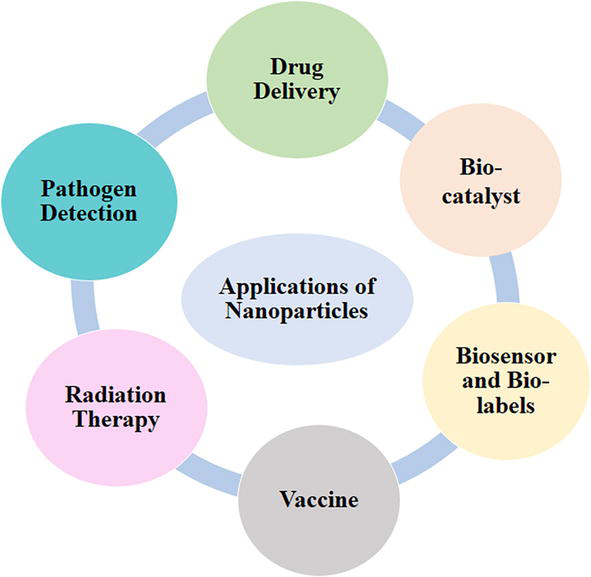
Functionalized nanoparticles with various targeting moieties have a wide range of uses in biomedicine, including diagnosis, targeting, drug/nucleic acid administration, imaging, and therapy. Furthermore, NPs are used as sensitive probes in Raman scattering and imaging applications using the surface-enhanced Raman scattering approach [4]. The application of NPs in photothermal therapy, radiation therapy, computed tomography, biosensors, and other domains has greatly boosted their potential in biomedical fields [5] as shown in Figure 1. NPs-based biosensors with excellent sensitivity and selectivity are being developed due to their intrinsic electrical and optical properties, as well as their ability to conjugate with various biomolecules. Over the last decade, NPs-based biosensors have received a lot of interest in the diagnosis of numerous diseases. Recently, several NPs-based biological assays for the detection and quantification of analytes in urinary samples have been highlighted, with an emphasis on protein analysis. Such NP-based assays are beneficial in the diagnosis of a variety of ailments, including renal problems, cancer, and heart disease [6].
Figure 1.
Some important applications of nanoparticles in medicinal chemistry.
It is now widely accepted that nanotechnologies have the potential to be useful in a number of industries, including pharmaceutical research, water purification, and information and communication technologies and the development of stronger and lighter materials. Nanotechnologies produce and manipulate materials at the nanoscale by scaling up from small groups of atoms or by reducing or refining bulk materials [7].
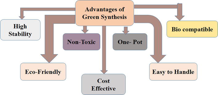
The concept of “green chemistry” for “sustainable development” has received a lot of attention in the recent decade. Sustainable development is defined as development that fulfills current demands while taking into account future generations’ abilities to meet their own [8]. Green chemistry is a growing area that advocates for the implementation of concepts aimed at minimizing the usage and manufacture of harmful chemical compounds [9]. As a result, greener methods reduce the environmental impact of industrial activities. Researchers created these technologies to propose potential solutions to the costly processes and harmful compounds discovered when using classic synthesis methods [10]. Green nanomaterial synthesis is the optimal way for lowering the risk level of nanotechnology by minimizing the negative repercussions of its creation and use. Figure 2 highlight the primary advantages of green chemistry approaches.
Figure 2.
Advantages of green synthesis.
In this chapter, authors have discussed the various biological properties of transition metals and their oxides. We have also explained their synthesis methods and different applications in medicinal chemistry like, antibacterial, antifungal, and antioxidant to stops the different chain reactions occurring in biological systems.
It has been marketed that silver nanoparticles (AgNPs) are a superior antibacterial agent that can fight infections-causing bacteria both in vitro and in vivo. The antibacterial properties of AgNPs extend to both Gram-negative and Gram-positive bacteria, including strains that are multidrug resistant. AgNPs have a number of simultaneous mechanisms of action, and they work synergistically against harmful bacteria like Escherichia coli and Staphylococcus aureus when paired with antibiotics or other antibacterial agents like chemical compounds. AgNPs are highly suited for usage in medical and healthcare goods because of their characteristics, which allow them to successfully treat or prevent infections. A new generation of powerful antibacterial drugs [11].
The exploration of silver nanoparticles (AgNPs) can be found in various products within the medical and healthcare sectors, including surgical and food handling tools, clothing, cosmetics, dental products, catheters, and dressings. The improved antibacterial activity of Ag at the nanoscale has proven to be most beneficial [12].
Numerous plant diseases pose a persistent danger to agricultural production. Despite a large increase in pesticide application, disease losses continue to be a major problem. Losses are predicted to be 21.5% for wheat, 22.5% for maize, and 30% for rice globally [12]. Fungi, which account for around 80% of plant infections, are the predominant group of pathogens in cereal crops [13]. According to recent estimates, there are 6.2 million different species of fungi, demonstrating how common they are [14]; because it works well against 650 different bacteria, silver can be employed in the plant protection industry [15].
2.2 Gold nanoparticles
Gold nanoparticles (AuNPs) are renowned for having outstanding physical and chemical properties. The optical properties of AuNPs are among the most researched physical characteristics in the biological field. Interestingly, while the color of gold in its bulk form is yellow, the color of finely separated AuNPs can vary according on their size, from violet to wine red. These color fluctuations in AuNPs can be linked to surface plasmon resonance (SPR) and or LSPR, two photophysical processes. Surface plasmon polaritons, sometimes referred to as plasmon waves, travel across a thin metal surface and cause SPR. Plasmon oscillation in a metal nanocrystal is known as LSPR. Applications for effective biological sensing can be made with both LSPR and SPR. It is believed that LSPR is responsible for the color shift from bright yellow (in bulk) to various size-dependent dazzling colors. A plasmonic phenomenon results from the shrinkage of particle size below the incident electromagnetic radiation’s wavelength [4].
AuNPs as a whole are nontoxic; however, the stabilizing capping agents and chemical byproducts employed to make AuNPs can make the AuNP solution poisonous. For instance, even at a nanomolar concentration, cetyl trimethyl ammonium bromide (CTAB), which is used to stabilize gold nanorods, is hazardous. However, toxicity can be decreased by switching out a harmful capping agent for a suitable biocompatible one or by altering CTAB to stop it from dissolving. It was discovered that citrate-coated AuNPs are noncytotoxic at low concentrations in a paper on the cytotoxicity of these particles. The size and concentration of AuNPs both affect toxicity. From the standpoint of their biomedical engineering applications, it is crucial to investigate how AuNPs interact with the components of biological medium. It has been discovered that AuNPs with a size of less than 2 nm destroy biological cells by oxidatively harming the mitochondrial structure.
2.3 Copper nanoparticles
Copper is one of the substances that is the most common on the planet. It has had a huge impact on history due to its many qualities, which include excellent electrical and thermal conductivity, remarkable corrosion resistance, and improved malleability. It has been used as jewelry, swords, and currency from the early fourteenth century [16]. Copper, which is necessary for the metabolism of living things, is present in more than 30 different types of proteins. Numerous enzymes with copper as a component support a number of physiological functions, including oxygen transport and iron homeostasis [17]. In addition, copper is present in numerous human organs, bones, and skin [18]. When taken in quantities that are too great for the body to handle, copper turns poisonous and can cause hemolysis, jaundice, abdominal pain, nausea, and, in extreme circumstances, death [19]. Tap water is a typical source of copper poisoning because the pipe used for water delivery either is made of copper alloys or contains copper [20]. Contrarily, human copper insufficiency is a fairly rare disorder.
Numerous studies in both humans and animals have demonstrated that a copper deficit can result in issues with the connective tissues, osteoporosis and other bone diseases, as well as a higher risk of infection [21]. Anemia and poor fetal development can result from copper shortage, according to a different study [22]. Copper nanoparticles (CuNPs) have attracted interest from the general public because of their capacities in the mechanical, electrical, magnetic, and thermal realms. They have also been used in water treatment, surgical tool coatings, and heat transfer systems to stop the growth of microbes [23]. Using copper has the advantage of being inexpensive and widely accessible, which lowers the cost of purchasing CuNPs. One of the disadvantages of CuNPs is their susceptibility to oxidation when exposed to water environments. Copper transforms into CuO and Cu2O and transforms into Cu2+ during preparation, making it challenging to continue synthesizing CuNPs in an ambient environment [24]. Some general properties of silver, gold, and copper nanoparticles have been presented in Table 1.
Silver nitrate solution (0.001 M) and sodium borohydride (0.1 M) were utilized as a metal salt precursor and a reducing agent, respectively, for the chemical production of AgNPs. Stabilizing agent polyethylene glycol 80 was utilized at a concentration of 50 mM [30].
3.2 Synthesis of AgNPs by green route
3.2.1 By phytochemical method
By reducing a silver nitrate solution at 0.001 M in the presence of the Lysiloma acapulcensis extract, AgNPs were prepared. 2.5 mL of Lysiloma acapulcensis aqueous extract was mixed with 2.5 mL of AgNO3 in a 1:1 ratio for 2 min at room temperature. The reaction solution was then made with distilled water to a final volume of 10 mL, and the solutions were exposed to white light for 15, 30, and 60 min. UV–Vis spectroscopy with a wavelength range of 300–600 and a resolution of 1 nm was used to track the development of the synthesis. A blank was created using the aqueous extract. There were three duplicates of each experiment. With a resolution of 400–4000 cm−1, FT-IR spectroscopy analysis was used to identify the functional group of biomolecules present in the Lysiloma acapulcensis aqueous extract at room temperature [30].
3.2.2 By using microorganisms
In this mechanism of synthesis, bacteria break down Ag+ ions to their elemental form (Ag0), which builds up outside the cell and produces AgNPs. Depending on the culture medium used to cultivate the bacteria, the extracellularly generated AgNPs might be hexagonal, spherical, triangular, circular, disc-shaped, or cuboidal. The reducing agent for the biogenic reduction of Ag+ to Ag0 is either a tiny soluble secreted enzyme or a protein found on the bacterial cell wall. Outside of the cell, Bacillus licheniformis, Bacillus pumilus, and Bacillus persicus all produce AgNPs that range in size from 72 to 92 nm [31]. AgNPs produced during extracellular production can be easily recovered via high-speed centrifugation (10,000 to 12,000 rpm), where they are retained in a pellet that can be redissolved in any favored solvent.
3.2.2.1 Mechanism of AgNP production
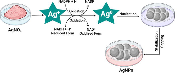
Bottom-up strategies used by bacteria to synthesize AgNPs from small building blocks like atoms and molecules using redox reactions have been shown in Figure 3. The biogenic reduction of silver ions takes place at or near ambient pressure, temperature, and pH. Silver ions are trapped, reduced, and capped, and crystals are stabilized as part of the procedure. According to certain theories, in the event of intracellular synthesis, silver ions are brought into the cell for reduction into elemental silver [32].
Figure 3.
Mechanism of AgNP production by bacteria mediated via NADH dependent nitrate reductase enzyme created with BioRender.com [32].
Some bacteria produce a transmembrane proton gradient that is actively supported by Na and Ag ions from the extracellular environment. The uptake of silver ions inside the cell and the initiation of AgNP synthesis are caused by a number of silver-binding membrane proteins that draw silver ions and use the energy from adenosine triphosphate hydrolysis to do so. The cell secretes extracellular polymeric materials made of proteins and polysaccharides that can bind to and capture ions. Reducing substances including proteins, enzymes, carbohydrates, and amino acids that are also secreted from the cells work to lower the trapped ions outside the cells. Only a few studies have demonstrated the importance of nicotinamide adenine dinucleotide (NAD) + hydrogen (H) NADH-dependent reductase in the synthesis of AgNPs, where NADH-H+ contributes its electrons to silver ions, which are then reduced to their elemental form and accumulate as AgNPs [32].
3.3 Synthesis of AuNPs by traditional route
3.3.1 By chemical method
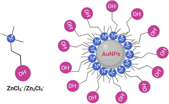
Choline chloride and ZnCl2, which have comparable physical and chemical properties, were combined to create the quaternary ammonium-based room temperature ionic liquids (QAILs). The QAILs were prepared by mixing choline chloride and ZnCl2 in a molar ratio of 1:1 (QAIL-I) and 1:2 (QAIL-II), which have similar physical and chemical properties. The QAILs were subjected to an overnight hoover treatment at 90°C to get rid of moisture and volatile contaminants before use. The liquids can be heated to at least 190°C and are at least as thermally stable as AlCl3-based liquids. Deionized water was used to dissolve HAuCl4 in order to create a 10.0 mmol l−1Au3+ solution. A series of QAIL stabilized AuNP colloids were created by adding 0.2 mL of Au3+ solution drop-by-drop into 10.0 mL of QAILs at various temperatures, with the Au3+ content held constant at 0.2 mmol l−1. These freshly generated AuNP colloids were then immediately supplemented with an additional 0.2 mL of Au3+ while being kept at the same temperature. For 15 min for each specimen, the QAILs and AuNP colloids were heated in an oil-bath heating reactor while being rapidly agitated in a glass beaker. For additional measurements, the AuNPs were centrifuged to methanol with a dilution ratio of 1:4 after the QAIL-AuNP colloid specimens had been separated with methanol [33].
3.3.1.1 Synthesis mechanism
The two-step process of nucleation and subsequent development of the particles is the most widely acknowledged mechanism for the formation of metal nanoparticles. The metal cores are gradually accumulated by the freshly reduced atoms, and occasionally cluster stabilizers participate in the reduction of some of the metal ions to zero valency metal nanoparticles. An organic cation and an inorganic anion form the structure of the QAIL given in Figure 4 [33].
Figure 4.
Molecular structures of QAILs and stabilized AuNPs created with BioRender.com [33].
3.4 Synthesis of AuNPs by green route
3.4.1 By phytochemical method
The plant leaf (Magnolia kobus) broth solution was made by boiling 5 g of fully cleaned, finely chopped leaves in 100 mL of pH 7.0 phosphate buffered saline for 5 min and then decanting the solution. 1 mM KAuCl4 was added to the leaf broth to reduce the Au3+ ions. By conducting the reaction in a water bath at 95°C with reflux, the effects of temperature on the synthesis rate and particle size/shape of the produced AuNPs were investigated. By repeatedly centrifuging the AuNPs solution at 15,000 rpm for 20 min and redispersing the pellet in deionized water, the AuNPs solution was purified [34].
3.4.2 By using microorganisms
Pseudomonas aeuroginosa is obtained from the Department of Microbiology and Immunology in Egypt. Burns are the source of two clinical samples of bacterial isolates used in this investigation. The isolates were identified as Pseudomonas aeruginosa ATCC 90271 by microbiology and biochemistry and were utilized as a reference strain [35].
Pseudomonas aeruginosa control strains, and the two isolates were used. At 37°C, the bacteria were incubated while growing aerobically in a 50 mL nutritional broth medium. It was shaken for 24 h at 150 rpm. After the incubation, the overnight bacterial culture was centrifuged at 5000 rpm for 5 min to get the supernatants. In order to create AuNPs, 50 mL of cell-free supernatant was combined with hydrogen tetrachloroaurate to produce a final concentration of gold ions of 1 mM. The resulting solution was then incubated at 37°C for 24 h. The cell-free supernatant containing nanoparticles was recovered after 24 h of incubation [35].
3.5 Synthesis of CuNPs by traditional route
3.5.1 By chemical method
Copper(II) sulfate pentahydrate was used as a precursor salt in the chemical reduction procedure to prepare the Cu nanoparticles, and starch was used as a capping agent. The first step in the preparation process is to add 120 mL of starch (1.2%) solution to a 0.1 M copper(II) sulfate pentahydrate solution while vigorously stirring for 30 min. The synthesis solution is mixed rapidly and continuously as 50 mL of a 0.2 M ascorbic acid solution is added in the second stage. The resulting solution was then progressively supplemented with 30 mL of a 1 M sodium hydroxide solution while being heated at 80°C for 2 h. The color of solution was changed from yellow to brown. After the reaction was finished, the solution was removed from the heat and allowed to settle for the night. The next day, the supernatant solution was carefully thrown away. Filtration was used to separate the precipitates from the mixture, and the excess starch linked to the nanoparticles was removed by washing the precipitates three times with deionized water and ethanol. Precipitates of brown color were produced, and they were dried at room temperature. Nanoparticles were stored in glass vials for later examination after drying [36].
3.6 Synthesis of CuNPs by green route
3.6.1 By phytochemical method
The Kigelia Africana fruits were properly washed, cut into slices, and sun-dried for 5 days to ensure that all moisture was removed. After that, the fruits were pulverized using a mechanical grinder. In order to perform the extraction, 50 g of the powdered plant fruit were added to a 1000 mL beaker along with 500 mL of ethanol. To achieve thorough extraction, the beaker holding the ethanol and powdered fruit was kept for 48 h while being repeatedly stirred with a magnetic stirrer every day. After filtering, the extract was put into an airtight container for storage [37].
3.6.2 By using microorganisms
Using pseudomonas fluorescens, copper sulfate (CuSO4) solution was treated separately with cell pellet and cell-free supernatant to prepare CuNPs. The culture was centrifuged at 7500 rpm for 20 min at 30°C after 48 h, and both the cell pellet and the cell-free culture supernatant were collected and used separately. 1 g of biomass was separately suspended in 20 mL of solutions containing 318, 750, and 1000 ppm (CuSO4). The preparation of CuNP was assessed after supernatant had been collected, centrifuged at 10,000 rpm for 20 min at 30°C. Varied concentrations of CuSO4 solution were mixed with varied volumes of cell-free supernatant, and the mixture was shaken in an incubator for 24–48 h at 30°C and 150 rpm [38].
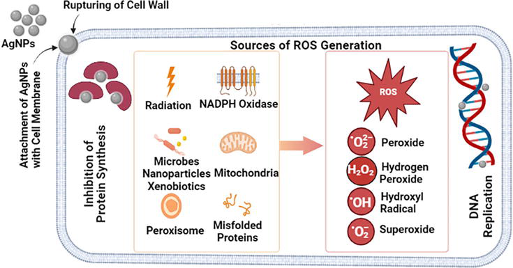
The precise cause of AgNPs’ antibacterial impact on bacteria is yet unclear. We describe a potential mechanism of AgNP action in Figure 5 that could lead to antibacterial activity. AgNPs continually release silver ions, which might be a method for destroying microorganisms. Since silver ions are more intimately related to sulfur proteins and are likewise attracted to them electrostatically, they can readily cling to the cytoplasmic membrane and cell wall. Silver ions attaching to the cell wall or cytoplasmic membrane boost the permeability of the cell, which ultimately results in cell breakdown, and disrupt the bacterial envelope at the same time. Free silver ions disable respiratory enzymes as they enter cells, producing reactive oxygen species that stop the synthesis of adenosine triphosphate. The primary species that initiates deoxyribonucleic acid (DNA) alteration and cell membrane rupture is reactive oxygen species (ROS). Phosphorus and sulfur are crucial elements in DNA. Even yet, the interaction of AgNPs with the sulfur and phosphorus in DNA can make it difficult to replicate DNA and support new cells, or even cause bacteria to die. Because the silver ions can prevent protein production, the ribosome can occasionally get denaturated in the cytoplasm [39].
Figure 5.
Action mechanism of AgNPs against the bacteria and the biofilm created with BioRender.com [39].
4.1.2 Mechanism of action against fungus
The size, shape, and coating agents have a significant impact on the antifungal activity of biogenic AgNPs. Deciphering a single mechanism of action is particularly challenging due to the large diversity of biogenic AgNPs. For this reason, the majority of the present study has concentrated on figuring out how chemically synthesized AgNPs work, which is attributed to the AgNPs adhering to the surface of the fungus due to electrostatic attraction as presented in Figure 6. Ag+ is dynamically released from extracellular AgNP accumulation, enters the cell, and increases the intracellular concentration as well as the intracellular manufacture of AgNPs. No cell receptors or membrane channels for the absorption of silver have been identified to yet. However, it has been discovered that Ag+ is imported through the high-affinity copper transporter (Ctr1) [40].
Figure 6.
Action mechanism of AgNPs against fungi [40].
4.1.3 Mechanism of action against cancer cells
Apoptosis, or programmed cell death, is avoided by cancerous cells, which keep multiplying. The development of cancer therapies is mostly focused on the aforementioned characteristics of cancer cells. To effectively combat cancer, plant-based nanosized silver is gaining popularity. There are two signaling pathways—intrinsic and extrinsic—that can be activated to cause apoptosis. Apoptosis, which is a limiting process in malignant cells, is triggered by DNA damage or extreme cell stress [41].
Beginning with various steps of apoptotic protein activation, DNA damage, mitochondrial disintegration, the development of an apoptosome, and finally cell shrinkage, apoptosis is a natural process in all living things. These turn into the most crucial targets to be used in cancer therapy. When applied to certain target locations, AgNP exhibits anticancer action. According to recent studies, AgNPs primarily affects ROS, upping oxidative stress and DNA synthesis. Normal cellular homeostasis, which is essential for cell survival, is maintained by ROS [42].
4.2 Applications of AuNPs
4.2.1 Mechanism of action against bacteria
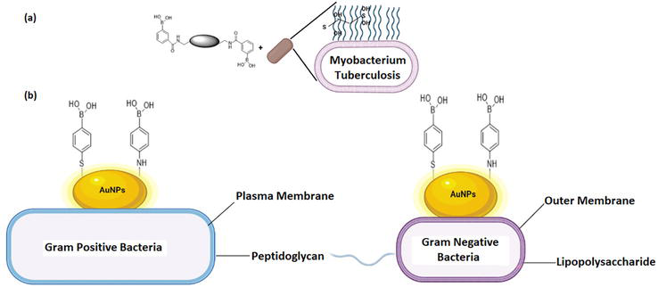
In order to prepare effective functionalized metallic nanoparticles, it is important to take into account that Gram-positive and Gram-negative bacteria have different cell wall and membrane structures [43]. The boronic acid group of phenylboronic acid is highly bound by the glycolipid-containing bacterial cell membrane presented in Figure 7(a) [44]. As a result of interaction with lipoteichoic acid and lipopolysaccharide of Gram-positive and Gram-negative bacteria, respectively, AuNPs were changed with thiol- and amine-tethered phenylboronic acids. Nanoparticles have been prepared and modified using natural substances derived from bacteria, fungus, and plants [46]. For instance, sildenafil citrate, geniposidic acid, 3,5-dimethylphenol, palmitic acid, borneol, 2-hexyl-1-octanol, and -terpinyl acetate were used as phytochemicals in an aqueous leaf extract from Acalypha indica that was used to synthesize spherical AuNPs with a 20 nm size shown in Figure 7(b) [45].
Figure 7.
(a) Multimeric boronic acid to target cell-envelope glycans of Mycobacterium tuberculosis [44] (b) functionalization of the AuNPs by amine- and thioltethered phenylboronic acids to target Gram-positive and Gram-negative bacteria [45].
4.2.2 Antifungal activity
AuNPs produced from cyanobacteria have been shown to have antifungal action; however, this activity was nanoparticle-size dependent. For instance, AuNPs of size 7 nm were more effective than those of size 15 nm. Candida was more effectively controlled by 25 nm particles than by 30 nm particles in this situation. Additionally, the entire AuNPs test had demonstrated action based on nanoparticle dose or concentration. The primary reason for the antifungal effect of AuNPs was the potential interaction with fungal protein. The metabolically active H+- adenosine triphosphatase enzyme in fungi is interacted with by AuNPs, which changes the enzyme’s usual alignment and causes a loss of fungi activity. This disturbance in metabolic activity prevents fungi from absorbing nutrients, which ultimately results in fungi cell death. Furthermore, the efficient antifungal activity may have been caused by a unique interaction involving shapes and sizes between AuNPs and the components of the plasma membrane, such as sulfur-containing proteins or phosphorus of bases in DNA. AuNPs impair normal processes like as synthesis, replication, and repair, which results in cell death [47].
4.2.3 Antioxidant activity
Recently, AuNPs have been considered a top choice for delivering a range of drugs to their intended locations. These payloads range in size from tiny drug molecules to bigger macromolecules including proteins, ribonucleic acid, and DNA [48]. The oxidative stress brought on by ROS such hydroxyl, epoxyl, peroxylnitrile, superoxide, and singlet oxygen plays a significant role in a variety of pathological disorders, such as inflammatory processes, atherosclerosis, aging, cancer, and neurological illnesses. Nucleic acid damage and enzyme inactivation are caused by excess ROS or oxidative stress that affects the host’s antioxidant system [49].
4.3 Applications of CuONPs
4.3.1 Antibacterail activity
The size of nanoparticles is important in many applications, including medicine delivery. The surface area that may be targeted for medication release increases with decreasing particle size. Smaller nanoparticles have greater mobility and the capability to enter and move about within the bacterial cell compartments, which makes their average size crucial for copper oxide nanoparticles (CuONPs’) potential antibacterial action. As a result, they are powerful antibacterial agents. However, various types of anionic compounds, such as organic acids, albumins, surfactants, polymers, and others, can easily prevent electrostatic adhesion from occurring in a solution. This phenomenon influences how different biomolecules, such as proteins and carbohydrates, which can adsorb on nanoparticles and produce a corona with different surface properties from the original nanoparticles, interact with the nanoparticles. This is most likely the cause that in biological fluids, formulations containing anionic polyelectrolytes, and surfactant-containing formulations, CuONPs can rapidly lose their antibacterial copper oxide efficacy [50].
One putative antibacterial mechanism is the release of free Cu2+ ions from CuONPs, which may interact with cell membrane proteins. The concentration of free Cu2+ ions in the aqueous solution around the CuONPs is, however, incredibly low due to its exceedingly poor solubility. Although the solubility of CuO varies with pH, it is around 3 × 10⁵ M in pure water. This does not adequately explain the antibacterial action of CuONPs, which increases with concentration whereas CuO solubility remains constant at set pH and temperature [51].
4.3.2 Antifungal activity of CuONPs
Microorganisms can grow with harmful metals through certain mechanisms, which can result in the synthesis of nanoparticles as a consequence of reduction mechanisms. This field of nanobiotechnology is still in its infancy. 25 mL of CuSO4. 5H2O (10 mM) was combined with a cell-free supernatant after being heated at 100°C for 15 min. A color change was then noticed since these bacteria have a tendency to create enzymes that diminish the poisonous metal, resulting in the formation of CuO nanoparticles. Additional characteristics using XRD and TEM revealed that the average size of nanoparticle, which may have antibacterial properties, is 61.7 nm [52].
Copper nanoparticles (CuONPs) in the 10–190 nm range were produced using a fungal-mediated, cell-free extract of Trichoderma asperellum, and IR spectroscopy revealed that the amide and aromatic groups of the secondary metabolite act as an encapsulating or reducing agent. This nanoparticle was also used to study in vitro photothermal-induced therapy utilizing human lung carcinoma-A549 cancer cells. The fungus Agaricus bisporus produced CuNPs with a size range of 2–10 nm that exhibited antifungal activity [52].
4.3.3 Antioxidant activity
Complete Freund’s adjuvant, a substance that mimics the progression of arthritic disease in humans, showed improved antioxidant enzymes and a decline in pro-inflammatory markers. In rats with induced arthritis, anti-inflammatory and anti-arthritic potentials were seen. Radical scavenging of 2,2-diphenyl-1-picryl-hydrazyl-hydrate (DPPH) is used to evaluate the DPPH radical-scavenging operation; the Shimada technique is applied. A 5 mL sample solution was mixed with an aliquot of DPPH methanol solution (20, 40, 60, and 80 g/mL) in 2 mL at various concentrations. After thoroughly shaking the mixture, it was let to stand at room temperature in the dark for 30 minutes. The absorbance was then measured in a spectrophotometer at 517 nm. The reaction mixture’s lower absorbance showed a higher ability to scavenge free radicals [53].
4.4 Silver, gold, and copper nanoparticles used in biomedical applications
Silver, gold, and copper nanoparticles play crucial roles in advancing biomedical applications. Their unique properties, such as antimicrobial activity, optical properties, and biocompatibility, make them versatile tools in various fields within biomedicine. As research continues to unveil their potential benefits and address associated challenges, these nanoparticles could revolutionize diagnostics, therapy, and healthcare delivery, ultimately leading to improved patient outcomes and enhanced medical technologies.
Different forms of nanoparticles, their size ranges, and application in biomedical field have been presented in Table 2.
5. Challenges and prospective applications of green nanoparticles
The synthesis of green NPs and their applications is an area of significant interest due to the growing demand for sustainable and environmentally friendly technologies. Green NPs refer to NPs that are synthesized using eco-friendly and biocompatible methods, often utilizing natural sources and reducing the use of hazardous chemicals. Here are some challenges and perspectives for the synthesis of green NPs and their applications:
5.1 Challenges
Standardization of synthesis methods: Green nanoparticle synthesis methods often involve natural extracts, biomolecules, and bio-inspired approaches [67]. Standardizing these methods to ensure consistent nanoparticle size, shape, and properties can be challenging due to the variability of natural sources.
Control over nanoparticle properties: Achieving precise control over the size, shape, and composition of green nanoparticles can be difficult, which may impact their performance in various applications [68].
Scale-up and production: Many green synthesis methods are conducted in small batches [69]. Scaling up these methods to produce nanoparticles on a larger industrial scale while maintaining their green and sustainable nature presents technical and economic challenges.
Characterization techniques: Accurately characterizing green nanoparticles and understanding their properties require specialized techniques that might not be readily available or well-established.
Stability and shelf life: Ensuring the stability and shelf life of green nanoparticles can be challenging, as they may be susceptible to aggregation, degradation, or changes in their properties over time [70].
Regulatory considerations: New synthesis methods and applications for nanoparticles may raise regulatory concerns related to safety, toxicity, and environmental impact. Understanding and addressing these concerns is crucial for their successful implementation [71].
5.2 Perspectives
Biomedical applications: Green nanoparticles have promising applications in drug delivery, imaging, and targeted therapy due to their biocompatibility and reduced toxicity compared to traditional nanoparticles [72].
Agriculture and food industry: Green nanoparticles can be used for crop enhancement, pest control, and food preservation. They could replace conventional chemical-based approaches, reducing environmental impacts [73].
Environmental remediation: Green nanoparticles can be utilized for water purification, air filtration, and remediation of polluted environments, offering sustainable solutions to address pollution and contamination [74].
Energy conversion: Green nanoparticles can play a role in enhancing the efficiency of solar cells, catalytic processes, and energy storage devices, contributing to renewable energy technologies [75].
Electronics and optoelectronics: Green nanoparticles could find applications in flexible electronics, displays, and sensors due to their unique optical and electronic properties [76].
Nanomedicine: Green nanoparticles could be used in disease diagnosis, therapeutics, and imaging, opening up new possibilities for personalized medicine [77].
Collaborative research: Collaborations between scientists, engineers, biologists, chemists, and environmental experts are essential to address the multidisciplinary nature of green nanoparticle synthesis and applications [78].
Education and awareness: As green nanoparticles are relatively new, educating researchers, industries, and the public about their benefits, limitations, and responsible use is crucial for their successful integration into various sectors [79].
Overall, there are challenges associated with synthesizing green nanoparticles and applying them in various fields; the potential benefits in terms of sustainability, reduced environmental impact, and innovative applications make this area of research promising. Addressing the challenges through continued research, technological advancements, and collaborative efforts can lead to the development of practical and impactful green nanoparticle-based solutions.
Authors concluded that the research on the medicinal applications of Ag, Au, and CuNPs showed promising potential in various fields of medicine. Synthesis of Cu, Au, and AgNPs by using phytochemicals, microorganisms, and less toxic chemicals has been considered an effective and green approach. AgNPs possess strong antimicrobial properties and have been extensively studied for their potential in combating bacteria, viruses, and fungi. AgNPs have shown potential in cancer therapy due to their ability to induce apoptosis (cell death) in cancer cells and inhibit tumor growth. However, further research and clinical trials were needed to establish their efficacy and safety. AuNPs have been studied for their diagnostic and therapeutic applications in medicine. In cancer therapy, AuNPs have been explored as photothermal agents, using their ability to convert light into heat to destroy cancer cells selectively. CuNPs have also been studied for their antioxidant properties, indicating potential applications in reducing oxidative stress and inflammation-related conditions.
Authors are thankful to the manager of INTECHOPEN for providing opportunity to write a chapter for publication under book title “Advances in Green Chemistry: Catalysis and Renewable Feedstocks for Sustainable Chemical Synthesis.”
Authors declared that they have no potential conflict of interest.
References
1.Pryshchepa O, Pomastowski P, Buszewski B. Silver nanoparticles: Synthesis, investigation techniques, and properties. Advances in Colloid and Interface Science. 2020;284:102246
2.Yang S-E, Liu P, Zhang Y-J, Guo Q-N, Chen Y-S. Effects of silver nanoparticles size and shape on light scattering. Optik. 2016;127(14):5722-5728
3.Manivannan K, Cheng C-C, Anbazhagan R, Tsai H-C, Chen J-K. Fabrication of silver seeds and nanoparticle on core-shell Ag@ SiO2 nanohybrids for combined photothermal therapy and bioimaging. Journal of Colloid and Interface Science. 2019;537:604-614
4.Bansal SA, Kumar V, Karimi J, Singh AP, Kumar S. Role of gold nanoparticles in advanced biomedical applications. Nanoscale Advances. 2020;2(9):3764-3787
5.Klębowski B, Depciuch J, Parlińska-Wojtan M, Baran J. Applications of noble metal-based nanoparticles in medicine. International Journal of Molecular Sciences. 2018;19(12):4031
6.Utreja P, Verma S, Rahman M, Kumar L. Use of nanoparticles in medicine. Current Biochemical Engineering. 2020;6(1):7-24
7.Al-Khattaf FS. Gold and silver nanoparticles: Green synthesis, microbes, mechanism, factors, plant disease management and environmental risks. Saudi Journal of Biological Sciences. 2021;28(6):3624-3631
8.Hekmati M, Hasanirad S, Khaledi A, Esmaeili D. Green synthesis of silver nanoparticles using extracts of Allium rotundum l, Falcaria vulgaris Bernh, and Ferulago angulate Boiss, and their antimicrobial effects in vitro. Gene Reports. 2020;19:100589
9.Khalaj M, Kamali M, Costa MEV, Capela I. Green synthesis of nanomaterials-a scientometric assessment. Journal of Cleaner Production. 2020;267:122036
10.Pal K, Chakroborty S, Nath N. Limitations of nanomaterials insights in green chemistry sustainable route: Review on novel applications. Green Processing and Synthesis. 2022;11(1):951-964
11.Bruna T, Maldonado-Bravo F, Jara P, Caro N. Silver nanoparticles and their antibacterial applications. International Journal of Molecular Sciences. 2021;22(13):7202
12.Savary S, Willocquet L, Pethybridge SJ, Esker P, McRoberts N, Nelson A. The global burden of pathogens and pests on major food crops. Nature Ecology & Evolution. 2019;3(3):430-439
13.Shuping D, Eloff JN. The use of plants to protect plants and food against fungal pathogens: A review. African Journal of Traditional, Complementary and Alternative Medicines. 2017;14(4):120-127
14.Baldrian P, Větrovský T, Lepinay C, Kohout P. High-throughput sequencing view on the magnitude of global fungal diversity. Fungal Diversity. 2022;114(1):539-547
15.Salomoni R, Léo P, Montemor A, Rinaldi B, Rodrigues M. Antibacterial effect of silver nanoparticles in Pseudomonas aeruginosa. Nanotechnology, Science and Applications. 2017;10:115-121
16.Ismail M. Green synthesis and characterizations of copper nanoparticles. Materials Chemistry and Physics. 2020;240:122283
17.Malhotra N, Ger T-R, Uapipatanakul B, Huang J-C, Chen KH-C, Hsiao C-D. Review of copper and copper nanoparticle toxicity in fish. Nanomaterials. 2020;10(6):1126
18.Božić Cvijan B, Korać Jačić J, Bajčetić M. The impact of copper ions on the activity of antibiotic drugs. Molecules. 2023;28(13):5133
19.Wang P, Yuan Y, Xu K, Zhong H, Yang Y, Jin S, et al. Biological applications of copper-containing materials. Bioactive Materials. 2021;6(4):916-927
20.Manne R, Kumaradoss MMRM, Iska RSR, Devarajan A, Mekala N. Water quality and risk assessment of copper content in drinking water stored in copper container. Applied Water Science. 2022;12(3):27
21.Chen J, Jiang Y, Shi H, Peng Y, Fan X, Li C. The molecular mechanisms of copper metabolism and its roles in human diseases. Pflügers Archiv-European Journal of Physiology. 2020;472:1415-1429
22.Collins JF. Copper nutrition and biochemistry and human (patho) physiology. In: Advances in Food and Nutrition Research. Vol. 96. Academic Press; 2021. pp. 311-364
23.Nieto-Maldonado A, Bustos-Guadarrama S, Espinoza-Gomez H, Flores-López LZ, Ramirez-Acosta K, Alonso-Nuñez G, et al. Green synthesis of copper nanoparticles using different plant extracts and their antibacterial activity. Journal of Environmental Chemical Engineering. 2022;10(2):107130
24.Ermini ML, Voliani V. Antimicrobial nano-agents: The copper age. ACS Nano. 2021;15(4):6008-6029
25.Crisan CM, Mocan T, Manolea M, Lasca LI, Tăbăran F-A, Mocan L. Review on silver nanoparticles as a novel class of antibacterial solutions. Applied Sciences. 2021;11(3):1120
26.Bai X, Wang Y, Song Z, Feng Y, Chen Y, Zhang D, et al. The basic properties of gold nanoparticles and their applications in tumor diagnosis and treatment. International Journal of Molecular Sciences. 2020;21(7):2480
27.Chen H, Zheng X, Chen Y, Li M, Liu K, Li X. Influence of copper nanoparticles on the physical-chemical properties of activated sludge. PLoS One. 2014;9(3):e92871
28.Pareek V, Gupta R, Panwar J. Do physico-chemical properties of silver nanoparticles decide their interaction with biological media and bactericidal action? A review. Materials Science and Engineering: C. 2018;90:739-749
29.Kart HH, Yildirim H, Kart SO, Çağin T. Physical properties of Cu nanoparticles: A molecular dynamics study. Materials Chemistry and Physics. 2014;147(1-2):204-212
30.Garibo D, Borbón-Nuñez HA, de León JND, García Mendoza E, Estrada I, Toledano-Magaña Y, et al. Green synthesis of silver nanoparticles using Lysiloma acapulcensis exhibit high-antimicrobial activity. Scientific Reports. 2020;10(1):12805
31.Elbeshehy EK, Elazzazy AM, Aggelis G. Silver nanoparticles synthesis mediated by new isolates of Bacillus spp., nanoparticle characterization and their activity against bean yellow mosaic virus and human pathogens. Frontiers in Microbiology. 2015;6:453
32.Javaid A, Oloketuyi SF, Khan MM, Khan F. Diversity of bacterial synthesis of silver nanoparticles. BioNanoScience. 2018;8:43-59
33.Huang W, Chen S, Liu Y, Fu H, Wu G. The controlled synthesis of stable gold nanoparticles in quaternary ammonium ionic liquids by simple heating. Nanotechnology. 2010;22(2):025602
34.Li Y, Wu T-Y, Chen S-M, Ali MA, AlHemaid FM. Green synthesis and electrochemical characterizations of gold nanoparticles using leaf extract of Magnolia kobus. International Journal of Electrochemical Science. 2012;7(12):12742-12751
35.Singh PK, Kundu S. Biosynthesis of gold nanoparticles using bacteria. Proceedings of the National Academy of Sciences, India Section B: Biological Sciences. 2014;84:331-336
36.Khan A, Rashid A, Younas R, Chong R. A chemical reduction approach to the synthesis of copper nanoparticles. International Nano Letters. 2016;6:21-26
37.Alao II, Oyekunle IP, Iwuozor KO, Emenike EC. Green synthesis of copper nanoparticles and investigation of its anti-microbial properties. Advanced Journal of Chemistry-Section B. 2022;4(1):39-52
38.Shantkriti S, Rani P. Biological synthesis of copper nanoparticles using Pseudomonas fluorescens. International Journal of Current Microbiology and Applied Sciences. 2014;3(9):374-383
39.More PR, Pandit S, Filippis AD, Franci G, Mijakovic I, Galdiero M. Silver nanoparticles: Bactericidal and mechanistic approach against drug resistant pathogens. Microorganisms. 2023;11(2):369
40.Mussin J, Giusiano G. Biogenic silver nanoparticles as antifungal agents. Frontiers in Chemistry. 2022;10:1023542
41.Sathiya P, Geetha K. Fruit extract mediated synthesis of silver oxide nanoparticles using Dimocarpus longan fruit and their assesment of catalytic, antifungal, antioxidant and cytotoxic potentials. Inorganic and Nano-Metal Chemistry. 2021:1-11
42.Jain N, Jain P, Rajput D, Patil UK. Green synthesized plant-based silver nanoparticles: Therapeutic prospective for anticancer and antiviral activity. Micro and Nano Systems Letters. 2021;9(1):5
43.Alavi M, Hamblin MR, Martinez F, Kennedy JF, Khan H. Synergistic combinations of metal, metal oxide, or metalloid nanoparticles plus antibiotics against resistant and non-resistant bacteria. Micro Nano Bio Aspects. 2022;1(1):1-9
44.Guy CS, Gibson MI, Fullam E. Targeting extracellular glycans: Tuning multimeric boronic acids for pathogen-selective killing of Mycobacterium tuberculosis. Chemical Science. 2019;10(23):5935-5942
45.Alavi M, Kowalski R, Capasso R, Douglas Melo Coutinho H, Rose Alencar De Menezes I. Various novel strategies for functionalization of gold and silver nanoparticles to hinder drug-resistant bacteria and cancer cells. Micro Nano Bio Aspects. 2022;1(1):38-48
46.Alavi M, Adulrahman NA, Haleem AA, Al-Râwanduzi ADH, Khusro A, Abdelgawad MA, et al. Nanoformulations of curcumin and quercetin with silver nanoparticles for inactivation of bacteria. Cellular and Molecular Biology. 2021;67(5):151-156
47.Mandhata CP, Sahoo CR, Padhy RN. Biomedical applications of biosynthesized gold nanoparticles from cyanobacteria: An overview. Biological Trace Element Research. 2022;200(12):5307-5327
48.Fawad AA. Brief review on applications of gold and silver nanoparticles in drug delivery and DNA interaction. World Journal of Physics. 2023;1(1):5
49.Mikhailova EO. Gold nanoparticles: Biosynthesis and potential of biomedical application. Journal of Functional Biomaterials. 2021;12(4):70
50.Bhavyasree P, Xavier T. Green synthesised copper and copper oxide based nanomaterials using plant extracts and their application in antimicrobial activity. Current Research in Green and Sustainable Chemistry. 2022;5:100249
51.Halbus AF, Horozov TS, Paunov VN. Strongly enhanced antibacterial action of copper oxide nanoparticles with boronic acid surface functionality. ACS Applied Materials & Interfaces. 2019;11(13):12232-12243
52.Harishchandra BD, Pappuswamy M, Antony P, Shama G, Pragatheesh A, Arumugam VA, et al. Copper nanoparticles: A review on synthesis, characterization and applications. Asian Pacific Journal of Cancer Biology. 2020;5(4):201-210
53.Thakar MA, Jha SS, Phasinam K, Manne R, Qureshi Y, Babu VH. X ray diffraction (XRD) analysis and evaluation of antioxidant activity of copper oxide nanoparticles synthesized from leaf extract of Cissus vitiginea. Materials Today: Proceedings. 2022;51:319-324
54.Abdellatif AA, Rasheed Z, Alhowail AH, Alqasoumi A, Alsharidah M, Khan RA, et al. Silver citrate nanoparticles inhibit PMA-induced TNFα expression via deactivation of NF-κB activity in human cancer cell-lines, MCF-7. International Journal of Nanomedicine. 2020;15:8479-8493
55.Oviya P, Francis AP, Mahalaxmi B. Fabrication of hemocompatible chitosan-biogenic silver nanocomposite for biomedical applications. Journal of Drug Delivery Science and Technology. 2023;87:104826
56.Qamar S, Karim S, Aslam S, Jahangeer M, Nelofer R, Nadeem AA, et al. Alginate-based bio-nanohybrids with unique properties for biomedical applications. Starch-Stärke. 2022:2200100
57.Herizchi R, Abbasi E, Milani M, Akbarzadeh A. Current methods for synthesis of gold nanoparticles. Artificial Cells, Nanomedicine, and Biotechnology. 2016;44(2):596-602
58.Kolathupalayam Shanmugam B, Rajendran N, Arumugam K, Rangaraj S, Subramani K, Srinivasan S, et al. Curcumin loaded gold nanoparticles–chitosan/sodium alginate nanocomposite for nanotheranostic applications. Journal of Biomaterials Science, Polymer Edition. 2023;34(7):875-892
59.Zhang NMY, Qi M, Wang Z, Wang Z, Chen M, Li K, et al. One-step synthesis of cyclodextrin-capped gold nanoparticles for ultra-sensitive and highly-integrated plasmonic biosensors. Sensors and Actuators B: Chemical. 2019;286:429-436
60.Akintelu SA, Folorunso AS, Folorunso FA, Oyebamiji AK. Green synthesis of copper oxide nanoparticles for biomedical application and environmental remediation. Heliyon. 2020;6(7):e04508
61.Roy S, Rhim J-W. Gelatin-based film integrated with copper sulfide nanoparticles for active packaging applications. Applied Sciences. 2021;11(14):6307
62.Mostafalu P, Sonkusale S. A high-density nanowire electrode on paper for biomedical applications. RSC Advances. 2015;5(12):8680-8687
63.Din MI, Rehan R. Synthesis, characterization, and applications of copper nanoparticles. Analytical Letters. 2017;50(1):50-62
64.Woźniak-Budych MJ, Langer K, Peplińska B, Przysiecka Ł, Jarek M, Jarzębski M, et al. Copper-gold nanoparticles: Fabrication, characteristic and application as drug carriers. Materials Chemistry and Physics. 2016;179:242-253
65.Covarrubias C, Trepiana D, Corral C. Synthesis of hybrid copper-chitosan nanoparticles with antibacterial activity against cariogenic Streptococcus mutans. Dental Materials Journal. 2018;37(3):379-384
66.Pavithra CL, Sarada BV, Rajulapati KV, Rao TN, Sundararajan G. A new electrochemical approach for the synthesis of copper-graphene nanocomposite foils with high hardness. Scientific Reports. 2014;4(1):4049
67.Tidke PR, Gupta I, Gade AK, Rai M. Fungus-mediated synthesis of gold nanoparticles and standardization of parameters for its biosynthesis. IEEE Transactions on Nanobioscience. 2014;13(4):397-402
68.Hui Y, Yi X, Hou F, Wibowo D, Zhang F, Zhao D, et al. Role of nanoparticle mechanical properties in cancer drug delivery. ACS Nano. 2019;13(7):7410-7424
69.Tighe CJ, Cabrera RQ , Gruar RI, Darr JA. Scale up production of nanoparticles: Continuous supercritical water synthesis of Ce–Zn oxides. Industrial & Engineering Chemistry Research. 2013;52(16):5522-5528
70.Zhang W. Nanoparticle aggregation: Principles and modeling. In: Nanomaterial: Impacts on Cell Biology and Medicine. 2014. pp. 19-43
71.Fard JK, Jafari S, Eghbal MA. A review of molecular mechanisms involved in toxicity of nanoparticles. Advanced Pharmaceutical Bulletin. 2015;5(4):447
72.McNamara K, Tofail SA. Nanoparticles in biomedical applications. Advances in Physics: X. 2017;2(1):54-88
73.Khodakovskaya M, Marmiroli M. Polymeric nanoparticles for sustainable plant agriculture and food industry. Frontiers in Plant Science. 2023;14:1183938
74.Rafeeq H, Hussain A, Ambreen A, Waqas M, Bilal M, Iqbal HM. Functionalized nanoparticles and their environmental remediation potential: A review. Journal of Nanostructure in Chemistry. 2022;12(6):1007-1031
75.Kumar A, Choudhary P, Kumar A, Camargo PH, Krishnan V. Recent advances in plasmonic photocatalysis based on TiO2 and noble metal nanoparticles for energy conversion, environmental remediation, and organic synthesis. Small. 2022;18(1):2101638
76.MacFarlane LR, Shaikh H, Garcia-Hernandez JD, Vespa M, Fukui T, Manners I. Functional nanoparticles through π-conjugated polymer self-assembly. Nature Reviews Materials. 2021;6(1):7-26
77.Perciani CT, Liu LY, Wood L, MacParland SA. Enhancing immunity with nanomedicine: Employing nanoparticles to harness the immune system. ACS Nano. 2020;15(1):7-20
78.Grabbe S, Landfester K, Schuppan D, Barz M, Zentel R. Nanoparticles and the immune system: Challenges and opportunities. Nanomedicine. 2016;11(20):2621-2624
79.Usmani A, Dash PP, Mishra A. Metallic nanoformulations: Green synthetic approach for advanced drug delivery. Materials Science: Advanced Composite Materials. 2018;2(2):1-4
Written By
Faisal Nawaz, Irum Jamil and Bushra Shakoor
Submitted: 03 August 2023Reviewed: 09 August 2023Published: 09 November 2023
 Open access peer-reviewed chapter